


1. Introduction Arguments
A 44-year-old male presents with shortness of breath, non-productive cough, and chest tightness to the ski clinic in Winter Park, Colorado. He flew from his home town, San Antonio, Texas, three days earlier. Symptoms began the day after arrival and progressively worsened over the course of the last 2 days. Shortness of breath is exacerbated by physical activity and relieved by rest. He tried his son’s albuterol inhaler with no improvement in his symptoms. He denied fevers, chills, and sick contacts. He is in excellent shape and has never experienced these symptoms before despite many similar ski trips. His past medical, family, and social histories are unremarkable, and he takes no medications. On arrival he appears generally healthy, but exhibits significantly labored breathing. He is afebrile and normotensive, but tachycardic (104 BPM) and hypoxic with an oxygen saturation of 61% on room air. His lung examination is significant for diffuse rales. Chest radiography was performed (Figure 1).
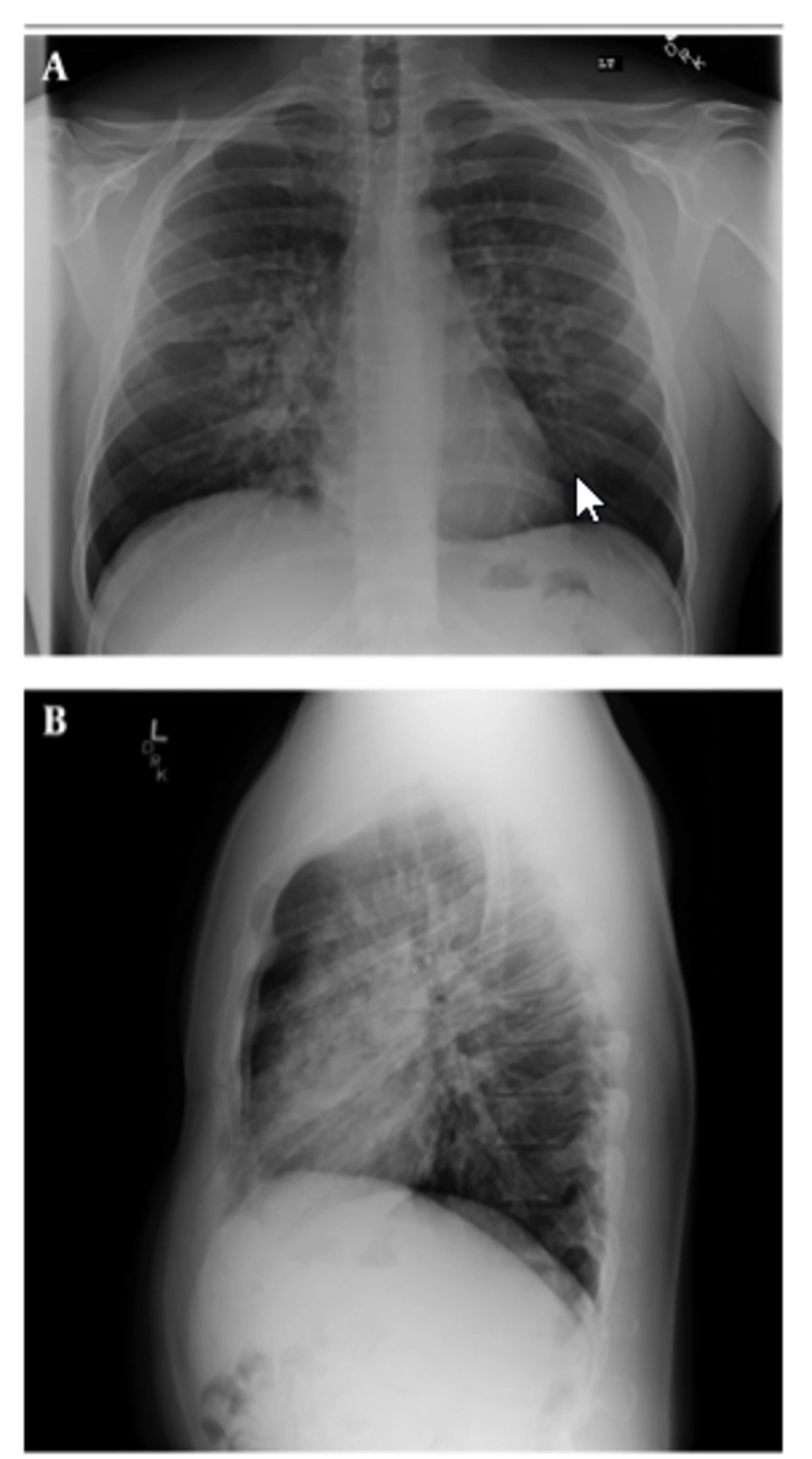
Figure 1.
Chest Radiography
2. Arguments Against
Based on the patient's history, physical examination, and radiographs, which one of the following is the most likely diagnosis?
- Atypical pneumonia
- Pulmonary embolism
- High altitude pulmonary edema
- Exercise induced bronchospasm
- Pneumothorax
3. Conclusions
The answer is C: High altitude pulmonary edema (HAPE) is the leading cause of altitude-related death (1). It is characterized by non-cardiogenic pulmonary edema occurring with rapid ascent to altitudes above 2,500 m (1-3). This patient travelled from San Antonio, TX with altitude of 235 m to Winter Park, CO with altitude of 2,800 m. Risk factors for development of HAPE include prior history of altitude illness, higher altitude, rapid ascent, male sex, cold ambient temperatures, pre-existing respiratory infections, and intense exercise (1-3). Estimates of HAPE incidence vary, but have been reported as high as 4% in un-acclimatized mountaineers above 4,600 m (2). Onset of HAPE typically occurs within 1-4 days of ascent to altitude (3). Symptoms include fatigue, dyspnea on exertion, decreased exercise tolerance, low-grade fever, and dry cough which sometimes progresses to become productive of pink, frothy, sputum (2, 3). On physical examination, patients with HAPE are tachycardic, tachypneic, and hypoxic, frequently with low-grade fevers (1-3). Pulmonary examination reveals inspiratory crackles, initially in the right middle lobe, and becoming more diffuse and bilateral later in the illness (1-3). Chest x-ray (CXR) classically shows patchy perihilar opacities, sometimes with interstitial edema and pleural effusions. Heart size is normal, which is consistent with no cardiogenic pulmonary edema (3). The cornerstones of HAPE treatment are immediate descent to lower altitude and supplemental oxygen (1-3). If descent is not possible or supplemental oxygen not available, pharmacotherapy with nifedipine may be considered as the next line of treatment, with the goal being to decrease pulmonary artery pressure (1, 3) Several studies have suggested benefit with long acting beta agonists (e.g. salmeterol) and with phosphodiesterase-5 inhibitors, but no strong evidence for either exists (1, 3). Similarly, HAPE is best prevented by gradual ascent to altitude (1-3). Pharmacologic prophylaxis can be considered, particularly in those with a history of HAPE, with nifedipine as the first-line agent and other potential options including phosphodiesterase-5 inhibitors, long acting beta agonists, and dexamethasone (1-3).
Atypical organisms account for up to 40% of community-acquired pneumonia (4). Patients usually present with fevers and productive cough (4). Lung examination may reveal rales and wheezing (4). Hypoxia is rare (4). CXR may reveal patchy or circumscribed opacities (4). Pulmonary embolism (PE) may present with a sudden episode of SOB at rest (5). CXR is non-diagnostic in patients with PE and most commonly shows atelectasis, non-specific parenchymal abnormalities, or pleural effusions (5). Less commonly, the classic signs of a peripheral wedge-shaped opacity may be seen (5). Exercise-induced bronchospasm causes dyspnea and chest tightness during or after exercise (6). Physical examination is usually unremarkable with occasional wheezing (6). CXR is usually normal or may show increased bronchial wall thickening or flattening of the diaphragm (6). Pneumothorax is a relatively common condition among adults and can present with dyspnea and pleuritic chest pain (5). Physical examination typically reveals decreased chest excursion as well as decreased breath sounds on the affected side (5). CXR usually demonstrates a visceral pleural line, beyond which no pulmonary markings are present and possible mediastinal shift (5, 7).
Table 1.Summary table
| Condition | Characteristics |
|---|---|
| Atypical pneumonia | Fever, productive cough; unilateral or bilateral heterogeneous, patchy, reticular, segmental, peribronchial opacifications on CXR a |
| Pulmonary embolism | Acute onset of cough, dyspnea, tachycardia, tachypnea, pleuretic chest pain, and hypoxia; CXR is usually normal, but a wedge-shaped opacity of the lung tissue may be present |
| High altitude pulmonary edema | Caused by rapid ascent to high altitude above 2,500m; cough, dyspnea, hypoxia; dense bilateral patchy opacities in variable locations |
| Exercise induced bronchospasm | Cough, dyspnea, and wheezing triggered by exercise; CXR is usually normal |
| Pneumothorax | Acute onset of dyspnea, pleuretic chest pain, diminished breath sounds; CXR may reveal displacement of visceral pleura and absence of pulmonary markings |
a Chest x-ray.

