1. Background
Flat foot is a common foot deformity which describes as a decrease in medial longitudinal arch (MLA) height of the foot (1). The difference of MLA height between individuals with normal and flat feet has an effect on various kinetic and kinematic parameters of locomotion (2) which might increase the risk of injury (3). On the other hand, there are some studies which have reported no significant difference between normal and flat footed subjects in their kinetic parameters and susceptibility to injury (4, 5).
Center of pressure (CoP) is one of the kinetic-related variables which can present the whole body balance dynamics. As the foot determines the base of support geometry and dynamic variability of CoP, foot problems can affect CoP path and parameters (6-9). Furthermore, as CoP is the mean of all pressures applied to the plantar surface of the foot, any deformity in foot shape can alter its path and parameters (8, 10, 11). Previous literature found a significant difference in CoP course and plantar pressure distribution between individuals with normal and flat feet during standing posture, walking and running (6-8, 10, 12, 13). A less plantar pressure in 4th and 5th metatarsal heads and heel region (6), a higher pressure distribution under the cuboid bone (7) and a more medially deviated CoP (8) were reported in individuals with flat feet compared to the normal ones during walking.
Fatigue is one of the major factors that could alter biomechanical parameters and risk of injury (14-16). Fatigue effect on CoP course and plantar pressure distribution in individuals with normal feet was studied previously in different situations of standing, walking and running (14, 16, 17). A more posterior displacement of CoP course was reported after fatigue during walking (16).
2. Objectives
Although the fatigue effect in normal subjects has been studied before, to our knowledge, there is no literature examining the fatigue effect on CoP measures between individuals with normal and flat feet. It was previously suggested that studying individuals with normal and flat feet under high load tasks or fatiguing condition could indicate the effect of dissimilar MLA shape more precisely (18). Therefore, we tested the hypothesis that a functional fatigue task changes the linear CoP parameters between the two groups of normal and flat footed subjects. Since individuals with flat feet often suffer from pain caused by fatigue caused by walking or long time standing during the day, the functional fatigue protocol was used in this study to mimic the kind of fatigue that happens in the real life and not just in controlled laboratory conditions. As walking is a frequent daily activity and the most common type of locomotion, CoP parameters were compared during a walking task.
3. Methods
3.1. Subjects
The experimental data used for the current analyses was adopted from our previous study. Subjects in this study participated as part of a larger study examining the fatigue effect on gait of individuals with flat feet (19). Thirty-four women, 17 with normal feet and 17 with flexible flat feet, were studied. A complete clinical screening was performed for all subjects to exclude any visible deformities in the lower extremity structure. Subjects had no history of lower limb surgery or any orthopedic, neurologic or rheumatologic disorders. Moreover, subjects had no pain in any parts of the body during the test. The number of subjects entered in each group was calculated using the sample size formula and the pilot data (α = 0.05 and β = 0.2). All participants were matched according to their height and weight. Participants were informed at the time of recruitment that the experiment would evaluate the fatigue effect on walking parameters. Each subject was free to leave the study at any time. All subjects signed an informed-consent form approved by the Iran University of Medical Sciences ethics committee. This study was also registered in the Iranian registry of clinical trials. The experiment was conducted from June to September 2010 in the rehabilitation research center of Iran University of Medical Sciences.
3.2. Foot Type Classification
Subjects were classified as normal or flat footed according to their arch height ratio (AHR). AHR is a reliable and valid method for classifying the foot posture in bilateral standing (20). This method assumes 1.5 SD below or above the mean value to identify the individuals with flat and normal feet (20). Using 1.5 SD rather than 1 SD below the mean, could provide samples with more extreme differences in their arch heights and probable less mechanical adaptation (2).
A custom made device according to McPoil et al.’s (20) study was used in this study to measure the AHR of subjects. AHR is defined as a division of dorsum height to the total foot length. Total foot length is a distance between the posterior aspect of the calcaneus bone to the tip of the longest toe. Dorsum height was measured at the midpoint of the total foot length. Prior to the main study, the intratester reliability of this device was assessed in 3 weight-bearing conditions at 3 different times. ICC values of more than 0.91 were obtained for all conditions (21).
3.3. Test Procedure
Each subject walked barefoot across the two force plates before and after a fatigue protocol. After a familiarization phase subjects started walking five steps away from the force plates and walked with their preferred walking speed over the force plates.
The fatigue protocol used in this study was a functional fatigue protocol including 5 sets of consequent, side to side lateral hops to the rhythm of a metronome (108 beep/min). There was 30s of walking as rest between each set. Failure to follow the metronome pace for 5 consecutive hops or inability to continue the test was considered as the end of each set. This protocol was based on a fatigue protocol developed in Hoch’s thesis (22). Functional fatigue protocols such as jumping or running consist several sets of stretch shortening cycles (SSC)s. Previous literature showed that SSCs are the natural function of muscles in real life (23). To quantify the amount of fatigue induced in each subject, a 15-grade Borg Scale was used. Borg scale is a tool to measure the rating of perceived exertion (24). The level 15 of the Borg Scale is correlated with at least 75% of VO2max and used to quantify the amount of fatigue in central fatigue protocols (25). Therefore, each subject had to mark at least level 15 of the Borg scale after the fatigue protocol to be considered fatigued.
Immediately after the fatigue protocol, subjects walked across the force plates for three times. All the three trials were performed in less than 60 seconds to prevent recovery from fatigue. The first appropriate trial in which each foot landed separately on each force plate was selected for the statistical analyses.
3.4. Data Extraction and Statistical Analysis
Unlike various CoP parameters in quiet standing posture, the CoP parameters in walking are few. CoP position data were obtained from a strain gauge Bertec 4060-10 force plate and a Bertec AM-6701 amplifier (Bertec Corp, Columbus, OH, USA) with a sampling frequency of 200 Hz. Data were filtered using a dual-pass second order Butterworth filter with a cut-off frequency of 15 Hz.
The independent variables of this study were foot type (normal and flat feet) and fatiguing condition (before and after fatigue). The dependent variables were all linear parameters extracted from CoP data. Linear parameters are easy to calculate and more understandable and interpretable than non-linear parameters. Standard deviation of CoP in mediolateral direction (SD of CoPx) and in anteroposterior direction (SD of CoPy), overall mean velocity of CoP and length of CoP construction line were extracted from CoP data of the left foot. In this study, the left side was the non-dominant foot in all subjects. Since less muscle torque and muscle thickness on the non-dominant side have been shown previously, non-dominant leg data was used to elucidate even a small fatigue effect (26, 27).
CoP construction line is a line attached from the initial to the final point of CoP excursion path. To avoid bias due to anatomical differences, the value of SD of CoPy and length of CoP construction line were normalized to individual foot lengths prior to the statistical analysis.
The normality of data distributions was checked using the Kolmogorov-Smirnov test with the Lilliefors correction. For normally distributed data, a repeated-measure ANOVA approach was employed. There was one within-subject factor (before and after fatigue) and one between-subjects factor (normal and flat feet). A Levene’s test of equality of error variances was used to test the assumption of homogeneity of group variances. A Box’s test of equality of covariance matrices test was also used to check the homogeneity of covariance matrices. Comparison of non-normally distributed data was performed with Mann-Whitney U-test and Wilcoxon Signed Rank test. All data were analyzed with the statistical software SPSS (SPSS Inc. Chicago, IL, USA). P values less than 0.05 were considered significant.
4. Results
The subjects’ demographic and AHR data are presented in Table 1. The shows shows the mean AHR values for the flat feet group were significantly lower than the normal group (an independent Student t test showed P = 0.001). Descriptive statistics of linear CoP parameters are presented in Table 2.
| Flat Feet | Normal | P Value | |
|---|---|---|---|
| Age | 24.05 ± 2.77 | 23.05 ± 2.81 | 0.43 |
| Height, cm | 162.00 ± 0.07 | 162.17 ± 0.05 | 0.46 |
| Weight, kg | 54.82 ± 8.43 | 55.23 ± 5.36 | 0.51 |
| AHR | 0.197 ± 0.011 | 0.248 ± 0.010 | 0.001a |
Abbreviation: AHR, arch height ratio.
aStatistically significant.
| Linear CoP Measures | Flat Feet Group | Normal Feet Group | ||
|---|---|---|---|---|
| Before Fatigue | After Fatigue | Before Fatigue | After Fatigue | |
| SD of CoPy, cm | 29.75 (1.66) | 29.12 (1.12) | 29.99 (1.76) | 29.22 (1.51) |
| SD of CoPx, cm | 0.70 (0.41) | 0.79 (0.29) | 0.81 (0.44) | 0.76 (0.40) |
| CoP construction line, cm | 94.21 (11.87) | 93.02 (10.09) | 94.73 (8.57) | 89.94 (11.68) |
| CoP mean overall velocity, cm/s | 41.89 (3.05) | 40.98 (4.72) | 40.20 (3.66) | 39.69 (2.89) |
Abbreviation: CoP, center of pressure.
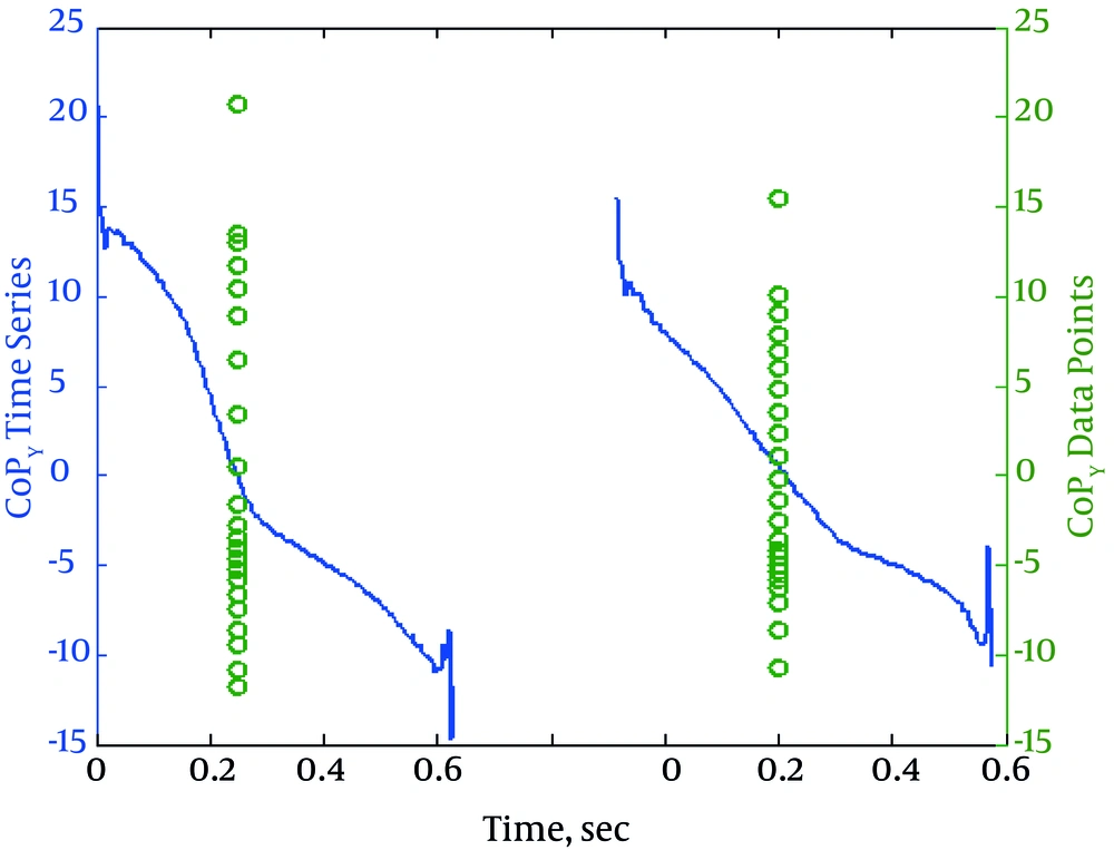
Three variables of SD of CoPy, length of CoP construction line and mean overall velocity of CoP had normal distribution, so a repeated measure ANOVA was used to analyze the data. The ANOVA results are presented in Table 3. No significant interaction effect was observed between fatigue (before and after) and group (foot type) for all variables (P > 0.05). The only significant difference was in within-subject effect for the SD of CoPy (F = 8.07, df = 1, P = 0.008). Fatigue resulted in lower SD of CoPy in both groups. Figure 1 shows the sample plots related to a subject with the higher SD of CoPy and a one with the lower SD of CoPy.
| CoP Measures | Group Effect | Fatigue Effect | Interaction Effect | ||
|---|---|---|---|---|---|
| F Ratio | P Value | F Ratio | P Value | P Value | |
| SD of CoPy | 0.04 | 0.84 | 8.07 | 0.008a | 0.99 |
| Overall Mean Velocity of CoP | 2.42 | 0.13 | 1.24 | 0.27 | 0.67 |
| Length of CoP Construction Line | 0.31 | 0.58 | 3.64 | 0.06 | 0.49 |
Abbreviation: CoP, center of pressure.
aStatistically significant.
SD of CoPx had a non-normal distribution, so non-parametric tests were used for analysis.
Mann-Whitney U test showed no significant differences between the two groups of normal and flat feet before fatigue (P = 0.41) and after fatigue (P = 0.44). Wilcoxon Signed Rank test also showed no significant difference within flat feet group (P = 0.21) and within normal group (P = 0.98), before and after the fatiguing condition.
5. Discussion
In the current study, changes of linear CoP parameters due to fatigue were studied between individuals with flat and normal feet during walking. Although the effect of fatigue on CoP excursion and plantar pressure distribution in normal subjects was studied before (14, 16, 17), there is no study examining fatigue effect on linear CoP parameters in individuals with flat feet.
The only significant finding was lower SD of CoPy in both groups of flat and normal footed subjects after the fatiguing condition. There was no significant between-subjects (group) effect for SD of CoPy. Consequently, despite the different shape of MLA, the biomechanical behavior of both groups was similar. SD of CoPy shows the dispersion of CoP values around the mean in the anteroposterior direction (Figure 1). It has been shown that center of mass (CoM) and CoP movements are related to each other in different situations of standing and locomotion (11, 28). Therefore, lower values of SD after fatigue mean fewer fluctuations of CoPy which probably indicates less movement of CoM. This reduced movement of CoM could be due to more contact of plantar surface of foot after fatigue especially in normal feet subjects. It has been shown that MLA is supported by the activity of extrinsic and intrinsic foot muscles (29-31). Also, it has been reported that fatigue of intrinsic foot muscles could result in more navicular drop and thus more foot contact (30). Therefore, this more foot-ground contact resulted in more centralized movement of CoM to anteroposterior axis of gait.
In this study, no significant finding was observed between groups in linear CoP measures. Similar results have been previously reported for the ground reaction force between individuals with normal and flat feet. It has been shown that fatigue resulted in within group changes but no between group changes for vertical ground reaction force (19). This similar response to fatigue in individuals with flat and normal feet indicates the same biomechanical behavior despite their different MLA height. Furthermore, these findings show that linear CoP measures are not able to distinguish the gait behavior difference among individuals with normal and flat feet. Further research studying the nonlinear measures may explain these two group differences.
The main limitation of this study was analyzing the SD of CoPx without normalization to the foot width. As stated before, AHR was assessed for identifying individuals with normal and flat feet in this study. AHR is a common method of assessing MLA height by using dorsum height and total foot length (20). Therefore due to lack of the foot width anthropometric data, we were not able to normalize SD of CoPx to the subjects' foot width.
In conclusion, the results showed reduction of the CoPy fluctuations which might reduce the risk of injury by probably reducing CoM fluctuations; however, this relationship needs verification by further research. In addition, the biomechanical behavior was similar for both groups, despite the different foot shape among them. This suggests that while flat foot deformity is a deviation from normal foot shape, the kinetic response to fatigue is not influenced by it.
