1. Background
A large number of biomechanical studies of human locomotion deal with the comparison of over-ground and treadmill walking. A comprehensible analysis of the conclusions from such studies is not easy, if at all possible. While debate remains, many authors have recently established that the overall gait patterns between the two are biomechanically similar (1-3). Parvataneni et al., (4) showed that step, stride and joint angular kinematics are similar for treadmill and over-ground walking with the exception of the maximum hip flexion and knee extension angles which were both more prominent on treadmill relative to over-ground walking, but in these instances differed by less than 3 degrees. Lee and Hidler (1) reported that peak flexion and extension measures of the lower extremities did not differ between treadmill and over-ground walking. It has been accepted that lower limb kinematics in the sagittal plane are comparable between over-ground and treadmill locomotion (2, 5-7). Strathy et al., (8) established that knee joint angular kinematics in the coronal and transverse planes did not differ significantly between the two conditions. However, some previous studies have documented significantly greater hip range of motion and flexion angles during treadmill locomotion (2, 6). Interestingly, the majority of surveys (2, 7, 8) which have compared the human movement differences between treadmill and over-ground walking have only focused on the lower extremity muscles, while the role of trunk muscles in motor control has received less attention. Electromyographic studies have signified the role of the erector spinae muscle as an important core muscle in the organization of locomotor patterns during walking and other various rhythmic motor tasks (9-11). Moreover the critical role of core muscles in sport’s performance is widely accepted (12).
2. Objectives
It can be assumed that the pattern of muscle training and the level of muscle strengthening are different between treadmill and over-ground walking. Therefore this study was conducted to compare the activity pattern of selected trunk and lower extremity muscles in over-ground versus treadmill walking.
3. Materials and Methods
3.1. Design and Setting
This cross-sectional study was conducted to determine whether the trunk and lower extremity muscle activation pattern differed in treadmill vs. over-ground walking.
3.2. Samples and Sampling
Nineteen healthy men were included via simple sampling in this study. Inclusion criteria were as follows: male gender, being sedentary based on American college of sports medicine (ACSM) definition (less than 30 minutes of moderate intensity physical activity at least three times per week for a minimum period of three months), being within the age range of 20 - 40, lack of history of musculoskeletal problems, cardiac diseases, hypertension, chronic low back pain, back surgery or any known gait abnormality such as an orthopedic injury, lower limb pain, or neurological injury that would bias the results of this study. Also abnormality in gait pattern of the subjects was considered as the exclusion criterion.
3.3. Codes of Ethics
First, all participants were examined by the physicians and their demographic data such as, height, weight and body mass index were obtained. Second, enough information about the purposes of the study was given to the subjects and they were asked to sign the informed consent approved by the ethics committee of Tehran University of Medical Sciences. All experiments were conducted at the sports medicine research center of Tehran University of Medical Sciences.
3.4. Instrumentation
To compare selected trunk and lower limb muscle activation patterns during ground versus treadmill walking, the eight channel DataLog model surface EMG made by Biometrics was utilized. According to the manufacturer’s recommendations, 1000 Hz was chosen as the sampling rate of the device to record the data and the SX230 active electrodes were used. Center -to-center distance between the electrodes was 2 cm. In order to match the speed of walking on the ground and treadmill, the subjects started to walk with a self-selected speed in a certain distance with their own shoes, while the time was recorded by a digital chronometer. Therefore the suitable walking speed for every subject was determined individually. Finally, according to each individual’s normal walking pace, the walking speed of subjects was specified on the treadmill.
3.5. Process
With the previous studies in mind, in order to assess the activity of trunk as well as lower limb muscles; the following muscles were chosen: rectus abdominus, external oblique, longissimus, multifidus as well as vastus medialis, vastus lateralis and hamstrings, all from the right side of the subjects’ body. The best place for recording every muscle’s EMG activity was determined according to the related references (13). To increase the quality of electrical signal transmission from the subjects’ skin to the device, body hair at the electrode position was shaved and cleaned with alcohol and the electrodes were placed in the suitable area using special glue. Using a belt, the EMG device was placed on the subjects’ lumbar area. This device was wirelessly connected to the computer and the data were recorded on the software.
Locations of the electrodes were as follows: rectus abdominus (RA) muscle: The electrode was placed perpendicular to the horizon, 3cm lateral and 3cm superior to the umbilicus (13). External oblique (EO) muscle: The electrode was placed at a 45° oblique angle, midway between anterior superior iliac spine (ASIS) and the lowest part of the rib cage (13). Multifidus (MF) muscle: The electrode was placed perpendicular to the horizonin front of the fifth lumbar vertebra on an imaginary line between the right posterior superior iliac spine (PSIS) and the first lumbar intervertebral space. Longissimus (LO) muscle: The electrode was placed 4cm lateral to the first lumbar vertebral spinous process (13). Vastus medialis obliquus (VMO) muscle: The electrode was placed at a 55° oblique angle over the center of the muscle belly of the vastus medialis obliquus muscle, 2 cm medially from the superior rim of the right patella (13). Vastus lateralis (VL) muscle: The electrode was placed at two thirds of the imaginary line from the anterior superior iliac spine (ASIS) to the lateral side of the right patella in the direction of the muscle fibers (14). Hamstring (HAM) muscles: A general electrode placement was applied for the entire hamstring muscle group midway between the gluteal fold and the popliteal line on the posterior surface of the right knee in the center of the posterior thigh (13). A reference electrode was placed over the right medial malleolus.
Foot switch was placed under the subject’s right heal and the subjects walked over ground with their desired speed. The electromyographic data were recorded by the electromyogram for every person, and then the subjects walked over the treadmill (Technogym model) for 5 minutes with the same speed. The process of electromyographic data analysis was as follows:
1. Initial processing of EMG data: in this phase two separate files were provided for every subject, one for walking over the ground and the other for walking on the treadmill. To determine the noise level, Frequency spectrum was plotted and the maximum frequency of AC (50 - 60 Hz) as well as the low frequency (motional noise) was assessed. Since based on the logarithmic scale, this level of frequency was much smaller than the frequency content in original bandwidth (20 - 450 Hz active electrode), the noise was in an acceptable limit, and therefore there was no need to use a filter (15). After evaluating the different windows for EMG signal processing, a 50 ms window of RMS (root mean square) data was used to calculate the muscles’ electrical activity.
2. Isolation of gait cycles: in this phase, using foot switch data, RMS curves were isolated based on gait cycles.
3. Separation of gait cycles: this step was performed according to the seven phases of gait cycle which is described in kinesiology references. Inter-stage distances during the gait cycle were isolated based on the percentage of their occurrence. Based on this method the stance phase is divided into the following four stages: loading response, mid-stance, terminal stance and pre-swing. Loading response begins when the foot contacts the ground and ends with the contralateral toe off, when the opposite extremity leaves the ground. Mid-stance initiates with contralateral toe off and ends where the center of gravity is directly over the reference foot. Terminal stance begins when the center of gravity is over the supporting foot and ends when the contra-lateral foot contacts the ground. During terminal stance the heel rises from the ground. The pre-swing stage begins at contra-lateral initial contact and ends at toe off.
Swing phase is also divided to three stages: initial swing, mid-swing and terminal swing. Initial swing begins at toe off and continues until maximum knee flexion occurs. Mid-swing is the period from maximum knee flexion until the tibia is vertical or perpendicular to the ground. Terminal swing begins where the tibia is vertical and ends at initial foot contact.
4. Determination of the threshold of muscle activity: there are different methods to identify the active state of muscles; and the threshold value was used in this study. This means that if the amount of activity was higher than the certain percentage of the total range of RMS changes in the muscle, that muscle was assumed to be active. Considering the pilot study, the threshold of 20% was chosen to evaluate all data in this study.
5. Calculation of the dependent variables: using the data of previous stage, the mean amplitude as well as the duration of muscle activity was calculated for every stage of gait cycle in all subjects.
6. Description of the pattern of muscle activity: to provide a comprehensive profile of the muscle activity during walking over ground as well as on the treadmill, the frequency of activity states of muscles in each stage of the gait cycle was plotted as a bar graph. To harmonize the data among subjects, the percentage of action of each muscle during a gait cycle was considered for the duration of muscle activity. Wilcoxon test and interquartile range (IqR) were used to analyse the data and show the data scattering in this study.
4. Results
The duration of activity (the percentage of a gait cycle which the muscle is active) in selective trunk (RA, EO, MF and LO) and lower limb (VMO, VL, HAM) muscles has been shown in the Table 1.
| Trunk and Lower Limb Muscles | Over-Ground Median (IqR)a | Treadmill Median (IqR)a | P Valueb |
|---|---|---|---|
| Rectus abdominis(RA) | 100 (0) | 100 (0) | 0.317 |
| External oblique (EO) | 100 (0) | 100 (0) | 0.655 |
| Multifidus (MF) | 79 (27) | 82 (28) | 0.446 |
| Longissimus (LO) | 62 (14) | 76 (43) | 0.098 |
| Vastus medialis (VMO) | 46 (30) | 46 (28) | 0.931 |
| Vastus lateralis (VL) | 42 (24) | 40 (20) | 0.981 |
| Hamstring (HAM) | 71 (44) | 53 (43) | 0.212 |
aIqR, Interquartile range.
bUsing Wilcoxon test.
The amplitude of trunk and lower limb muscles activity was greater in walking on the treadmill than over-ground walking. This difference was significant for RA, MF, LO, VMO and VL muscles (Table 2).
| Trunk and Lower Limb Muscles | Over-Ground Median (IqR)a, mv | Treadmill Median (IqR)a, mv | P Valueb |
|---|---|---|---|
| Rectus abdominis(RA) | 3.0 (1.6) | 4.1 (5.1) | 0.005 |
| External oblique (EO) | 6.5 (1.9) | 8.2 (5) | 0.136 |
| Multifidus (MF) | 11.4 (7.6) | 17.0 (7.9) | 0.044 |
| Longissimus (LO) | 10.2 (4.8) | 14.5 (10.4) | 0.018 |
| Vastus medialis (VMO) | 12.8 (9.2) | 17.4 (24) | < 0.001 |
| Vastus lateralis (VL) | 13.5 (4.9) | 16.5 (8.3) | 0.005 |
| Hamstring (HAM) | 19.1 (15.8) | 22.4 (24) | 0.064 |
aIqR, Interquartile range.
bUsing Wilcoxon test.
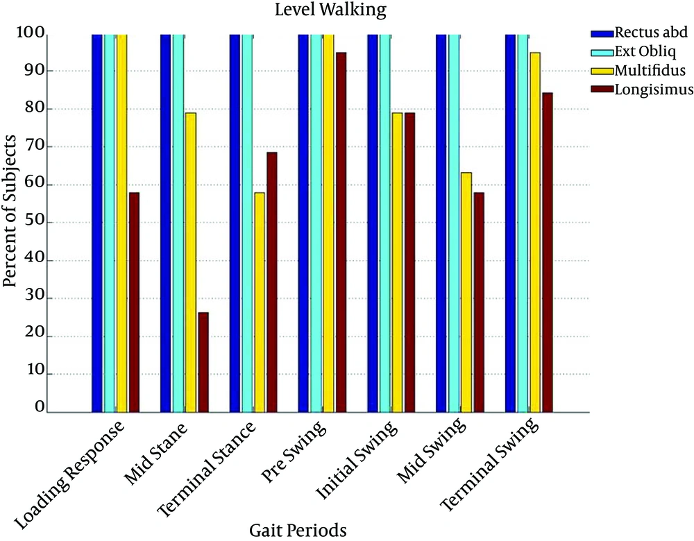
Among selected trunk muscles, RA and EO muscles were active in all subjects in the entire gait cycle, while MF was active in 100% of subjects during loading response and pre-swing stages and in 90% of subjects in terminal swing stage during the over-ground walking (Figure 1).
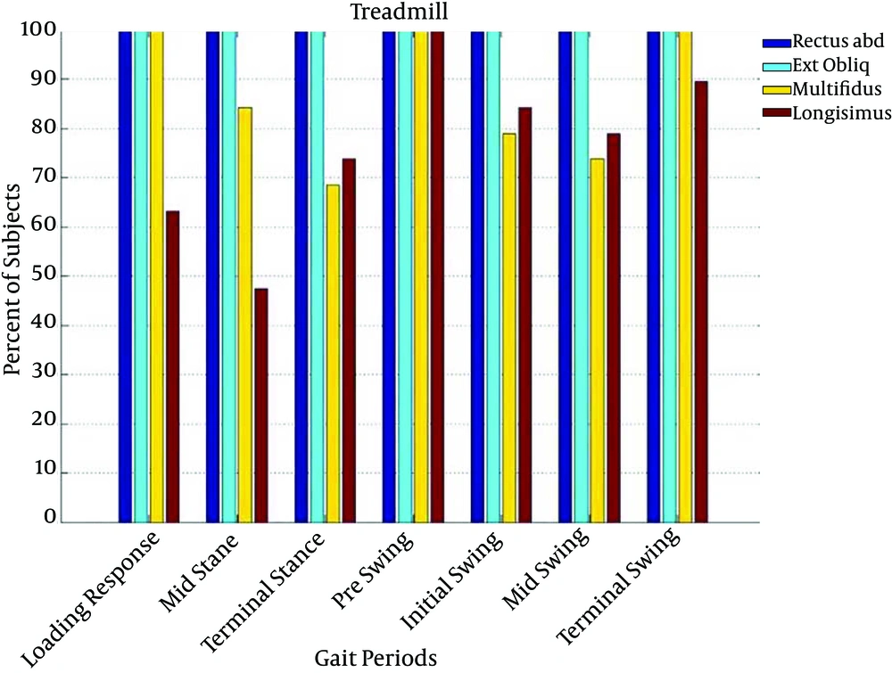
Likewise, during treadmill walking, RA and EO muscles were active in all subjects in the entire gait cycle and also MF was active in 100% of subjects during loading response, pre-swing and terminal swing stages (Figure 2).
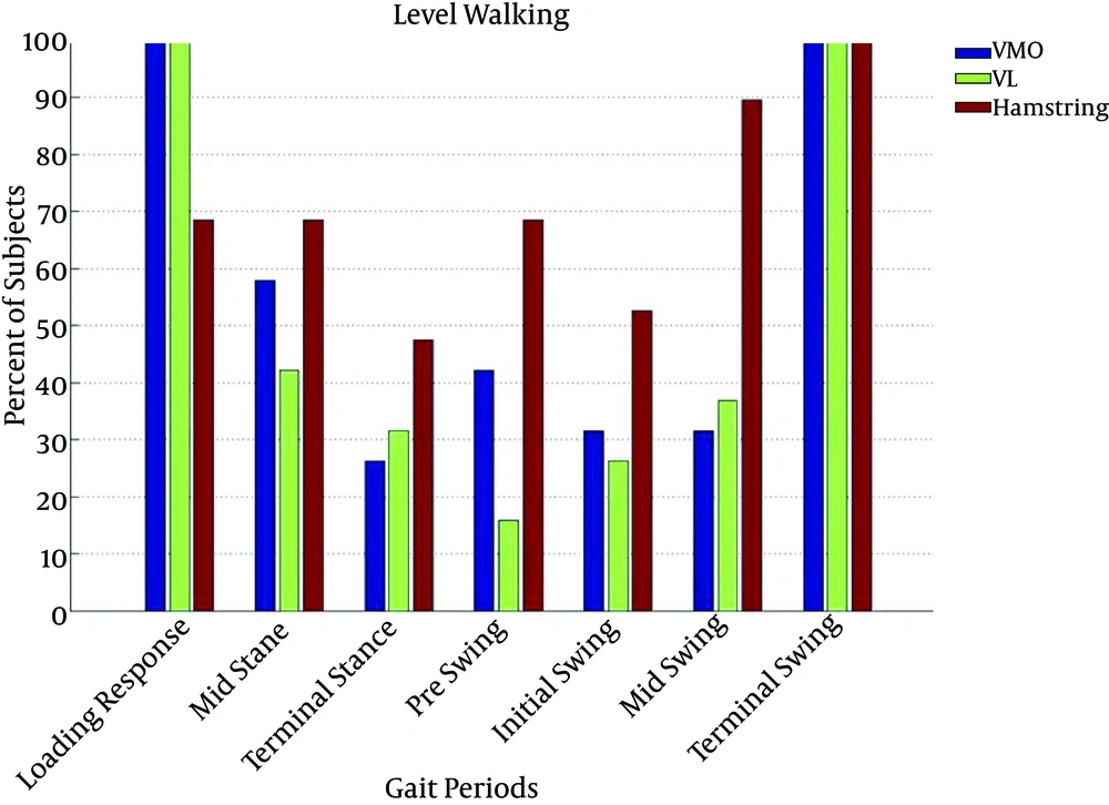
Among selected lower limb muscles, VMO and VL muscles were active in 100% of subjects in loading response and terminal swing stages and HAM muscles were active in all subjects in terminal swing stage during the over ground walking (Figure 3).
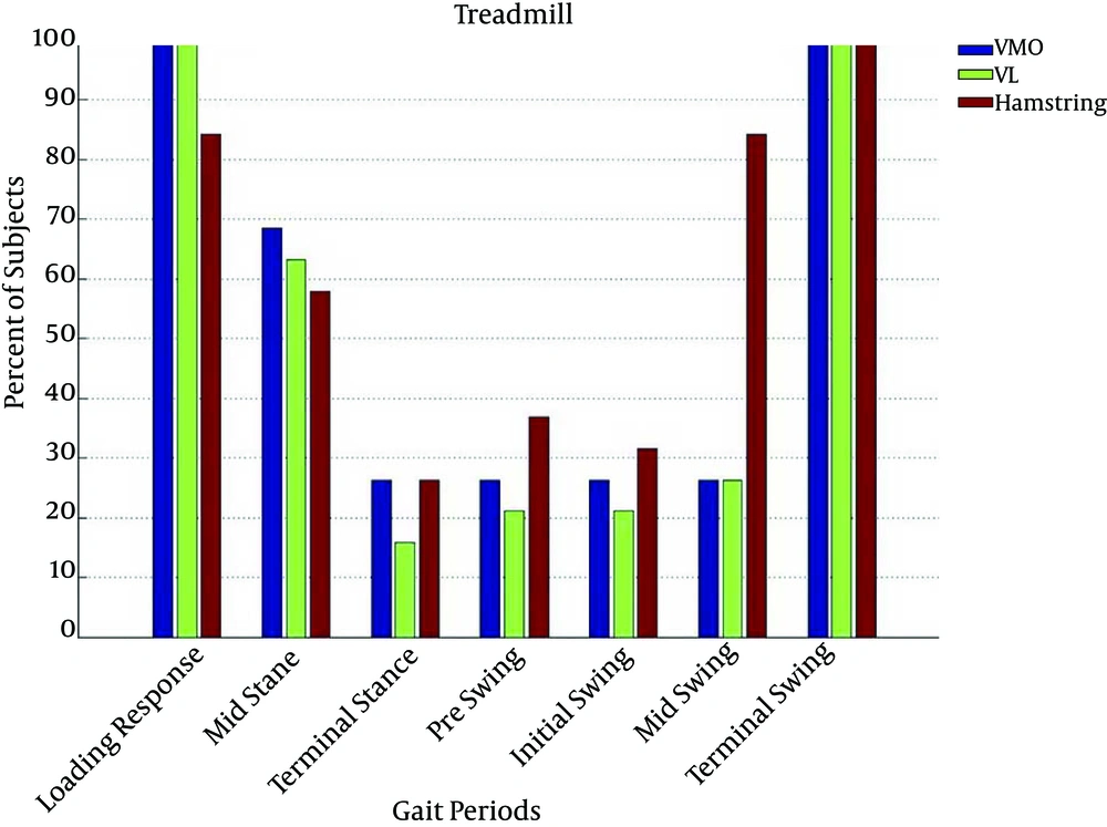
Equally, VMO and VL muscles were active in 100% of subjects during loading response and terminal swing stages and HAM muscle was active in all subjects in terminal swing stage during treadmill walking (Figure 4).
5. Discussion
Although numerous studies have been performed to compare the over-ground versus treadmill walking, there is still some controversy about their similarities and dissimilarities. However it is widely accepted that the overall patterns of muscle activation are sufficiently similar between the two modes, they can therefore be used interchangeably (1).
Though some studies have compared the amplitude of activity of lower limb muscles by surface EMG during over ground and treadmill walking (16, 17), to our knowledge none of them has evaluated the duration of activity as well as amplitude of both trunk and lower limb muscles and this study is probably the first which has been performed to compare all these parameters between over ground versus treadmill walking.
This study has found that the duration of electromyographic activity of trunk and lower limb muscles during over ground and treadmill walking are generally similar. Moreover RA and EO muscle activity throughout the gait cycle indicate their key role in spinal stabilization and balance during walking. Conversely, based on the results of this study, the amplitude of electromyographic activity of trunk and lower limb muscles was greater in walking on the treadmill than over-ground walking. This finding is consistent with those of Arsenault et al. who found the amplitude of activity in some lower limb muscles including rectus femoris, biceps femoris, soleus, vastus medialis and anterior tibialis is greater in walking on the treadmill than over-ground walking (16). Also the findings of the current study are in agreement with Nymark’s findings which showed that the amplitude of anterior tibialis and gastrocnemius activity is greater in walking on the treadmill than over ground walking (17). Lee and his colleagues reported an interesting pattern of electromyographic activity among hamstrings, vastus medialis, and adductor longus muscles where they found higher activity during over-ground walking in each of these muscles throughout early and mid-swing, while this relationship reversed at terminal swing (more activity during treadmill walking) (1). The findings in Lee’s study somehow mirror those of the present study that showed the amplitude of lower limb muscles activity was greater in walking on the treadmill than over ground walking particularly during terminal swing where VMO, VL and HAM muscles were active in all subjects. Generally these results come to agreement with the findings of other studies (18, 19), in which the duration and the amplitude of quadriceps muscle electromyographic activity were greater in treadmill than over-ground walking.
Based on this study the amplitude of trunk muscle activity was greater in walking on the treadmill than over-ground walking. This difference was more significant for MF (50%), LO (42%) and then RA muscles. Therefore it seems that, like the muscles of the lower extremities, trunk muscles are also more active during treadmill walking compared to over-ground walking. This result may be explained by the fact that treadmill walking differs with over-ground walking biomechanically. More passive hip extension as well as forward bending of trunk during treadmill walking compared to over ground walking (17) can explain why the pattern of electromyographic activity of trunk and lower limb muscles varies between these two modes of walking. However, according to this study and also previous surveys (2, 5-8), the patterns of muscle activity on both treadmill and over-ground walking are enough similar to use these two modes of walking interchangeably. Moreover it seems, RA and EO muscles which are active during the entire gait cycle, have a critical stabilizing role during walking. On the other hand, the higher amplitude of trunk muscles activity during treadmill walking compared to over ground can probably make this mode of walking appropriate for exercise prescription in patients who have core muscles weakness.
5.1. Conclusion
The patterns of trunk and lower limb muscle activity on both treadmill and over-ground walking are generally very similar. The amplitude of muscle activity is greater on treadmill walking compared to over-ground which may indicate more muscular function during treadmill walking.


