1. Introduction
The differential diagnosis for acute buttock pain in athletes is extensive (1). The differential includes strain of gluteal, hamstring, adductor and hip external rotator muscles, sacroiliac or hip joint disease, bony fractures/stress fractures, avulsion injuries, slipped capital femoral epiphysis, labral injuries, hip dislocation, lumbar radiculopathies, or nerve entrapment. Many of these processes can be ruled in or out based on mechanism of injury, presenting symptoms (including the presence or absence of neurologic symptoms), and physical examination. However, some of these processes can be difficult to delineate from one another without imaging, particularly those involving injury to the deep external rotators of the hip, as in this case. The exact mechanism for isolated injury of one of these deep hip muscular structures is unknown. The mechanism of injury of the quadratus femoris in a tennis player was speculated to be due to a strong eccentric contraction in a closed kinetic chain to try to control hip internal rotation during the end phase of serving (2). We present a previously unreported case of acute obturator internus and Obturator externus strain in an adolescent.
2. Case Presentation
A 14 year old high school quarterback presents 2 days after injuring his right buttock while playing football. He attempted to grab the football from the ground behind him after a high snap passed over his head and bent awkwardly as he was tackled to the ground. Immediately he felt a sharp pain deep in his right buttock. He was unable to continue playing. The pain was achy and constant, radiating to his right hip. He was able to walk with significant limping. He denied any numbness or weakness. There was no obvious bruising or swelling. Physical examination revealed normal vital signs. He was 175 cm tall and weighed 58 kg (BMI 18.9 kg/m2). His hip range of motion was limited mainly due to pain. He was only able to flex his back to reach his mid-thigh. Back extension was normal. Straight leg raise was to about 30 degrees on the right side. Patient was tender medial to the right ischial tuberosity. There was no edema or ecchymosis. Neurovascular exam was unremarkable.
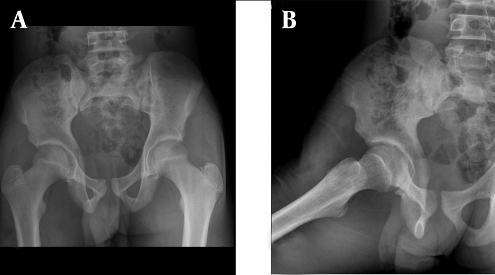
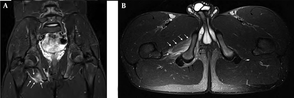
Plain radiography in the office was normal (Figure 1). MRI (including post-gadolinium images) was performed to evaluate bony, chondral, or labral abnormality on day three post-injury. MRI revealed high grade right obturator internus and obturator externus muscle strain involving the attachments to the obturator ring and ischium (Figure 2). Otherwise, MRI was unremarkable with no signs of joint effusion, chondral, or labral pathologies. The patient was advised to do relative rest for a few days and use acetaminophen as needed for pain. Then he began gentle hip range of motion exercises and partial weight bearing as tolerated for two weeks. He started physical therapy consisting of joint/soft tissue mobilization, aquatic therapy, electrical stimulation, ultrasound, iontophoresis, infra-red light therapy, hot/cold modalities, therapeutic exercise, functional movement of the hip, and gait training three weeks post injury. After completing a few weeks of formal physical therapy he transitioned to home exercises as instructed. Patient was symptom free during a follow up visit six weeks after the injury. His physical examination was unremarkable with normal, pain-free hip range of motion. He was cleared to return to full physical activity at that time.
3. Discussion
The deep external rotators of the hip include the piriformis, quadratus femoris, the inferior and superior gemelli, and external and internal obturators (3). Many of these muscles have complex origins and sit deep within the pelvis making them difficult to isolate on examination. The obturator internus originates from the rami surrounding the obturator foramen and the quadrilateral plate and leaves the pelvis through the lesser sciatic notch before inserting on the greater trochanter of the proximal femur (4). Based on EMG evaluation, its functions include hip extension, external rotation and abduction (5). The obturator externus originates from the external bony margin of the obturator foramen and passes like a sling under the femoral neck to insert on the piriformis fossa of the femur (4).
These deep muscles of the hip are either infrequently injured or misdiagnosed as hamstring or gluteal strains as occurred in a prior case report of a quadratus femoris strain (2). There have been very few case reports in the literature of acute obturator internus or externus strains and to the authors’ knowledge this is the first report involving both obturator internus and externus without other hip structure injuries (6, 7). The only prior literature describing strain of both the obturator internus and externus was seen on MRI after posterior hip dislocation and was associated with quadratus femoris strain, acetabular fracture and avulsion of the lesser trochanter (3).
MRI is the study of choice for confirming a diagnosis and ruling out bony abnormalities and should be used early to help delineate muscle involvement and therefore determine an appropriate rehab program (7). In the few case reports related to deep hip external rotator muscle strains, most have done well with initial rest followed by physical rehabilitation. Athletes have typically been able to return to full sport participation in 2 - 6 weeks.
Strain of the hip external rotator muscles is rare and should be in the differential diagnosis of proximal posterior thigh pain in athletes. MRI is useful in ruling out conditions requiring surgical intervention. Identifying the exact injured muscle with MRI can appropriately help modify the rehabilitation program.

