Deep Vein Thrombosis in Combined Ankle Arthroscopy for the Anterior and Posterior Ankle Impingement Syndrome

authors:

avatar Jorge Javier Del Vecchio 1 , * , avatar Anuar Emanuel Uzair 2 , avatar Jorge Pablo Batista 3 , avatar Mauricio Esteban Ghioldi 2 , avatar Enrique Baldessari 4 , avatar Blas Pezzini 4

Favaloro University, Buenos Aires, Argentine
Favaloro Foundation, Solís 461, Ciudad Autonoma de Buenos Aires, Argentine
Consultorios de Artroscopia Jorge Batista, Avenida Pueyrredon 2446 - Piso 1º - Ciudad Autonoma de Buenos Aires
Favaloro Foundation, Av. Belgrano 1746, Ciudad Autonoma de Buenos Aires, Argentine

how to cite: Del Vecchio J J, Emanuel Uzair A, Pablo Batista J, Esteban Ghioldi M, Baldessari E, et al. Deep Vein Thrombosis in Combined Ankle Arthroscopy for the Anterior and Posterior Ankle Impingement Syndrome. Asian J Sports Med. 2018;9(1):e55563. https://doi.org/10.5812/asjsm.55563.

Abstract

Introduction:

The ankle arthroscopy complication rates from 7.6% to 13.6% and deep vein thrombosis (DVT) is considered as a serious complication. The risk of DVT for patients with isolated foot and ankle conditions, even with plaster cast immobilization, and the possible benefits of mechanical and chemical prophylaxis are poorly studied. Nevertheless, some studies mentioned the risk factors such as injury severity, immobilization, obesity, nonweight-bearing, hindfoot surgery, and tourniquet time.

Case Presentation:

A 27-year-old male patient, smoker, and casual athlete with complaint of left recurrent painful ankle presented an anterior ankle impingement type B and a posterior impingement syndrome. In the immediate postoperative period, the patient developed a DVT as a complication of a combined ankle arthroscopy (anterior and posterior).

Conclusions:

It seems that the use of prolonged tourniquet in addition to repositioning the patient may increase the risk of complications such as VTE (venous thromboembolism) and the combination was not included in any published protocol. The current study recommended the use of the Calder antithrombotic recommendations associated with the Caprini risk assessment model in order to cover a greater at-risk population.

1. Introduction

Several studies showed that ankle injuries occur in sports up to 30%. Residual symptoms may occur in up to 50% of people. In this regard, ankle impingement is a major cause of chronic pain. Anterior and posterior impingements are well documented and generally considered as separate entities with different causal injury patterns (1).

Complications are well described. The ankle arthroscopy complication rates from 7.6% to 13.6%, according to different authors (2, 3). Neurological injury is the most frequently described within the spectrum of complications including vascular injuries and false aneurysms, infections and synovial fistula, invasive distraction related problems and stress fractures. Furthermore, instrument breakage, complex regional pain syndrome (CRPS), compartment syndrome, thromboembolic complications (4), and painful scars are reported (5).

Deep vein thrombosis (DVT) is considered as a serious complication, especially in lower extremities and has the potential to result in significant morbidity and possible mortality (6, 7). The risk of DVT for patients with isolated foot and ankle conditions, even with plaster cast immobilization, and the possible benefits of mechanical and chemical prophylaxis were poorly studied (8). Nevertheless, some studies mentioned risk factors as injury severity (9), immobilization (10), obesity (11), nonweight-bearing (12), hindfoot surgery (13), and tourniquet time (13).

The current report aimed at showing an infrequent complication (DVT) in combined ankle arthroscopy for anterior and posterior ankle impingement syndrome.

2. Case Presentation

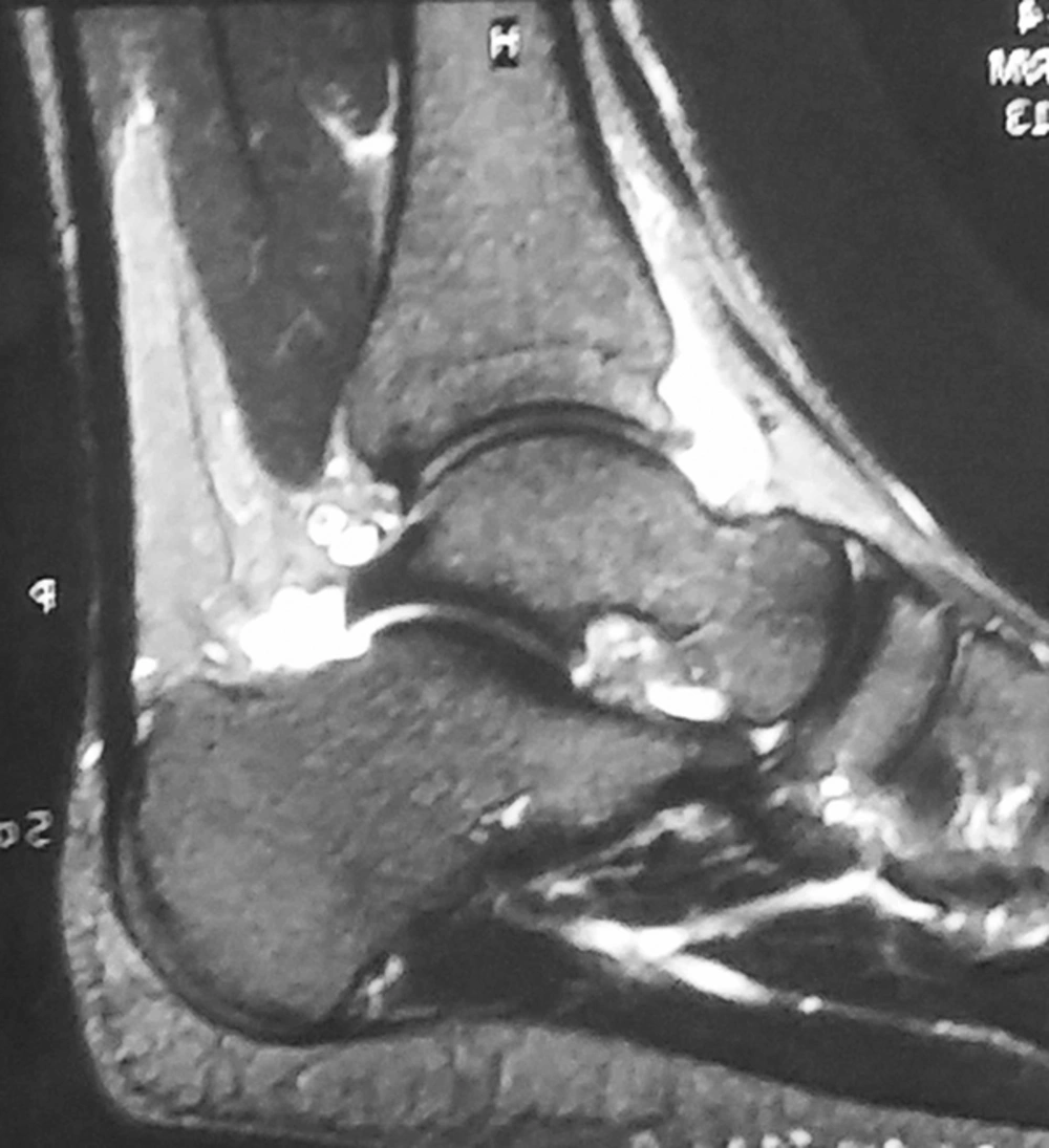
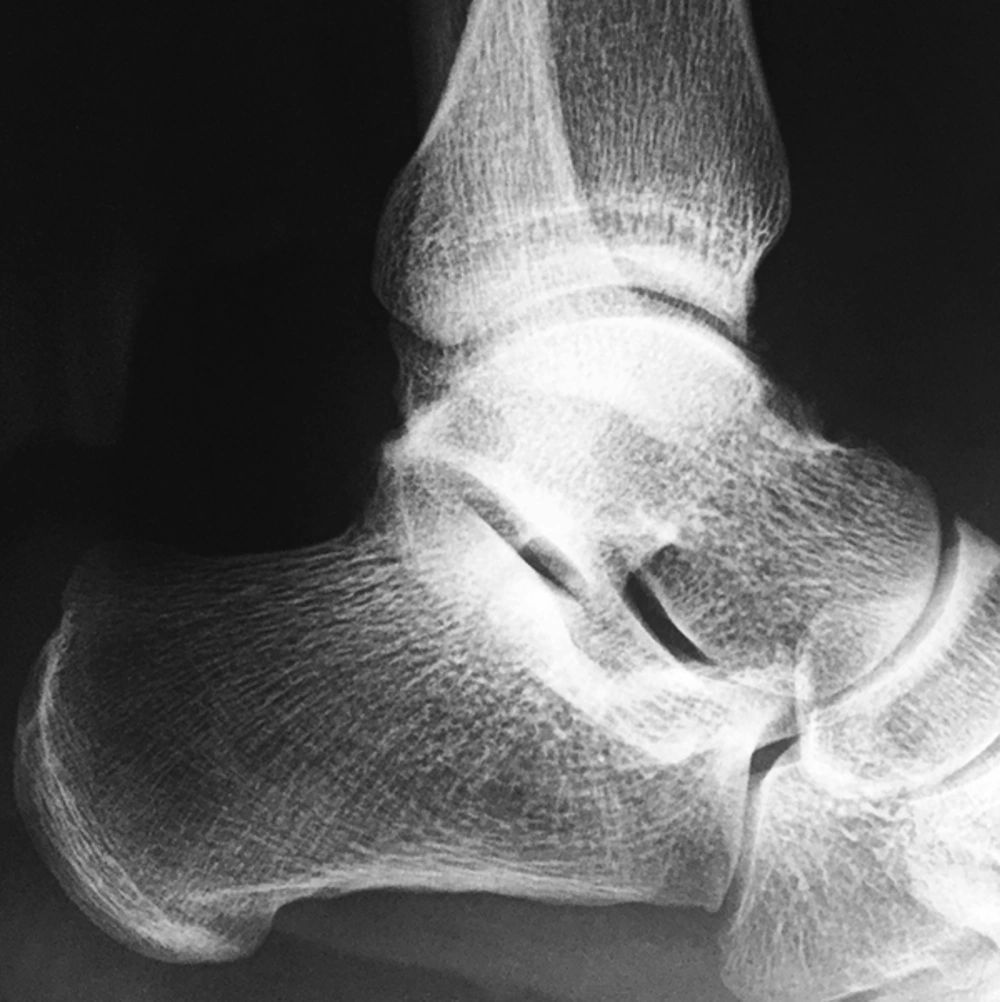
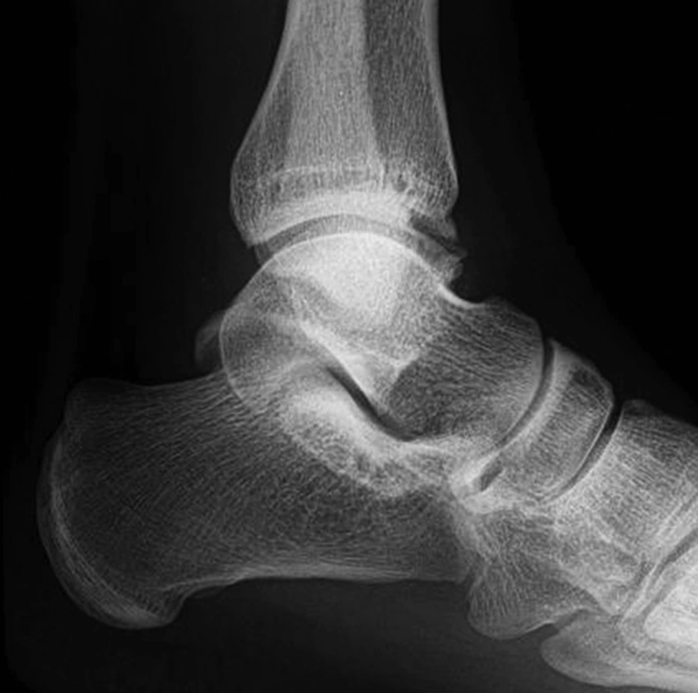
Informed consent was obtained from the study participant. A 27-year-old male patient, smoker, and recreational athlete referred to the outpatient orthopedics department complaining of left recurrent painful ankle. The patient had no specific risk factors for DVT preoperatively (family history, obesity, previous DVT, medical co-morbidities, or drugs, etc.). Hypercoagulable states were factor V leiden + and lupus anticoagulant -, as well as protein C or S deficiency, based on the evaluation of blood parameters. He previously had an ankle sprain while playing soccer. In the physical examination presented anterior (predominantly located anteromedially) and posterior ankle pain (positive ankle hyperflexion plantar test). The preoperatively AOFAS (the American orthopedic foot and ankle society score) (14) was 70. Radiological analysis showed an anterior ankle impingement type B (15) and a posterior impingement syndrome (posterior talar spur) (Figure 1).

Lateral X-Ray Showing AP Impingement

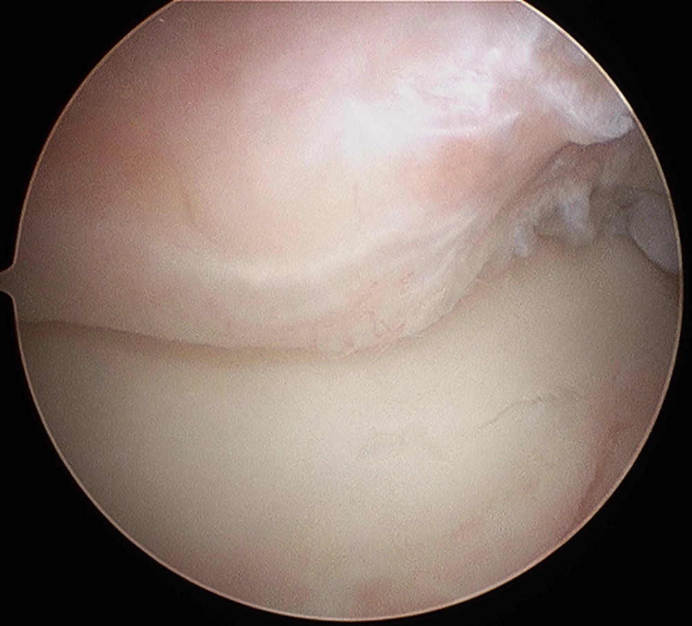
Magnetic resonance imaging (MRI) showed an ankle synovitis and a flexor hallucis longus tenosynovitis (Figure 2).

MRI; Ankle Synovitis and FHL Tenosynovitis

He had conservative treatment (non-steroidal anti-inflammatory drugs, rehabilitation, and cryotherapy) for 3 months. Due to the lack of response, surgery was performed.

2.1. Surgical Technique

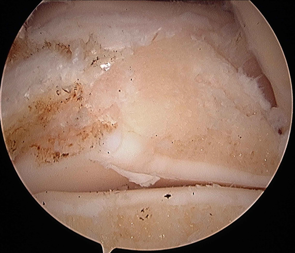
The patient received lumbar spinal anesthesia and placed in the supine position with a thigh tourniquet without any distraction technique. First, an anterior ankle arthroscopy was performed through the 2 standard anteromedial and anterolateral portals (5) to perform the debridement of osteophytes (Figure 3) and hypertrophic synovial in the anterior ankle (4.0 mm; 30 degrees; Storz). The duration of surgery was 25 minutes. Then, the patient was placed in prone position and a hindfoot endoscopy (16, 17) was performed to resect the talar spur (Figures 4 and 5) and tenosynovectomy; the duration was 22 minutes. The whole procedure lasted 65 minutes (including the patient’s change of decubitus). The tourniquet was deflated between the 2 procedures.

Anterior Arthroscopy Showing Anteromedial Spur
Posterior Endoscopy; Spur Resected
Postoperative Lateral X-ray; Proper Resection

After the surgical procedure, the patient was not immobilized and authorized to full weightbear according to pain control with elastic bandage. According to the Caprini DVT risk factor assessment score (score 1: minor surgery planned) (8, 18), it was decided not to use pharmacological DVT prophylaxis.

2.2. Outcome

At 72 hours postoperatively, the patient had pain in posterior region of left leg, without other clinical sign of DVT; then, Eco Doppler was performed and reported DVT in deep posterior tibial vein. The patient was admitted for 24 hours to be matched for anticoagulation protocols (low molecular weight heparin 60 mg/24 hours), and then, discharged with rivaroxaban (19) 15 mg/12 hours for 30 days, reducing the dose to 20 mg/24 hours until 60 days of treatment. Four weeks after surgery, he began the rehabilitation without symptoms. At the 12 months postoperative visit, the patient was satisfied with the results and was performing sports activity (AOFAS score: 91).

3. Discussion

VTE (venous thromboembolism) is highlighted as a particular risk following the orthopedic surgery or injury to the lower limb. However, most studies investigating VTE were conducted on patients undergoing major orthopedic surgeries at or above the knee (4, 20).

The risk of VTE in patients with isolated foot and ankle conditions, even with plaster cast immobilization, and the possible benefits of mechanical and chemical prophylaxis were poorly studied (8).

The Caprini risk assessment model (RAM) was derived over a decade ago based on a combination of clinical experience and published data for risk-stratified plastic and reconstructive surgery patients for VTE risk (21). The Caprini RAM was 1st reported in 1980, and then, it was regularly updated to a relatively mature model in 2010 (18) (eg, supplementary file Appendix 1 shows the Caprini assessment and supplementary file Appendix 2 the suggested prophylaxis). Calder et al., published on VTE following the isolated foot and ankle surgery and proposed guidelines for VTE prevention (14). The most recent review by the American college of chest physicians (ACCP) also recommended against chemical prophylaxis in lower leg injuries requiring immobilization (22).

In the current report, the patient was authorized to walk with full weight bearing with elastic bandage, and had a low-risk of thrombosis according to the Caprini protocol; therefore, no anticoagulants were indicated.

Although the presented surgical technique had the disadvantage of repositioning the patient (from supine to prone), some studies mentioned that ankle arthroscopy and hindfoot endoscopy can be performed without adding endovascular complications (23). In the current study, the median total operative time, including the time needed for switching the position from supine to prone, was 94 minutes (ranged 72 to 168 minutes) and the result included 3 bilateral cases. However, the authors did not experience complications caused by changing the patient’s position. Nevertheless, the results of the current study suggested that simultaneous arthroscopic and endoscopic surgery can enable professional athletes with an intractable combination of AAIS (anterior ankle impingement syndrome) and PAIS (posterior ankle impingement syndrome) to return to athletic activity as soon as possible (23). Authors agree with the disadvantage proposed by Miyamoto et al., in relation to repositioning the patient, but it is thought that combined anterior and posterior ankle arthroscopy has other disadvantages such as longer surgical time with the aggravation of the use of tourniquet associated (13).

Vascular changes due to tourniquet may also include direct vascular injury, hyperemia on tourniquet deflation, and increased incidence of deep venous thrombosis, pulmonary embolism, and cardiac arrest (24, 25). Solis and Saxby found a direct relationship between the DVT and the duration of the tourniquet use. However, there are still no data regarding the removal and replacement of the tourniquet during the same procedure (13).

Anterior and posterior impingement can be treated arthroscopically or by open debridement with good results (1), but the arthroscopic technique is related to minor complications. Recently, Song et al. (26) presented a new technique to treat anterior and posterior impingements in dorsal decubitus by means of 3 portals. This, by avoiding turning the patient, might prevent complications avoiding long surgeries.

In conclusion, it seems that the use of prolonged tourniquet in addition to repositioning the patient may increase the risk of complications such as VTE and this combination is not included in any published protocol. Finally, authors also recommend the combination of the Calder antithrombotic recommendations (8) associated with the Caprini RAM (21), aimed at encompassing a higher risk population.

References

  • 1.

    Tol JL, Verheyen CP, van Dijk CN. Arthroscopic treatment of anterior impingement in the ankle. J Bone Joint Surg Br. 2001;83(1):9-13. [PubMed ID: 11245545].

  • 2.

    Ferkel RD, Karzel RP, Del Pizzo W, Friedman MJ, Fischer SP. Arthroscopic treatment of anterolateral impingement of the ankle. Am J Sports Med. 1991;19(5):440-6. [PubMed ID: 1962707]. https://doi.org/10.1177/036354659101900504.

  • 3.

    Unger F, Lajtai G, Ramadani F, Aitzetmuller G, Orthner E. [Arthroscopy of the upper ankle joint. A retrospective analysis of complications]. Unfallchirurg. 2000;103(10):858-63. [PubMed ID: 11098746].

  • 4.

    Barber FA, Click J, Britt BT. Complications of ankle arthroscopy. Foot Ankle. 1990;10(5):263-6. [PubMed ID: 2187776].

  • 5.

    van Dijk CN. Ankle Arthroscopy. Springer; 2014. p. 81-94.

  • 6.

    Ferkel RD, Small HN, Gittins JE. Complications in foot and ankle arthroscopy. Clin Orthop Relat Res. 2001;(391):89-104. [PubMed ID: 11603693].

  • 7.

    Henderson I, La Valette D. Ankle impingement: combined anterior and posterior impingement syndrome of the ankle. Foot Ankle Int. 2004;25(9):632-8. [PubMed ID: 15563385]. https://doi.org/10.1177/107110070402500907.

  • 8.

    Calder JD, Freeman R, Domeij-Arverud E, van Dijk CN, Ackermann PW. Meta-analysis and suggested guidelines for prevention of venous thromboembolism (VTE) in foot and ankle surgery. Knee Surg Sports Traumatol Arthrosc. 2016;24(4):1409-20. [PubMed ID: 26988553]. https://doi.org/10.1007/s00167-015-3976-y.

  • 9.

    Shibuya N, Frost CH, Campbell JD, Davis ML, Jupiter DC. Incidence of acute deep vein thrombosis and pulmonary embolism in foot and ankle trauma: analysis of the National Trauma Data Bank. J Foot Ankle Surg. 2012;51(1):63-8. [PubMed ID: 22196459]. https://doi.org/10.1053/j.jfas.2011.10.017.

  • 10.

    Kock HJ, Schmit-Neuerburg KP, Hanke J, Rudofsky G, Hirche H. Thromboprophylaxis with low-molecular-weight heparin in outpatients with plaster-cast immobilisation of the leg. Lancet. 1995;346(8973):459-61. [PubMed ID: 7637478].

  • 11.

    Riou B, Rothmann C, Lecoules N, Bouvat E, Bosson JL, Ravaud P, et al. Incidence and risk factors for venous thromboembolism in patients with nonsurgical isolated lower limb injuries. Am J Emerg Med. 2007;25(5):502-8. [PubMed ID: 17543652]. https://doi.org/10.1016/j.ajem.2006.09.012.

  • 12.

    Felcher AH, Mularski RA, Mosen DM, Kimes TM, DeLoughery TG, Laxson SE. Incidence and risk factors for venous thromboembolic disease in podiatric surgery. Chest. 2009;135(4):917-22. [PubMed ID: 19017868]. https://doi.org/10.1378/chest.08-1631.

  • 13.

    Solis G, Saxby T. Incidence of DVT following surgery of the foot and ankle. Foot Ankle Int. 2002;23(5):411-4. [PubMed ID: 12043985]. https://doi.org/10.1177/107110070202300507.

  • 14.

    Kitaoka HB, Alexander IJ, Adelaar RS, Nunley JA, Myerson MS, Sanders M. Clinical rating systems for the ankle-hindfoot, midfoot, hallux, and lesser toes. Foot Ankle Int. 1994;15(7):349-53. [PubMed ID: 7951968]. https://doi.org/10.1177/107110079401500701.

  • 15.

    Parma A, Buda R, Vannini F, Ruffilli A, Cavallo M, Ferruzzi A, et al. Arthroscopic treatment of ankle anterior bony impingement: the long-term clinical outcome. Foot Ankle Int. 2014;35(2):148-55. [PubMed ID: 24174431]. https://doi.org/10.1177/1071100713510912.

  • 16.

    van Dijk CN, Scholten PE, Krips R. A 2-portal endoscopic approach for diagnosis and treatment of posterior ankle pathology. Arthroscopy. 2000;16(8):871-6. [PubMed ID: 11078550]. https://doi.org/10.1053/jars.2000.19430.

  • 17.

    van Dijk CN, de Leeuw PA, Scholten PE. Hindfoot endoscopy for posterior ankle impingement. Surgical technique. J Bone Joint Surg Am. 2009;91 Suppl 2:287-98. [PubMed ID: 19805591]. https://doi.org/10.2106/JBJS.I.00445.

  • 18.

    Bahl V, Hu HM, Henke PK, Wakefield TW, Campbell DJ, Caprini JA. A validation study of a retrospective venous thromboembolism risk scoring method. Ann Surg. 2010;251(2):344-50. [PubMed ID: 19779324]. https://doi.org/10.1097/SLA.0b013e3181b7fca6.

  • 19.

    Lassen MR, Ageno W, Borris LC, Lieberman JR, Rosencher N, Bandel TJ, et al. Rivaroxaban versus enoxaparin for thromboprophylaxis after total knee arthroplasty. N Engl J Med. 2008;358(26):2776-86. [PubMed ID: 18579812]. https://doi.org/10.1056/NEJMoa076016.

  • 20.

    Bjornara BT, Gudmundsen TE, Dahl OE. Frequency and timing of clinical venous thromboembolism after major joint surgery. J Bone Joint Surg Br. 2006;88(3):386-91. [PubMed ID: 16498018]. https://doi.org/10.1302/0301-620X.88B3.17207.

  • 21.

    NICE guidelines. Venous thromboembolism: reducing the risk. 2015. Available from: http://www.nice.org.uk/guidance/cg92.

  • 22.

    Clanton TO, Schon LC. Athletic Injuries to the Soft Tissues of the Foot and Ankle. In: Mann RA, Coughlin MJ, editors. Surgery of the Foot and Ankle. St. Louis: Mosby; 1993. p. 1095-224.

  • 23.

    Miyamoto W, Takao M, Matsui K, Matsushita T. Simultaneous ankle arthroscopy and hindfoot endoscopy for combined anterior and posterior ankle impingement syndrome in professional athletes. J Orthop Sci. 2015;20(4):642-8. [PubMed ID: 25797332]. https://doi.org/10.1007/s00776-015-0712-x.

  • 24.

    Olivecrona C, Lapidus LJ, Benson L, Blomfeldt R. Tourniquet time affects postoperative complications after knee arthroplasty. Int Orthop. 2013;37(5):827-32. [PubMed ID: 23417522]. https://doi.org/10.1007/s00264-013-1826-4.

  • 25.

    Zaidi R, Hasan K, Sharma A, Cullen N, Singh D, Goldberg A. Ankle arthroscopy: a study of tourniquet versus no tourniquet. Foot Ankle Int. 2014;35(5):478-82. [PubMed ID: 24375673]. https://doi.org/10.1177/1071100713518504.

  • 26.

    Song B, Li C, Chen Z, Yang R, Hou J, Tan W, et al. Combined Anterior and Dual Posterolateral Approaches for Ankle Arthroscopy for Posterior and Anterior Ankle Impingement Syndrome. Foot Ankle Int. 2016;37(6):605-10. [PubMed ID: 26892889]. https://doi.org/10.1177/1071100716632042.