

DISCUSSION
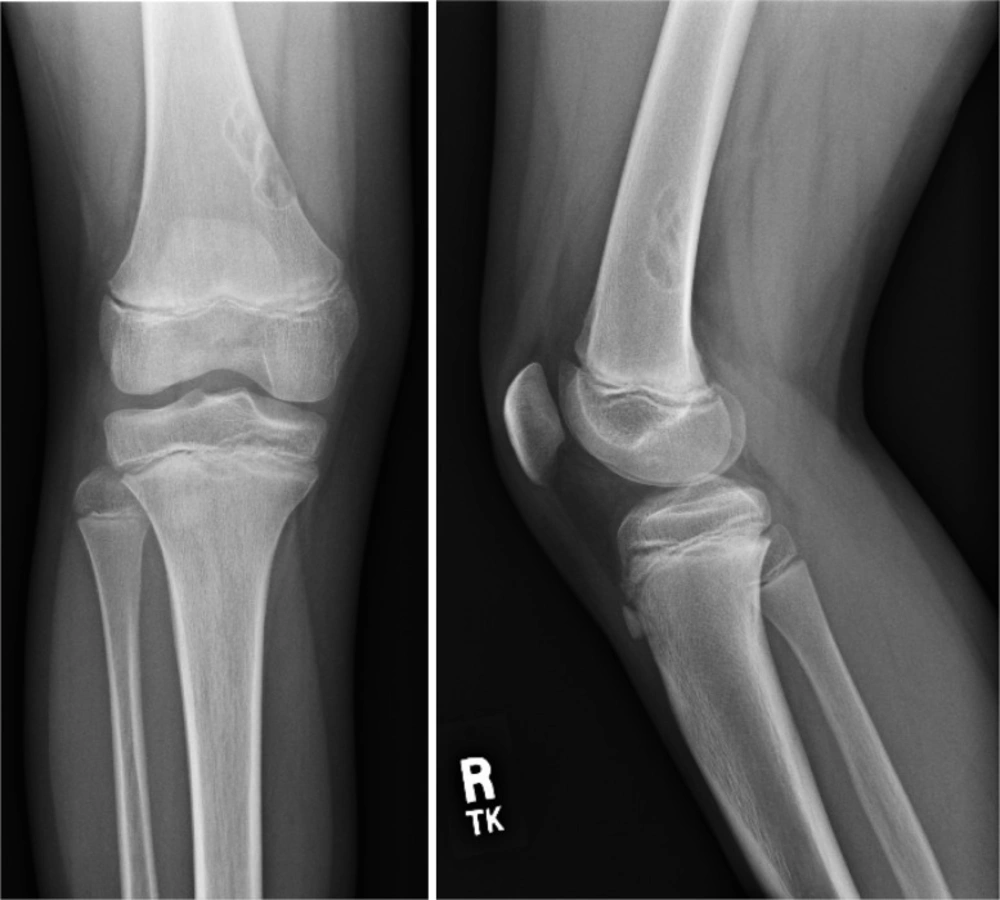
Plain radiographs of the right knee; a) anteroposterior view; (b) Lateral view
The correct answer is B. Nonossifying fibromas are common benign bone lesions seen almost exclusively in young patients under 30 years of age [1]. They are usually asymptomatic and found incidentally on radiographs [2]. They are typically located in the metaphysis or diaphysis of long bones, most commonly the femur and tibia. Furthermore, these lesions are characteristically cortically-based and eccentrically located within the medullary cavity. As seen in the radiograph above, the lesion should have well-defined borders, with no signs of periostitis, cortical breakthrough, soft tissue mass, or edema around the lesion [1].
Enchondromas are one of the most common benign bone tumors and can often be incidental findings on routine knee radiographs and MRI studies [1]. These are intramedullary lesions, typically more centrally located than eccentrically, and are usually found in the metaphysis or diaphysis of long bones. Additionally, there is no related pain, no soft tissue mass and no cortical disruption or periosteal reaction [1, 3].
Osteoid osteomas account for approximately 10% of all benign bone tumors, usually occur between 5 and 25 years of age, and are found predominantly in males (2:1 – 3:1 male to female ratio) [4]. The radiographic appearance is characteristically of a well-circumscribed, 1 to 2 cm lesion with a central osteoblastic nidus surrounded by a sclerotic border in the proximal femur or tibia[2]. They are clinically associated with pain which is worse at night and is relieved with NSAIDs [4].
Salter-Harris type II fracture refers to a physeal injury in which the fracture extends along the growth plate then out through a portion of the metaphysis, producing a triangular shaped metaphyseal fragment referred to as the “Thurston Holland sign.” This is the most common form of physeal injury and has historically been associated with a good prognosis, though recent data now suggest that this injury is associated with a greater risk of growth impairment than previously thought [5].
