


1. Background
The anterior cruciate ligament (ACL) is one of the most commonly injured ligaments of the knee (1). ACL ruptures usually need reconstruction, because with chronic instability, patients (up to 90 percent) will have meniscal damage and chondral lesions when reassessed 10 or more years after the initial injury (2-4). The middle third of the patellar tendon of the patient, along with a bone plug from the shin and the kneecap were used in the patellar tendon autograft. Occasionally referred to by some surgeons as the "gold standard" for ACL reconstruction, it is often recommended for high-demand athletes and patients whose jobs do not require a significant amount of kneeling (5). The other commonly used graft is the hamstring graft. This graft is usually enfolded to increase the size and strength of the graft. For this capability of the enfolding, they usually are fixed to the femur by suspensory mechanisms, which are superior to other methods of fixation to bone (6). We conducted a study to evaluate various biomechanical parameters of BTB graft when it is used in enfolded configuration. We have also compared these biomechanical properties with the conventional method of BTB graft preparation. If acceptable results are achieved, then the suspensory fixation methods of Endobuttons could be applied to BTB graft.
2. Methods
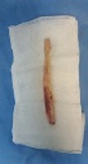
Eight fresh frozen human BTB grafts were prepared. Cadavers were between 35 and 70 years old and were all male. De-freezing was done by immersing the grafts in 37° centigrade normal saline. Then the grafts were randomly divided into two separate groups each containing 4 grafts. In the first group, grafts were prepared as routine with 25 by 10 millimeter bone blocks at each end. They were also trimmed to pass through a number 10 sizer. Two holes of 2 millimeters diameter were created in both bone blocks and 4 number 2 fiber-wires were passed through them. In the second group after the grafts were trimmed to 10 millimeters they were prepared with a newly invented method. In this method, the tibial side bone block was removed with dimensions of 10 by 30 millimeters. Two holes with 2 millimeters diameter were created 1 centimeter apart from each other. The femoral bone block was removed with dimensions of 9 by 15 millimeters (Figure 1). One 1.5 millimeter hole was created in the junction of one third to two thirds of bone block and a number 2.0 fiber wire was passed through it. Then the bone block is passed through the loop of an Endobutton (Smith and Nephew) and the bone block was folded on itself at the ligament-osseous junction. At this time the previously created hole was positioned on the ligament itself and using the No 2.0 fiber wire (which had been passed previously) the bone block was stitched to the ligament itself (Figures 2 and 3).

In enfolded method the patellar bone block and tibial bone blocks are 15 and 30 millimeters in length respectively.

The patellar side bone block is able to bend on itself at the bone ligament junction.

Graft is enfolded on itself after passing through the loop of Endobutton
After preparing the grafts, they were assembled in a universal testing machine (Santam 5 cap) for testing the mechanical properties of the grafts (Figure 4). Grafts were loaded by 1 millimeter per second tensile force and cycling of 0.05 HZ for 100 cycles. Maximum tensile strength, failure load and failure mode were derived and the values compared in each group. stress-strain curves for each graft were drawn by computer.

Graft is assembled on tension machine.
For statistical analysis between the two groups, SPSS 17 / Win was used for non-parametric Mann Whitney U statistical methods. In the current study, P-values of less than 0.05 was considered as significant.
3. Results
Table 1 shows failure loads in classic and enfolded groups. Despite the lower values for the enfolded group, difference was not significant (P = 0.25). Figure 5 shows the stress strain graph in the cases with maximum failure loads of both groups. As it is depicted in this figure, the curves have similar characteristics for yielding point and elastic domain. Also, results showed little difference in values for stiffness in both groups, which is reflected in this graph by a little bit more gradient curve in the classic group (285.25 N/meter in classic group versus 268.75 N/ per meter in new method). Difference was not significant (P = 0.1).
| Group | Number | Minimum (Newton) | Maximum (Newton) | Mean (Newton) | Standard Deviation | Result |
|---|---|---|---|---|---|---|
| Classic | 4 | 1610 | 1719 | 1660 | 51.4 | P-value = 0.25 |
| Enfolded | 4 | 1410 | 1672 | 1579 | 117.3 |
a Cases with maximum failure loads.

Stress-strain curve in cases with maximum failure loads
In all cases of classic group failures happened through the junction of ligament and the bone itself. In the enfolded group, failures occurred through the tendino-osseous junction except for one case. Only in the excepted case failure happened through ligament itself.
4. Discussion
In this study ultimate strength point, yielding point and failure mode of enfolded graft preparation were compared with standard BTB graft preparation. Although the grafts in classic group had a higher value for the failure load and yielding points, the differences were negligible. More interestingly, failure mode was similar in both groups.
Some additional issues concerning experimental protocol should be addressed. The use of fresh human cadaver bone in place of young human bone was a significant substitution made in the testing. The lower density of cadaveric bone may lead to premature severing (or partial severing) of the tendons in both groups. The lowered stiffness and strength of the cadaveric tendons may reduce the stiffness and failure loads of the tested constructs. The mechanical properties of the graft are affected by preservation, storage, and sterilization. Incorporation and remodeling of the graft further alter its properties. However, the clinical results between non irradiated allografts and autografts are the same (7-9). As this study was a parametric one, it is reasonable to accept that the tendon properties were affected equally in both groups, maintaining the validity of the comparison.
This study has shown that enfolded graft preparation has non-significant lower biomechanical properties compared to classic preparation methods. Here, we discuss that the clinical outcome of ACL reconstruction is not merely dependent on the shape of graft but also it is dependent on the overall stiffness of graft –fixation device complex (10). In the study of Shen et al, the stiffness of the flexor tendon graft connected to the Endobutton CL-BTB (Smith & Nephew) was inferior to that of the Endobutton CL (11). Smith et al on a study on canine models has shown that the suspensory graft fixation had a better result in terms of ligamentization compared to interference screw (12).
During the tension of grafts, failure occurred in both groups at the tendon-bone junction. Because of the configuration of the graft in the enfolded group, the maximum force was exerted at the tendon-bone junction. In the classic method this force was applied on bone itself but the results of failure mode were the same in both groups. In clinical setting and during graft tensioning the tensile force on the grafts is much lower than what is exerted during testing by tensile machine (120 N versus 1579). Rapid bone to bone healing without interference screw would justify its use in ACL reconstructions.
This could be a reason for the lower ultimate failure strength in this group. One may consider this weakness as a drawback for the utilization of this technique in ACL reconstruction but if we take a closer look at the mean failure load of the enfolded technique, we will realize that this failure point is much lower than what is exerted during surgery for graft tensioning (120 N versus 1579). Rapid bone to bone healing without an interference screw would justify its use in ACL reconstructions.
4.1. Conclusion
Suspensory fixation methods of Endobuttons is a common method for ACL reconstruction with hamstring autograft. However there was controversy using it as a BTB graft preparation method. Based on our findings, enfolded method of BTB graft preparation had acceptable biomechanical characteristics. This method is comparable to the conventional methods. We recommended using this method among the other graft preparation methods in patients who need ACL reconstruction and the suspensory fixation methods of Endobuttons could be easily applied to BTB graft.
