


1. Background
Non-contact anterior cruciate ligament (ACL) injury often occurs during decelerating movements such as single-leg landing after jumping (1). The timing and magnitude of the peak vertical ground reaction force (pVGRF), which represents the landing impact force, might increase the risk of ACL injury (2-5). During single-leg landing, ACL strain was shown to peak at the instant of the pVGRF (2). The pVGRF correlates positively with the proximal tibial anterior shear force and the knee extension internal moment, both of which increase ACL strain (3). A prospective study demonstrated that the pVGRF during double-leg drop landing is 20% higher in female athletes who had sustained ACL injuries than in those who had not (4). Research has shown that deceasing the pVGRF magnitude and controlling the time from initial contact (IC) to the pVGRF (time to pVGRF) are important to avoid the excessive ACL strain that leads to ACL injury during landing (5, 6).
The pVGRF changes based on the sagittal joint angle of the lower extremity during landing (7-10). During single-leg landing, smaller knee and hip flexion angles (9, 10) and smaller ankle dorsiflexion angles (11, 12) generate larger pVGRF magnitudes. It has been reported that the pVGRF is smaller during a landing when subjects consciously bend the knee and hip than during a natural landing (8, 13).
During landing, the co-contraction index (CCI) between the quadriceps and hamstrings tends to correlate with the sagittal joint angle of the lower extremity in healthy subjects and patients with reconstructed ACLs (14, 15). In healthy individuals, the CCI between the quadriceps and hamstrings after landing is significantly greater when the knee flexion angle is restricted to 0° - 25° compared to 25° - 50° or 50° - 75° (14). Compared to healthy subjects, patients with reconstructed ACLs have smaller knee flexion angles and a higher CCI between the quadriceps and hamstrings during landing (15).
Feed-forward activation, a type of neuromuscular control, occurs during the flight phase (16, 17). For instance, during double-leg landing, activation of the lower extremity muscles occurs approximately 80 ms before foot contact (16). Studies have demonstrated that the quadriceps and hamstrings contract simultaneously during the flight phase (18, 19). Feed-forward co-contraction during the flight phase is believed to contribute to control of the joint angle and the pVGRF during landing (16, 20). However, the correlations between the CCI, sagittal joint angles, and the pVGRF from the flight phase to the landing phase have not been analyzed.
2. Objectives
The purposes of this study were to clarify how the sagittal joint angle correlates with the pVGRF during the landing phase and how the CCI during the flight phase correlates with the sagittal angle during the landing phase in a single-leg jump-landing task. We hypothesized that the pVGRF is negatively correlated with the knee flexion angle during the landing phase. We furthermore hypothesized that the knee flexion angle during the landing phase is negatively correlated with flight-phase CCI.
3. Methods
3.1. Subjects
The inclusion criteria were as follows: physically active, at least 18 years of age, and without a history of serious injury or surgery involving the lower extremities or lumbar region. Seven women and seven men participated in this study. The subjects’ characteristics (mean ± standard deviation) were as follows: age, 22.6 ± 2.7 years; height, 164.4 ± 7.6 cm; body weight, 58.9 ± 8.9 kg; and body mass index, 21.7 ± 2.3 kg/m2. The subjects usually participated in sports such as soccer, handball, tennis, volleyball, and rugby that involve jump-landing and abrupt turns and stops. The dominant leg was defined as the leg used to kick a ball for the maximum distance (21). One of the 14 subjects was left-leg dominant. The sample size was calculated to be 14 subjects using G*power statistical software (alpha = 0.05, power = 0.8, two-tailed) with reference to the correlation coefficient between pVGRF and the knee flexion angle during single-leg jump landing (9, 22). All subjects read and signed an approved informed consent document before data collection. This study was approved by the university’s institutional review board.
3.2. Equipment Protocol
All subjects were clothed in identical athletic attire comprising spandex shirts, shorts, and shoes with no air cushions (step101, Lucky Bell, Kobe, Japan). They performed active static stretching (23) and no-load ergometer pedaling for 5 minutes as a warm-up before measurements. To obtain kinematic data, reflective markers (diameter, 14 mm) were attached to the anterior superior iliac spine (ASIS), posterior superior iliac spine (PSIS), greater trochanter (GT), lateral joint space (LJS) at the midpoint of the knee, lateral malleolus of the fibula, and head of the fifth metatarsal based on the landmarks reported in a previous study (24). A 25-cm-high step (RBK-BO001, Reebok, Canton, MA, USA) was placed 60 cm from the center of the force plate (22). The height of the force plate surface was 5 cm above the ground level. The lower extremity joint angles during jump landing were recorded using a high-speed camera (120 Hz, GC-P100, JVC, Yokohama, Japan). The high-speed camera was placed with the lens 335 cm from the center of the force plate, perpendicular to the plane of motion, to record sagittal plane joint angles (22, 25). The height of the camera was 1 m from the center of the lens to the floor. Videographic and force plate data were time synchronized using a synchronizer (PTS-110/2 LED, DKH, Tokyo, Japan).
3.3. Procedures
All subjects performed a single-leg jump-landing task with their dominant leg. They stood on the step on their dominant leg with the other knee bent at up to 90°, neutral hip rotation, arms crossed, and hands inserted into the opposite axillae to eliminate the effect of arm movement. All subjects jumped forward without intentional upward action and landed as naturally as possible on the center of the force plate and maintained balance for 5 seconds. A trial was deemed unacceptable if the arms left the chest, part of the sole of the foot fell outside the force plate at landing, the foot moved or slid after landing, and/or if the sole of the opposite foot touched the force plate or floor. All subjects practiced three single-leg jump-landing tasks before data collection.
The VGRF data were recorded using a force plate (260AA6, Kistler Instrument AG, Winterthur, Switzerland) at a sampling rate of 1000 Hz using the software (IFS-4J/3J, DKH). The VGRF data were filtered through a fourth-order Butterworth low-pass digital filter at a cut-off frequency of 50 Hz and normalized by body weight. Extracted VGRF data included the magnitude of the pVGRF and the time to pVGRF. The IC was defined as the moment when VGRF exceeded 10 N (26). The pVGRF has demonstrated acceptable test-retest reliability during single-leg jump-landing (27).
The sagittal joint angles were measured on extracted video frames. The angles of anterior pelvic tilt, hip flexion, knee flexion, and ankle dorsiflexion were measured as the sagittal joint angles at the timing of the pVGRF. The sagittal joint angles that were indicated by markers in each frame were measured using ImageJ (National Institutes of Health, Bethesda, MD, USA). Anterior pelvic tilt was defined as the angle formed between a line joining the ASIS and PSIS. The hip flexion angle was defined as the angle added to the anterior pelvic tilt to thigh angle from a vertical line relative to the floor. The knee flexion angle was defined as the angle obtained by subtracting a line joining the GT and LJS at the midpoint of the knee and lateral malleolus of the fibula from a line extending between the GT and LJS. The ankle dorsiflexion angle was defined as the angle formed between a line joining the LJS with the lateral malleolus of the fibula and the head of the fifth metatarsal. Although the anterior pelvic tilt and ankle dorsiflexion angles may appear to be in ankle dorsiflexion or anterior pelvic tilt positions, they may have shifted in the plantar flexion or posterior tilt directions compared to the stationary standing position. Thus, these two angles were subtracted from the standing position. The sagittal joint angles using two-dimensional analysis have demonstrated acceptable intrarater and test-retest reliability during landing (28, 29).
The muscular activation data were obtained by surface electromyography (EMG; WEB-1000, Nihon Kohden, Tokyo, Japan) and recorded at 1000 Hz with band-pass filtering (20 - 500 Hz) on a personal computer (CC-700H, Nihon Kohden) using a receiver (ZR-100H, Nihon Kohden). The skin was cleaned with alcohol before surface electrodes (ZB-150H, Nihon Kohden) for EMG recordings were applied. The specifications of the electrodes were as follows: size, 25 × 34.5 × 12 mm3; weight, 10 g; electrode size, 10 × 2 mm2; interelectrode distance, 5 mm. The surface electrodes were applied to the vastus medialis (VM) and semitendinosus (ST) according to the method of the Surface ElectroMyoGraphy for the Non-Invasive Assessment of Muscles project (30).
Prior to collecting the data during the landing task, EMG data were recorded during maximum voluntary isometric contraction (MVIC) of both the VM and ST (30). The MVIC was performed for 3 seconds. The root-mean-square (RMS) amplitudes were calculated using a 100-ms window, and the highest averaged RMS amplitudes were defined as the MVIC.
The RMS amplitudes during the landing task were normalized by MVIC (%MVIC). The VM and ST activations during the flight phase were analyzed using the %MVIC data. The flight phase was defined as 100 ms before landing to the IC (20, 22, 31, 32). Co-contraction between the VM and ST during the flight phase was extracted and calculated using the co-contraction index (CCI), as recommended by Falconer and Winter (19, 33). The following equation was used:
Iant is the area of the total antagonistic activity and is calculated using the following equation:
Where t1 and t2 denote the period where the ST activations were less than the VM activations, whereas t2 and t3 denote the period where the VM activations are less than ST activation; Itotal is the integral of the sum of ST and VM activations during the landing:
The EMG (%MVIC) data at 100 ms and 50 ms before the IC were also extracted. EMG is a reliable method for assessing the reproducibility of both quadriceps and hamstrings muscle activation during landing (34).
3.4. Statistical Analysis
The correlations between the VGRF variables, sagittal joint angles, and CCI were assessed using Spearman’s rank correlation coefficient, rho (ρ). The a priori α level was 0.05. Data were analyzed using SPSS V. 21.0 (IBM Corp, Armonk, NY, USA).
4. Results
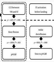
The results for the kinetics and kinematics variables during single-leg jump-landing are presented in Table 1. The knee flexion angle at the pVGRF was negatively correlated with the pVGRF (ρ = -0.609; P = 0.021) (Table 2). The knee flexion angle at the pVGRF was negatively correlated with the CCI between the VM and ST during the flight phase (ρ = -0.627; P = 0.016) (Table 3).
| Variable | Medium Value (Interquartile Range) |
|---|---|
| GRF | |
| pVGRF (%BW) | 371.7 (316.8 - 399.9) |
| Time to pVGRF (ms) | 40.5 (36.0 - 54.3) |
| Joint angle (°) | |
| Hip flexion | 34.1 (26.1 - 36.5) |
| Knee flexion | 44.2 (35.7 - 47.7) |
| Ankle dorsiflexion | 7.4 (4.7 - 15.1) |
| Muscular activation (%) | |
| VM at 100 ms before-landing | 40.9 (26.2 - 70.4) |
| VM at 50 ms before-landing | 66.3 (35.8 - 103.6) |
| ST at 100 ms before-landing | 27.1 (15.6 - 39.2) |
| ST at 50 ms before-landing | 37.9 (21.4 - 62.1) |
| CCI between VM and ST | 78.9 (66.2- 87.1) |
Abbreviations: BW, body weight; CCI, co-contraction index; IC, initial contact; pVGRF, peak vertical ground reaction force; ST, semitendinosus; Time to pVGRF, time from initial contact to pVGRF; VM, vastus medialis.
aThe EMG of each muscle was expressed as a percentage of the EMG value during the maximum voluntary isometric contraction.
Abbreviations: pVGRF, peak vertical ground reaction force; Time to pVGRF, time from initial contact to pVGRF.
aP < 0.05.
bP < 0.01.
Abbreviations: CCI, co-contraction index; IC, initial contact; ST, semitendinosus; VM, vastus medialis.
aThe EMG of each muscle was expressed as a percentage of the EMG value during the maximum voluntary isometric contraction.
bP < 0.05.
Although the hip flexion angle at the pVGRF was negatively correlated with the CCI between the VM and ST during the flight phase (ρ = -0.550; P = 0.042), it was not correlated with the pVGRF (ρ = -0.290; P = 0.923). Moreover, although the ankle dorsiflexion angle was positively correlated with the time to pVGRF (ρ = 0.660; P = 0.010), it was not correlated with the pVGRF (ρ = -0.530; P = 0.051).
5. Discussion
This cross-sectional study analyzed the correlations between single-leg jump-landing kinetics and kinematics in healthy athletes, and the results suggested that the flight-phase CCI is indirectly correlated with the pVGRF via the sagittal joint angles.
There was a significant negative correlation between the knee flexion angle at the pVGRF and the pVGRF magnitude. There was also a significant negative correlation between the knee flexion angle at the pVGRF and the CCI during the flight phase. The present study demonstrated that the knee flexion angle during the landing phase decreases as the CCI between the VM and ST increases during the flight phase. Furthermore, the pVGRF increases as the knee flexion angle decreases during the landing phase. These findings support the hypothesis of the present study.
Many experimental studies have clarified that increasing the knee flexion angle is important for impact absorption during landing (14, 35, 36). Theoretically, an increase in the knee flexion angle is beneficial for impact absorption because the negative work, which indicates the impact absorption energy, increases. In a double-leg landing task, a soft landing with knee flexion angle ≥ 90°, compared to a stiff landing by restricting the knee flexion angle to ≤ 90°, decreases the pVGRF, and the knee flexion angle and negative work affect impact absorption (7). In a single-leg jump-landing task, the maximum knee flexion angle and knee flexion excursion have been shown to affect impact absorption (8, 36). Similarly to previous studies, the present study demonstrated that a larger knee flexion angle at the pVGRF is important for impact absorption.
The present study also showed a negative correlation between the knee flexion angle at the pVGRF and the CCI between the VM and ST during the flight phase. The results suggest that excessive co-contraction of the VM and ST during the flight phase may decrease the knee flexion angle during the landing phase. It has been shown in single-leg landing tasks that the CCI between the quadriceps and hamstrings during the landing phase is significantly greater when the knee flexion angle is restricted to 0° - 25° compared to 25° - 50° or 50° - 75° (14). Compared to a preferred landing, a soft landing shows a greater knee flexion angle and a lower CCI between the vastus lateralis and biceps femoris during the landing phase (37). As described here, the CCIs analyzed in previous studies all pertain to the landing phase. Thus, the results from the present study are new data demonstrating the correlation between the CCI during the flight phase and the sagittal joint angle during the landing phase.
Feed-forward muscle activation prepares the lower extremities for impact immediately after landing (38). However, the correlation between the co-contraction between the VM and ST during the flight phase and the impact and lower extremity joint angles during landing had not been clarified. The results of the present study suggest that the co-contraction between the VM and ST during the flight phase is indirectly correlated with the pVGRF via the sagittal angles (Figure 1).

Correlations between the co-contraction index during the flight phase and impact absorption during single-leg jump-landing. The results suggest that the co-contraction between the VM and ST during the flight phase is indirectly correlated with the pVGRF via the sagittal angles. CCI, co-contraction index; pVGRF, peak vertical ground reaction force; ST, Semitendinosus; VM, Vastus medialis.
To the best of our knowledge, no other studies have demonstrated the correlation between the flight-phase CCI and the knee flexion angle at the pVGRF in single-leg jump-landing. In a drop vertical jump, which is a different task than that performed in the present study, knee flexion at initial contact decreases with increasing co-contraction of the quadriceps and hamstrings prior to landing (21). Although the task and the timing of measuring the knee flexion angle are different from the present study, comparable results were obtained. Thus, to the best of our knowledge, there are no reports that contradict the results of the present study.
Although the present study demonstrated a correlation between the flight-phase CCI and the hip flexion angle at the pVGRF, there was no correlation between the hip flexion angle and the pVGRF. A previous study also did not find a correlation between the hip flexion angle at the pVGRF and the pVGRF during single-leg jump-landing (9). Similar results were also reported in single-leg lateral jump-landing (22). Although hip flexion is considered to be important for impact absorption, the extent of its contribution is smaller than that of knee flexion (7). On the other hand, it has been reported that the pVGRF and the maximum hip flexion angle during landing are correlated (10), indicating that the correlation between the hip flexion angle and the pVGRF differs depending on the measurement timing. These findings highlight the importance of focusing on knee flexion rather than hip flexion for the timing of the pVGRF during the landing phase.
This study did not find a correlation between ankle dorsiflexion and the pVGRF. In landing tasks with the toes, dorsiflexion of the foot appears rapidly after contacting the ground in plantar flexion (7), indicating that the ankle could be in plantar flexion or dorsiflexion depending on the measurement timing. It has been shown in single-leg jump-landing, which is the same task used in the present study, that ankle plantar flexion is observed at the pVGRF when the height or distance of the jump is different, unlike the present study’s results (9). However, many studies have elucidated the effects of ankle kinematics on impact absorption (11, 12), indicating the importance of focusing on various parameters such as measurement timing, as well as moment (7) and displacement during landing (39).
In impact absorption, it is mechanically important to extend the time to pVGRF. The present study showed that the time to pVGRF is extended as the ankle dorsiflexion angle increases. It has been suggested that the time to pVGRF extends when consciously landing with the toes compared to landing with the heel (13). The present study also demonstrated that ankle dorsiflexion at the pVGRF is related to impact absorption.
The time from reaching maximum VGRF after the toes touch the ground to an actual ACL injury is extremely short, at approximately 40 ms (1, 2). It is therefore theoretically difficult to control the sagittal angle and to decrease VGRF simply through voluntary and reflexive feed-back control after landing. Furthermore, it is thought that the muscle activation prepares for impact absorption during the flight phase (38). Based on these reasons, the present results can be regarded as data that emphasize the importance of focusing on the flight-phase CCI, from the perspective of impact absorption.
A soft landing while consciously bending the hip and knee is recommended to decrease the pVGRF (7, 8, 13, 40). In a study in which subjects were instructed to land softly, the knee flexion angle increased and the landing-phase CCI and pVGRF decreased (37). The results of the present study suggested that controlling excessive co-contraction between the VM and ST during the flight phase increases the knee flexion angle during the landing phase. To promote better impact absorption, it may be important to provide instructions to avoid excessively increasing the CCI from the flight phase to landing and stiffening the joints.
5.1. Limitations
The present study had some limitations. First, because the CCI is the area of the EMG waveforms from the VM and ST that overlaps, the intensity of the co-contraction could not be ascertained. It has been reported that co-contraction enhances joint stability (41, 42) and that co-contraction is necessary for landing depending on the height of the fall (43). However, the effects of co-contraction were not assessed in the present study. Next, the CCI between the knee extensor and flexor muscles is affected by sex, age, and motor skill (44), but these factors were not considered. Last, the knee moment and strain of the ACL were not measured. Therefore, the direct effect that the VGRF variables and sagittal joint angle have on the risk of ACL injury remains unclear.
5.2. Conclusions
The CCI between the vastus medialis and semitendinosus during the flight phase may be related indirectly to a greater VGRF during single-leg jump-landing. In soft landing instructions to reduce the landing impact, it may be necessary to provide guidance that specifies avoiding excessive increases in the CCI from the flight phase until landing so that the joints do not become stiff.
