


1. Introduction
Due to increased life expectancy, pelvic organ prolapse (POP) is becoming a gynecological concern. Cervical elongation (CE) can occur in a company with POP (1). It is still being determined whether CE is an independent condition or always accompanies POP (2). Cervical elongation was also observed in uterus-preserving surgeries for POP and case reports and case series following various hysteropexy operations, ranging from 14% to 63% (1, 2). The presence of CE can affect the length of the surgery, change the operation technique and inform the surgeon about the difficulties along the way. Therefore, it should be evaluated preoperatively (3, 4). There is still no clear definition of CE (3). In some studies, cervical length > 3.38 cm and a cervix-to-corpus ratio > 0.79 are considered CE (3, 4). Also, it can be predicted via the difference between point C (cervix) and point D (posterior fornix and the attachment of the uterosacral ligament) in POP-Q assessment (5). Measuring CE using POP-Q has limited diagnostic value. Moreover, according to Alay et al. study, only about half of the patients with CE were diagnosed preoperatively (6).
The cervix comprises supra-vaginal and intra-vaginal parts with the same length, and CE can affect both parts equally (7). Cervical elongation of the supra-vaginal portion of the cervix is associated with POP, unlike the intra-vaginal portion, which is congenital and related to chronic cervicitis (7, 8). Antovska SV showed that POP-Q was ineffective in recognizing supra-vaginal cervical elongation (9).
In this case study, we are reporting the limitations of POP-Q in diagnosing supra-vaginal elongation.
2. Case Presentation
A 70-year-old woman, gravida 3, live 3, was referred to our center with a chief complaint of a protruding mass out of the vagina and urinary incontinence for the last six months. The previous year, she had failed prolapse surgery, including sacrospinous suspension, performed in a referral center by an expert pelvic surgeon.
2.1. Clinical Findings
Physical examination revealed hypermobility of the urethra and fourth-degree uterovaginal prolapse. The patient's current POP-Q exam, pelvic exam, and previous examination based on documents are illustrated in Table 1. We did not identify an elongated cervix on our pelvic exam or before the first prolapse repair.
2.2. Therapeutic Intervention
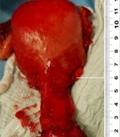
She was scheduled for transvaginal hysterectomy, high uterosacral suspension, anterior and posterior colporrhaphy, and trans-obturator tape for stress incontinence. Considering her comorbidities, including diabetes and coronary heart disease, she was not a good candidate for abdominal approaches for prolapse suspension. Because she was sexually active, obliterative POP procedures were not appropriate choices. Evaluation under anesthesia and after hysterectomy revealed an enlarged supra-vaginal cervix of about 6 centimeters (Figure 1).

Elongated supra vaginal cervix of about 6 centimeters
2.3. Follow-up and Outcomes
Postoperatively, she recovered well. There has been no recurrence to date.
3. Discussion
In 40% of women with POP, CE is associated with the degree of uterine descent. A surgeon should perform a cervical length evaluation preoperatively to choose the appropriate procedure. CE is considered a relative contraindication for uterine-preserving surgery in POP (7). Patients mostly prefer uterine-preserving surgeries rather than vaginal hysterectomies. However, surgeons believe CE can affect the patient's postoperative outcome, recurrence, and satisfaction (6). POP-Q examination differs CE from apical prolapse of the uterine (10). But POP-Q has low sensitivity and specificity (6). Cervical length (CL) calculated with POP-Q correlates with cervical length, but it doesn't give us an accurate measure. The anatomical cervical length is measured from the internal OS (the junction of the endometrium to the endocervical stroma) to the external OS (the junction of the endocervical stroma to the vaginal epithelium) on the removed uterus. POP-Q defines cervical length as the difference between points C and D. Given that point C is the most dependent portion of the intra-vaginal cervix and the anatomical location of the intra-vaginal cervix compared to the vaginal axis differs from patient to patient, and Point C can have a wide variation. Therefore CL is differently measured via POP-Q and anatomically (10). The Antovska SV study mentioned that the POP-Q examination could not identify supra-vaginal elongation between patients with POP and without CE (9).
Johnson et al. claimed that a Transvaginal sonogram gives a restricted evaluation of the cervix due to poor tissue discrimination from the vagina; in any case, sonovaginography is valuable at showing the portio vaginalis and outside cervical os and measuring cervical length (11).
Schulten et al. found that body mass index, smoking, and POP-Q point Ba are risk factors for pelvic organ prolapse recurrence after sacrospinous hysteropexy or vaginal hysterectomy with uterosacral ligament suspension (12). Our patient had none of these risk factors.
Hyakutake et al. reported a two-fold increase in cervical length compared with preoperative measurements in 62.5 % of cases during the first year following sacrospinous hysteropexy (13). Cervical elongation in our patient could have occurred after sacrospinous suspension.
Preoperative cervical length assessment is imperative before decision-making for pelvic organ prolapse surgical repair. In this case, POP-Q and bimanual examination showed POP and the failure of the previous surgery; however, there was no CE according to our preoperative POP-Q. However, after the hysterectomy, supra-vaginal elongation of 6 cm was seen. Based on this case and the studies mentioned above, it is visible that POP-Q is not reliable for diagnosing CE.
3.1. Conclusions
POP-Q is not effective at recognizing supra-vaginal elongation.
