The Role of CA19-9 and CA125 in Differentiation of Ovarian Dermoid Cyst and Ovarian Malignancy: A Case-Control Study

authors:

avatar Sima Shamshiri Khamene ORCID 1 , avatar Izat Mohammad Khawajah 1 , avatar Amir Ali Mahboobipour ORCID 2 , avatar Zahra Ataei Jafari 3 , avatar Elham Feizabad ORCID 4 , avatar Khadijeh Adabi 5 , avatar Zahra Rezaei 6 , 7 , *

School of Medicine, Tehran University of Medical Sciences, Tehran, Iran
Tracheal Diseases Research Center, National Research Institute of Tuberculosis and Lung Diseases, Shahid Beheshti University of Medical Sciences, Tehran, Iran
Faculty of Health, Liverpool Jhon Moores University, Liverpool, England
Department of Obstetrics and Gynecology, Yas hospital, Tehran University of Medical Sciences, Tehran, Iran
Division of Female Pelvic Medicine and Surgery, Department of Obstetrics and Gynecology, Yas Hospital, Tehran University of Medical Sciences, Tehran, Iran
Vali-e-Asr Reproductive Health Research Center, Imam Complex, Family Health Research Institute, Tehran University of Medical Sciences, Tehran, Iran
Division of Infertility, Department of Obstetrics and Gynecology, Yas Hospital, Tehran University of Medical Sciences, Tehran, Iran

how to cite: Shamshiri Khamene S, Mohammad Khawajah I, Mahboobipour A A, Ataei Jafari Z, Feizabad E, et al. The Role of CA19-9 and CA125 in Differentiation of Ovarian Dermoid Cyst and Ovarian Malignancy: A Case-Control Study. Fertil Gynecol Androl. 2023;3(1):e137144. https://doi.org/10.5812/fga-137144.

Abstract

Background:

An ovarian dermoid cyst is one of the most prevalent benign tumors in women of reproductive age. They can get complicated or be mistaken for malignant tumors. Most of the time, their pathology will not be discovered until after surgery.

Objectives:

Finding the role of CA19-9 and CA125 tumor markers in diagnosing the dermoid cyst before surgery and distinguishing it from the malignant type.

Methods:

A case-control study was conducted on all cases of dermoid cysts and malignant tumors (both diagnosed by pathology) from 2019 to 2021. Required information such as age, level of tumor markers, size, and whether it is unilateral or bilateral were extracted, and the tumor marker levels were analyzed in benign and malignant groups.

Results:

In this study, 95 cases of ovarian cysts and 85 control of malignant ovarian tumors were included. The mean level of CA19-9 and CA125 in the patients with dermoid cysts was 32.26 ± 5.45 units/mL and 31.37 ± 4.77 units/mL, respectively. In the malignant ovarian tumor group, the average level of CA19-9 and CA125 was equal to 9.56 ± 3.54 units/mL and 233.79 ± 54.57 units/mL, respectively. CA19-9 level was significantly higher in the dermoid cyst group (P-value < 0.001). CA125 level was significantly elevated among the malignant group (P-value < 0.001). It was observed that there was a significant relationship between the level of CA19-9 with the size of the cyst and the level of CA125 with the age of the patients. No significant relationship was found between the level of tumor markers and whether they were unilateral or bilateral.

Conclusions:

Regarding the correlation between the cyst size and CA19-9 in the comparison of the two groups, this tumor marker may be a suitable diagnostic marker in benign ovarian cysts along with ultrasound.

1. Background

The annual incidence rate of the ovarian dermoid cyst or mature cyst teratoma is 10 in 100,000 women (1). Ovarian cancer is the fifth leading cause of cancer-related deaths in women due to a lack of diagnostic tools resulting in late diagnosis (2, 3). Adnexal masses have different etiologies. They may present with various symptoms, such as abdominal and pelvic pain, bloating, or increasing abdominal size (4). Most ovarian cysts are benign (5), and the most common one is the dermoid cyst or mature cystic teratoma (MCT) (6).

Mature cystic teratomas are more common in reproductive age (7). They may present with different findings in imaging, including Rokitansky nodules, calcifications, palm tree-like protrusion, diffuse or regional high amplitude echoes called intratumoral fat, and dot-dash sign (8). The modality choice of diagnosis is transvaginal ultrasonography (9-11). Surgical resection of the cyst is often the treatment of choice, especially in uncomplicated cases. The incidence of malignant transformation in mature cystic teratoma has been reported to be 1 - 3%, mostly occurring in postmenopausal women (12, 13).

Tumor markers such as CA19-9 and CA125 may be used to manage ovarian cancer (14). CA125 is the best and one of the most common (15) tumor markers for ovarian cancer. However, diagnosing ovarian cancer is a combination of physical examination, ultrasonography, and CA125 measurement (16, 17). CA125 and CA19-9 can be helpful in diagnosing mature cystic teratoma (18). These markers may also be useful for malignancy prediction in some borderline tumors alongside other tumor markers like HE4, NLR (19, 20), and CEA (21).

2. Objectives

The aim of this study was to assess the role of tumor markers CA19-9 and CA125 in diagnosing the dermoid cyst before surgery and distinguishing it from the malignant type in Yas Hospital from 2019 to 2021.

3. Methods

We conducted a case-control study in patients with adnexal mass in Yas Hospital, Tehran, Iran, from 2019 to 2021. The study was approved by the Ethics Committee of Tehran University of Medical Sciences. The sample size for each group was calculated to be 80, using the reported values in previous studies10, the study power of 80%, and the two-tailed significance level of 5%. Patients’ information was collected considering ethical aspects using patient file numbers without mentioning names. The cyst size and being unilateral or bilateral, was determined with ultrasound imaging. Transabdominal (3.5 to 5 MHz) and, if needed, transvaginal ultrasonography (5 to 7 MHz transducer) were performed in one center by an attending radiologist. The level of tumor markers was also measured in the laboratory of Yas Hospital using blood serum or plasma. Tumor markers were determined by radioimmunoassay. The cut-off values for CA19-9 and CA125 were 37 and 35 U/mL, respectively. Patients’ symptoms were collected based on what they expressed in their history. Statistical analyses were done with SPSS-25 (IBM SPSS Statistics for Windows, Version 25.0. Armonk, NY: IBM Corp; 2017). The results were stated as frequency (%) or mean ± standard deviation (SD) for categorical and numerical variables. We used median and range for data that were not normally distributed. Chi-square or Fisher’s exact tests were used for comparisons. The mean differences in numerical variables between the two groups were compared by independent t-test. For numerical variables, Pearson’s correlation and linear regression were used. A P-value of less than 0.05 was considered statically significant.

4. Results

After pathology confirmation postoperatively, 95 cases with dermoid cyst and 85 cases with ovarian cancer were included. The average age of patients with the dermoid cyst was 33.23 ± 5.23 years, and the average age of ovarian cancer was 36.24 ± 4.53 years. Patients with dermoid cyst were significantly younger than malignant cases (P-value < 0.001). Our investigation of the symptoms at the first visit showed that abdominal pain was the most common symptom in the dermoid cyst group; however, the most frequent symptom was pelvic pain in the malignancy group (Table 1). The mean cyst diameter was 4.25 ± 1.31 cm in dermoid cysts and 8.56 ± 1.82 cm in malignant mass. Cyst diameter was significantly larger in malignant cases (P-value = 0.023).

Table 1.

Symptoms in Each Group a

Symptoms Dermoid CystOvarian CancerP-Value
Abdominal pain57 (59.6)77 (90)< 0.001
Menstrual disorders21 (21.7)7 (9)< 0.001
Incidental17 (18.7)1(1)< 0.001

The mean levels of CA19-9 and CA125 in the patients with dermoid cysts were 32.26 ± 5.45 units/mL and 31.37 ± 4.77 units/mL, respectively. More than 51% of them had a high level of CA19-9. In the malignant ovarian tumor group, the mean level of CA19-9 and CA125 was 9.56 ± 3.54 units/mL and 233.79 ± 54.57 units/mL, respectively. Interestingly, more than 77% of these patients had a high level of CA125. CA19-9 level was significantly higher in the dermoid cyst group (P-value < 0.001). CA125 level was significantly elevated in the malignant group (P-value < 0.001) (Table 2).

Table 2.

The Level of Tumor Markers in the Study Population a

Ovarian Dermoid Cyst (units/mL) (N = 95)Ovarian Cancer (units/mL) (N = 85)P-Value
CA19-9 32.26 ± 5.459.56 ± 3.54< 0.001
CA125 31.37 ± 4.77233.79 ± 54.57< 0.001

One of the factors expected to affect the tumor marker level is the presence of a mass in both ovaries simultaneously. For this reason, we separated the unilateral and bilateral cases. In the dermoid cyst group, 38 cases (35.2%) were right-sided, 49 cases (45.4%) were left-sided, and eight patients (19.4%) were bilateral. In the ovarian cancer group, 70 cases (82.4%) were one-sided, and the remaining cases were bilateral. Cyst location was not significantly different between both groups (P-value = 0.477).

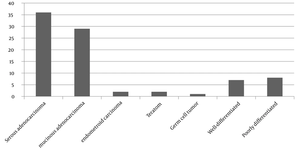
Among the cancerous patients, 36 people have serous adenocarcinoma, 29 people have mucinous adenocarcinoma, two people have endometroid carcinoma, two people have teratoma, one person has germ cell tumor, seven people have well-differentiated adenocarcinoma, and eight people have poorly differentiated carcinoma (most of the patients in this group are epithelial type) (Figure 1).

Pathology of ovarian cancers

In the correlation coefficient analysis, we measured the age and the level of tumor markers based on the symptoms, then whether the cyst was unilateral or bilateral. It was observed that there was a significant relationship between the level of CA19-9 with the size of the cyst (r = 0.192, P = 0.007) and the level of CA125 with the age of the patients (r = -0.237, P = 0.005). In the ANOVA analysis, no significant relationship was found between the level of tumor markers and whether they were unilateral or bilateral.

5. Discussion

Mature teratoma cyst or dermoid cyst is the most common teratoma, and ovarian neoplasm (10 - 25%) constitute 60% of all benign ovarian neoplasms (22). They are mostly unilateral (23). In our study, 80.6% of dermoid cysts were unilateral. This cyst grows slowly and is bilateral in 10% of cases (24). Malignant changes in this type of ovarian cyst are in about 1 - 2% of patients, and torsion is their most complication (25). Some radiologic features, like ultrasonography, can suggest MCT, but tumor markers alongside the imaging accommodate the diagnosis (8). The majority of signs and symptoms of ovarian cancer are nonspecific. Abdominal pain was the most frequent symptom (59.6%), followed by menstrual disorders in dermoid cyst cases in our study.

Lack of specific and sensitive biomarkers can lead to late diagnosis and even patient death (26). It showed that the mean level of CA19-9 was higher in the dermoid group than in the cancer group, and the mean level of CA125 was much lower in the dermoid group than in the cancer group. It is similar to Wang et al.’s study at Wenzhou Medical University. They found that CA19-9 was likely the most reliable tumor marker for diagnosing MCT (27). It also resembles Mughir and Al-Hilli’s study in 2019 in Iraq on 50 cases of ovarian dermoid cysts and 50 control patients. Their results show that the CA19-9 level in the case group was about four times higher than in the control group. In our study, there was a direct relationship between the size of the cyst and the level of CA19-9; likewise, in their study, there was a direct relationship between the size of the tumor and the level of CA19-9. In their study, most dermoid cysts are unilateral, and about 10 - 15% of them were bilateral, as well as our research (28).

In Prodromidou et al.’s study and colleagues, the meta-analysis showed that in patients with elevated CA19-9, there was an increased size in mature cystic teratoma. At the same time, there was no relationship between the patient’s age, bilaterality, site of the lesion, and the simultaneous elevation of CA-125 in patients with elevated CA19-9 (29). It is consistent with our findings that there was no correlation between CA19-9 and age or being unilateral/bilateral, but there is a significant correlation with size. In Lertkhachonsuk et al.’s study, CA125 was the best diagnostic tumor marker for borderline and malignant mucinous ovarian tumors, followed by CA19-9 and CEA (30). This is similar to our study that in the cancer group, 88% of patients have elevated CA125. Ali et al.’s study demonstrated that the combination of microRNA-204, CA125, and CA19.9 was the best test for the early detection of ovarian tumors and cancers, but in our study, elevated CA19-9 was more related to the dermoid cyst group and elevated CA125 more related to cancer group (26). Also, in Bagde et al.’s study LDH, BHCG, and CA19-9 did not significantly differ in malignant and benign cases (31).

In Sagi-Dain et al.’s study, they demonstrated that combining CA19-9 with CA125 compared with CA125 alone did not significantly affect sensitivity and specificity in differentiating malignant from benign adnexal masses. The mean CA19-9 levels were higher in metastatic carcinoma and could help differentiate metastatic tumors from primary ovarian malignancy (32). The strength point of this study is that all imaging assessments and tumor marker measurements are done in an academic referral center. Increased sample size and assessment of other tumor markers can be suggested for future studies to cover the weak points of our study. Most of the dermoid cysts in the present study were unilateral. According to the relationship of CA19-9 in the dermoid cyst group with cyst size, it can be said that the larger the cyst size, the higher the tumor marker level will be. The increase in the level of CA125 suggests the possibility of malignancy in the ovarian cyst alongside an increase in age and cyst size. Finally, CA19-9 can be used in diagnosis along with other diagnostic tools, especially ultrasound. This tumor marker has no diagnostic value in small cysts, but if the cyst size increases, it will help in a definitive diagnosis.

5.1. Conclusions

Regarding the correlation between the cyst size and CA19-9 by comparison of these two groups, this tumor marker may be a suitable diagnostic marker in benign ovarian cysts along with ultrasound. High levels of CA125 will be in favor of the malignant type.

Acknowledgements

References

  • 1.

    Li RY, Nikam Y, Kapurubandara S. Spontaneously Ruptured Dermoid Cysts and Their Potential Complications: A Review of the Literature with a Case Report. Case Rep Obstet Gynecol. 2020;2020:6591280. [PubMed ID: 32292616]. [PubMed Central ID: PMC7150697]. https://doi.org/10.1155/2020/6591280.

  • 2.

    Shetty M. Imaging and Differential Diagnosis of Ovarian Cancer. Semin Ultrasound CT MR. 2019;40(4):302-18. [PubMed ID: 31375171]. https://doi.org/10.1053/j.sult.2019.04.002.

  • 3.

    Gumusoglu E, Gunel T, Hosseini MK, Seymen N, Senol T, Sezerman U, et al. The importance of dysregulated miRNAs on ovarian cysts and epithelial ovarian cancer. Eur J Gynaecol Oncol. 2021;42(1):66-72. https://doi.org/10.31083/j.ejgo.2021.01.2167.

  • 4.

    Biggs WS, Marks ST. Diagnosis and Management of Adnexal Masses. Am Fam Physician. 2016;93(8):676-81. [PubMed ID: 27175840].

  • 5.

    Mobeen S, Apostol R. Ovarian Cyst. In: StatPearls, editor. StatPearls. Treasure Island, FL: StatPearls Publishing; 2020.

  • 6.

    Gupta A, Jha P, Baran TM, Maturen KE, Patel-Lippmann K, Zafar HM, et al. Ovarian Cancer Detection in Average-Risk Women: Classic- versus Nonclassic-appearing Adnexal Lesions at US. Radiology. 2022;303(3):603-10. [PubMed ID: 35315722]. https://doi.org/10.1148/radiol.212338.

  • 7.

    Abhilasha N, Bafna UD, Pallavi VR, Rathod PS, Krishnappa S. A review of squamous cell carcinoma arising in mature cystic teratoma of the ovary. Indian J Cancer. 2016;53(4):612-4. [PubMed ID: 28485364]. https://doi.org/10.4103/0019-509X.204763.

  • 8.

    Lee FK, Horng HC, Wang PH. Tumor markers and mature cystic teratoma of the ovary. J Chin Med Assoc. 2018;81(6):497-8. [PubMed ID: 28882729]. https://doi.org/10.1016/j.jcma.2017.08.002.

  • 9.

    Srisajjakul S, Prapaisilp P, Bangchokdee S. Imaging features of unusual lesions and complications associated with ovarian mature cystic teratoma. Clin Imaging. 2019;57:115-23. [PubMed ID: 31212220]. https://doi.org/10.1016/j.clinimag.2019.05.013.

  • 10.

    Dede M, Gungor S, Yenen MC, Alanbay I, Duru NK, Hasimi A. CA19-9 may have clinical significance in mature cystic teratomas of the ovary. Int J Gynecol Cancer. 2006;16(1):189-93. [PubMed ID: 16445632]. https://doi.org/10.1111/j.1525-1438.2006.00284.x.

  • 11.

    Li Q, Wang Q, Gao HY, Chen JM. Expression and clinical significance of tumor markers in ovarian mature cystic teratoma. Clin Exp Obstet Gynecol. 2016;43(3):397-400. https://doi.org/10.12891/ceog2128.2016.

  • 12.

    Mabuchi Y, Ota N, Kobayashi A, Tanizaki Y, Minami S, Ino K. Identical twins with mature cystic teratomas treated with laparoscopic surgery: Two case reports. Mol Clin Oncol. 2017;6(2):276-8. [PubMed ID: 28357110]. [PubMed Central ID: PMC5351738]. https://doi.org/10.3892/mco.2016.1118.

  • 13.

    Chiang AJ, Chen MY, Weng CS, Lin H, Lu CH, Wang PH, et al. Malignant transformation of ovarian mature cystic teratoma into squamous cell carcinoma: a Taiwanese Gynecologic Oncology Group (TGOG) study. J Gynecol Oncol. 2017;28(5):e69. [PubMed ID: 28657230]. [PubMed Central ID: PMC5540728]. https://doi.org/10.3802/jgo.2017.28.e69.

  • 14.

    Liu Y, Li DH, Xu H, Fan YJ, Malla S, Yan HQ, et al. The clinical significance of CA19-9 in ovarian mature cystic teratoma. Clin Exp Obstet Gynecol. 2016;43(4):522-5. https://doi.org/10.12891/ceog3003.2016.

  • 15.

    Chandra A, Pius C, Nabeel M, Nair M, Vishwanatha JK, Ahmad S, et al. Ovarian cancer: Current status and strategies for improving therapeutic outcomes. Cancer Med. 2019;8(16):7018-31. [PubMed ID: 31560828]. [PubMed Central ID: PMC6853829]. https://doi.org/10.1002/cam4.2560.

  • 16.

    Pan C, Stevic I, Muller V, Ni Q, Oliveira-Ferrer L, Pantel K, et al. Exosomal microRNAs as tumor markers in epithelial ovarian cancer. Mol Oncol. 2018;12(11):1935-48. [PubMed ID: 30107086]. [PubMed Central ID: PMC6210043]. https://doi.org/10.1002/1878-0261.12371.

  • 17.

    Hamd-Ghadareh S, Salimi A, Fathi F, Bahrami S. An amplified comparative fluorescence resonance energy transfer immunosensing of CA125 tumor marker and ovarian cancer cells using green and economic carbon dots for bio-applications in labeling, imaging and sensing. Biosens Bioelectron. 2017;96:308-16. [PubMed ID: 28525848]. https://doi.org/10.1016/j.bios.2017.05.003.

  • 18.

    Yesilyurt H, Seckin B, Aktulay A, Ozyer S. Age-stratified analysis of tumor markers and tumor characteristics in adolescents and young women with mature cystic teratoma. J Chin Med Assoc. 2018;81(6):499-504. [PubMed ID: 28826726]. https://doi.org/10.1016/j.jcma.2017.07.005.

  • 19.

    Seckin KD, Karsli MF, Yucel B, Bestel M, Yildirim D, Canaz E, et al. The utility of tumor markers and neutrophil lymphocyte ratio in patients with an intraoperative diagnosis of mucinous borderline ovarian tumor. Eur J Obstet Gynecol Reprod Biol. 2016;196:60-3. [PubMed ID: 26683535]. https://doi.org/10.1016/j.ejogrb.2015.10.025.

  • 20.

    Chen X, Fang C, Zhu T, Zhang P, Yu A, Wang S. Identification of factors that impact recurrence in patients with borderline ovarian tumors. J Ovarian Res. 2017;10(1):23. [PubMed ID: 28376898]. [PubMed Central ID: PMC5379723]. https://doi.org/10.1186/s13048-017-0316-5.

  • 21.

    Zhang W, Jia S, Xiang Y, Yang J, Jia C, Leng J. Factors associated with misdiagnosis of frozen section of mucinous borderline ovarian tumor. J Int Med Res. 2019;47(1):96-104. [PubMed ID: 30198356]. [PubMed Central ID: PMC6384450]. https://doi.org/10.1177/0300060518795582.

  • 22.

    Uysal F, Balsak D, Uysal A, Incebiyik A, Muratli A, Yildirim Y. Clinical, Diagnostic and Anatomical Aspects of Mature Cystic Teratomas. Austin J Radiol. 2014;1(1):3.

  • 23.

    Adam Osman A, Tahtabasi M, Gedi Ibrahim I, Issak Hussein A, Mohamud Abdullahi I. Ovarian Torsion Due to Mature Cystic Teratoma During the Early Postpartum Period: A Rare Case Report. Int Med Case Rep J. 2021;14:333-8. [PubMed ID: 34045903]. [PubMed Central ID: PMC8146748]. https://doi.org/10.2147/IMCRJ.S310071.

  • 24.

    Cho HY, Kim K, Jeon YT, Kim YB, No JH. CA19-9 elevation in ovarian mature cystic teratoma: discrimination from ovarian cancer - CA19-9 level in teratoma. Med Sci Monit. 2013;19:230-5. [PubMed ID: 23539074]. [PubMed Central ID: PMC3628710]. https://doi.org/10.12659/MSM.883865.

  • 25.

    Lee JY, Shin W, Kim JS, Park JH, Cho S. Combination of clinical and laboratory characteristics may serve as a potential diagnostic marker for torsion on mature cystic teratomas. Obstet Gynecol Sci. 2018;61(3):386-94. [PubMed ID: 29780782]. [PubMed Central ID: PMC5956123]. https://doi.org/10.5468/ogs.2018.61.3.386.

  • 26.

    Ali FT, Soliman RM, Hassan NS, Ibrahim AM, El-Gizawy MM, Mandoh AAY, et al. Sensitivity and specificity of microRNA-204, CA125, and CA19.9 as biomarkers for diagnosis of ovarian cancer. PLoS One. 2022;17(8):e0272308. [PubMed ID: 35921382]. [PubMed Central ID: PMC9348731]. https://doi.org/10.1371/journal.pone.0272308.

  • 27.

    Wang YQ, Xia WT, Wang F, Zhuang XX, Zheng FY, Lin F. Use of cancer antigen 125, cancer antigen 19-9, and the neutrophil-to-lymphocyte ratio to diagnose mature cystic teratoma with torsion. Int J Gynaecol Obstet. 2017;137(3):332-7. [PubMed ID: 28273351]. https://doi.org/10.1002/ijgo.12139.

  • 28.

    Mughir AK, Al-Hilli NM. Correlation of Mature Cystic Teratomas With CA19-9. J Med Surg Pract. 2021;7(2):177-89.

  • 29.

    Prodromidou A, Pandraklakis A, Loutradis D, Haidopoulos D. Is There a Role of Elevated CA 19-9 Levels in the Evaluation of Clinical Characteristics of Mature Cystic Ovarian Teratomas? A Systematic Review and Meta-analysis. Cureus. 2019;11(12):e6342. [PubMed ID: 31938630]. [PubMed Central ID: PMC6952044]. https://doi.org/10.7759/cureus.6342.

  • 30.

    Lertkhachonsuk AA, Buranawongtrakoon S, Lekskul N, Rermluk N, Wee-Stekly WW, Charakorn C. Serum CA19-9, CA-125 and CEA as tumor markers for mucinous ovarian tumors. J Obstet Gynaecol Res. 2020;46(11):2287-91. [PubMed ID: 32830422]. [PubMed Central ID: PMC7693209]. https://doi.org/10.1111/jog.14427.

  • 31.

    Bagde ND, Bagde MN, Lone ZA. Relationship between Serum Tumor Markers, CA-125, CEA, CA19-9, LDH, and βHCG with Histopathology and Age in Women with Ovarian Tumors. Asian Pacific Journal of Cancer Biology. 2020;5(4):167-71. https://doi.org/10.31557/apjcb.2020.5.4.167-171.

  • 32.

    Sagi-Dain L, Lavie O, Auslander R, Sagi S. CA 19-9 in evaluation of adnexal mass: retrospective cohort analysis and review of the literature. Int J Biol Markers. 2015;30(3):e333-40. [PubMed ID: 25704505]. https://doi.org/10.5301/jbm.5000139.