1. Background
Sarcocystis parasites are coccidian protozoan organisms with worldwide distribution and one of the most common parasites in domestic livestock that can cause severe infection in some hosts such as cattle and sheep (1, 2). Also, some species of the parasite transmit to humans and cause clinical presentations. Sarcocystis parasites have significant health importance, especially in certain parts of the world that animal breeding is a main occupation, thus impose economic losses. Reduced milk production, abortion, and sometimes death occurs in intermediate hosts such as cows, sheep, goats, and pigs (2). Sarcocystis parasites need two hosts to complete their life cycle. In its life cycle, there is a definitive host and an intermediate host. The sexual life cycle occurs in the definitive host that often belongs to carnivorous animals that accidentally ingest oocysts. The asexual part of the life cycle takes place in the intermediate host (mostly herbivores) (3). The dogs and cats play an important role in the spreading of the parasite in the environment. They can excrete up to 200 million infective oocysts in the course of disease. Animals remain asymptomatic and rarely have mild diarrhea. Humans are infected by eating schizonts (asexual form) of Sarcocystis hominis and S. suihominis in undercooked beef, pork, mutton, and raw or poorly cooked meat products (hamburgers, sausages, and hot dogs) (1, 3). People who voluntarily ate Sarcocystis-infected meat excreted Sarcocystis oocysts in feces after eight days or more. The severity of clinical signs depends on the type of meat consumed (beef or pork) and Sarcocystis species causative agent (4).
The Sarcocystis can act as an opportunistic organism in immunocompromised patients, which can cause intestinal or muscular disturbances, but epidemiology and clinical impact of human infections by Sarcocystis are largely unknown, and reports continue to be rare (5). The prevalence of sarcocystosis in slaughtered animals has been studied in many regions around the world. The digestion technique of infected tissues is the most common and sensitive method for detecting infections with microscopic forms of Sarcocystis, which comprises taking pieces of the contaminated tissue and crushing it, then solubilizing it in a digestive solution containing chloric acid and pepsin, and subsequently staining with Giemsa. Contamination rates from 3% to 100% have been reported by different methods in different parts of the world (6, 7). The method of identification of organisms affects the rate of positivity, e.g., microscopic or molecular methods. Its frequency in livestock such as cattle, sheep, and goats has been reported up to 100% in some parts of Iran (8). Some carnivores are the definitive host for many species of Sarcocystis, but species transmitted by dogs are usually not transmitted by cats (9). Systematic analysis revealed that the total prevalence (95% confidence intervals) of Sarcocystis spp. in Iranian ruminants, according to published studies, was an average of 74.40% (10).
2. Objectives
To the best of our knowledge, there is no reliable study and published data about Sarcocystosis in Hormozgan abattoirs (Industrial and traditional), which investigate consumed meat contamination. Thus, we designed a descriptive cross-sectional study to observe the situation. A large amount of consumed meats in Hormozgan is imported from nearby provinces, and determination of Sarcocystis frequency in Hormozgan can help researchers to estimate Sarcocystosis in those regions and get more ideas for further investigations.
3. Methods
The study was done from September 2019 to January 2020 in Hormozgan. The population of the study was 400 samples of slaughtered animals belonging to Hormozgan, Fars, and Kerman provinces.
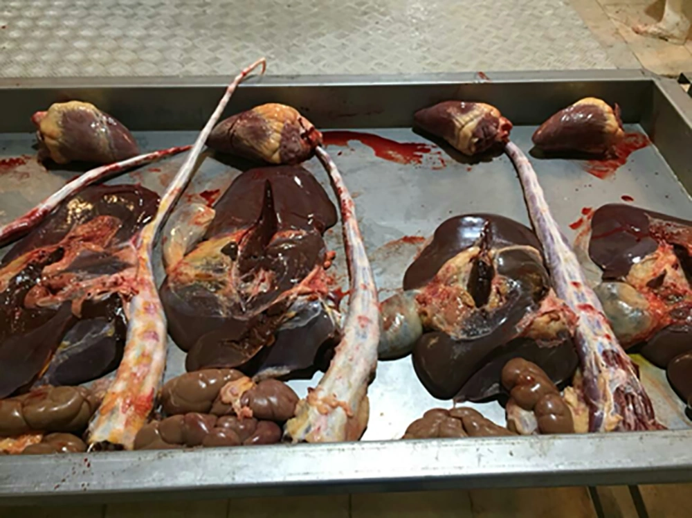
After taking and processing tissue specimens, 20 g of the tissues, i.e., heart, diaphragm, and esophagus, were crushed with an electric shredder and placed in 2 mL of digestion solution (Figure 1). To make a digestion buffer, it is necessary to solve 1.3 g of pepsin, 3.5 mL HCl and 2.5 g NaCl in 500 mL of distilled water at pH = 7.4. Meat samples were placed in the incubator at 40°C for 3 hours to release parasites and passed through a non-sterile gauze. The filtrated liquids were centrifuged at 2500 rpm for 5 min, and a direct smear was prepared from the precipitate. It is important to allow those smears to air- dry at least 1-hour before staining. Smears were fixed with methanol 96% (Germany, Merck) then stained with diluted Giemsa according to instructions for 15 minutes; after staining, slides were washed with tap water and allowed to air-dry completely. The stained slides were examined microscopically at magnifications of 400× and 1000×.
A minimum of 100 high-power fields must be examined. It should be noted that in cases that smears are strongly positive and especially when there was high contamination, plenty of parasites were readily detectable in magnification of 400×.
3.1. Statistical Analysis
The obtained data were analyzed via SPSS software ver. 22 (Chicago, IL, USA). Moreover, the chi-square test was used for the analysis of data. To control the confounder variables, the logistic regression analysis was implemented. The results would be considered statistically significant if the P value was less than 0.05 (P < 0.05).
3.2. Ethical Clearance
This descriptive cross-sectional study was approved by the Ethics Committee of Hormozgan University of Medical Sciences (HUMS) numbered IR.HUMS.REC.1397.143.
4. Results
In this study, a total of 400 samples were collected from slaughtered animals in two industrial and traditional slaughterhouses in Bandar Abbas, Iran, and transferred to the parasitology department laboratory. Slaughtered animals and carcasses were examined for macroscopic cysts, and all were negative. All samples were evaluated by the digestion method and direct smear microscopy. The slaughtered animals were 212 cows (53.0%), 152 goats (38.0%) and 36 sheep (9.0%). According to the information obtained, it was found that a large number of slaughtered cattle were imported to Bandar Abbas from other provinces.
The total frequency was 92.5%. Most of the imported cattle for meat consumption were from Kerman and Fars provinces. Therefore, at the time of sampling, important information, and animal characteristics such as sex, type of livestock, and breeding location were considered and recorded for each animal. Most of the samples belonged to animals bred in Hormozgan Province with 188 (47.0%), Kerman province with 38.3% (153), Fars 13.3% (49), and other provinces 2.5% (10). Hormozgan Province had the highest number of cows with 157 (74.1%), and Kerman province had the highest number of goats and sheep with 84 (55.26%) and 18 (50%), respectively (Table 1). The highest frequency of Sarcocystis in all three kinds of animals was detected in esophageal tissue, with 87.5% (350). Heart tissue with 72.8% (291) had the lowest frequency rate (Table 2).
| Province | Total | Others | Fars | Kerman | Hormozgan |
|---|---|---|---|---|---|
| Kind of animals | |||||
| Cow | 212 (53.0) | 4 (40.0) | 0 (0.0) | 51 (33.3) | 157 (83.5) |
| Goat, No | 152 (38.0) | 4 (40.0) | 42 (85.7) | 84 (54.9) | 22 (11.7) |
| Sheep | 36 (9.0) | 2 (20.0) | 7 (14.3) | 18 (11.8) | 9 (4.8) |
| The number of cattle according to province | 400 | 10 (2.5) | 49 (13.3) | 153 (38.3) | 188 (47.0) |
aValues are expressed as No. (%).
| Infected Tissue | Kinds of Cattlea | Totala | P Value | ||
|---|---|---|---|---|---|
| Sheep | Goat | Cow | |||
| Heart | < 0.001b | ||||
| Negative | 13 (36.1) | 64 (42.1) | 32 (15.1) | 109 (27.3) | |
| Positive | 23 (63.9) | 88 (57.9) | 180 (84.9) | 291 (72.8) | |
| Diaphragm | 0.009b | ||||
| Negative | 3 (8.3) | 22 (14.5) | 53 (25.0) | 78 (19.5) | |
| Positive | 33 (91.7) | 130 (85.5) | 159 (75.0) | 322 (80.5) | |
| Esophagus | 0.004b | ||||
| Negative | 5 (13.9) | 29 (19.1) | 16 (7.5) | 50 (12.5) | |
| Positive | 31 (86.1) | 123 (80.9) | 196 (92.5) | 350 (87.5) | |
| Total | 0.021b | ||||
| Negative | 2 (5.6) | 19 (12.5) | 10 (4.7) | 31 (7.75) | |
| Positive | 34 (94.4) | 133 (87.5) | 202 (95.3) | 369 (92.25) | |
aValues are expressed as No. (%).
bSignificant.
Statistics showed that total frequency had no significant relation with animal gender (P > 0.05). The breeding location was determined and considered during sampling. According to the gender variable, infection in male cows of Hormozgan Province with 163 (47.7%) is significantly higher than animals imported from other provinces. The total frequency in each province based on all three infected tissues includes Kerman 94.8%, Hormozgan 93.6%, Fars 83.7%, and 70% in other provinces, respectively. According to the results of the above-mentioned infection rates, Kerman Province had the highest level of contamination rate (94.8%). The results show that among all three provinces, the cows of Hormozgan with 151 (96.2%) had a significant frequency (P < 0.005). Using the chi-square test and comparing the total infections and by animal type of each province, goats in Kerman Province with 60.9% were significantly infected compared to goats of other provinces.
5. Discussion
In this study, we investigated the frequency of Sarcocystis parasites in domestic animals slaughtered in Bandar Abbas slaughterhouses. A total of 400 animals and 3 different tissues were studied. These animals consisted of 212 cows (53%), 152 goats (38%), and 36 sheep (9%), respectively. The carcasses of all the animals were examined for the presence of Sarcocystis macroscopic cysts, all of which were negative. However, microscopic examination by pepsin digestion showed a total frequency of 92.25% in these animals. Rate of infection indicates the high frequency of parasites in slaughtered animals that supply edible meat in Bandar Abbas. In other parts of the country, various studies have been conducted that show the high frequency of Sarcocystis in livestock in Iran, and our results are similar to many of these studies. Investigations in other provinces in Iran showed the reported frequency between 90% - 100% (6). In other parts of the world, Sarcocystis has been detected in 70 - 100 of examined meat samples (11).
In two studies carried out in Shahriar (Alborz Province) with a time interval, infection with macroscopic cysts was not reported (12). Also, studies in Isfahan and Kerman did not report any contamination with macroscopic cysts, or macroscopic cysts were restricted to a specific organ like the esophagus that is in agreement with our study (6, 13, 14). However, in some studies, macroscopic cysts have been reported at a low frequency (15, 16).
The results of this study showed that the total frequency of Sarcocystis has a significant relation with infected organ and livestock (animal) type (P < 0.005). The highest esophageal infection was reported in cows with 92.2%, the highest diaphragm infection in sheep with 91.7%, and the highest heart infection occurred in cows with 84.9%. In 2013, Alibeigi et al. (12), by direct microscopic method, detected the same results (12), but Mirzaei and Rezaei (17) identified the heart as the most infected organ in 2008. Some other studies indicated that all three examined organs were 100% infected.
Due to the geographical location and climatic conditions of Hormozgan Province, animal husbandry is not very prosperous in this province. Therefore, in order to supply edible meat, it is necessary to import livestock from other provinces of the country, especially neighboring provinces. The obtained information indicates that more than half of the animals slaughtered in Bandar Abbas slaughterhouses come from other provinces, e.g., Fars and Kerman. By reviewing the statistical results, it was found that most of the animals slaughtered in this study were related to Hormozgan Province with 47%, Kerman Province 38.3%, Fars Province 13.3%, and other provinces 2.5%, respectively.
Total Sarcocystis frequency had no significant statistical relation with animal gender. However, the frequency of Sarcocystis infection in male animals in Hormozgan Province was significantly higher than the livestock of other provinces based on the breeding location (P < 0.5). In the studies conducted in Kerman, Jahrom, and Tabriz, the frequency of infection was not significantly related to gender (2). In this study, the highest total infection based on breeding location is related to Kerman Province livestock with 94.8%, which is significantly higher than the frequency in the livestock of other provinces. These studies are consistent with our results. The key point of this study is that the current data of this study can be an estimate for the neighboring provinces because of a large number of slaughtered animals imported from neighbors. On the other hand, the lack of information about this parasite in Hormozgan was filled because there was no reliable and published data before.
5.1. Conclusions
The foodborne and zoonotic Sarcocystis parasite gives an idea to evaluate and examine fecal samples of people in future studies. Diagnostic limitations have impeded our ability to increase our understanding of the epidemiology and public health significance of Sarcocystis infection in humans. This study seems to be the first of its own kind carried out in meat samples of Hormozgan. This parasite also causes health, economic and pathological problems in humans and animals. Therefore, it is important to note that macroscopic methods implemented in abattoirs and slaughterhouses are not a good way to detect this parasite in livestock, and microscopic digestion methods should be used to obtain the correct results. The most important conclusion of this study is that more control should be applied over slaughtered livestock and that the people of the community who consume these meats should be given proper and complete training, such as full cooking or freezing of meat before consuming it.

