Isolation and Characterization of Exosomes Derived From Breast Cancer MDA-MB-231 Cell Line

authors:

avatar Zahra Rafighdoust ORCID 1 , avatar Javad Baharara 2 , * , avatar Mohammad Mahdi Forghanifard 3 , avatar Mohammad Amin Kerachian 4

Department of Biology, Mashhad Branch, Islamic Azad University, Mashhad, Iran
Department of Biology and Research Center for Animal Development Applied Biology, Mashhad Branch, Islamic Azad University, Mashhad, Iran
Department of Biology, Damghan Branch, Islamic Azad University, Damghan, Iran
Department of Medical Genetics, Faculty of Medicine, Mashhad University of Medical Sciences, Mashhad, Iran

How To Cite Rafighdoust Z, Baharara J, Forghanifard M M , Kerachian M A . Isolation and Characterization of Exosomes Derived From Breast Cancer MDA-MB-231 Cell Line. Gene Cell Tissue. 2021;8(1):e110505. https://doi.org/10.5812/gct.110505.

Abstract

Background:

Exosomes are membrane nanovesicles, 30 to 100 nm in diameter, secreted by most cell types. Besides playing many biological roles, especially in cell-cell communication, scientific proof indicated that the pathology of so many human cancers is closely related to many biologic elements in exosomes. They may serve as useful biomarkers for treatment, prognosis, and detection. Cancer cells produce more exosomes, inducing changes in target cells (near or distant from the tumor), such as metastasis and chemotherapy resistance. Therefore, isolation, identification, and analysis of these microvesicles seem essential.

Objectives:

The current study aimed at collecting and purifying microvesicles secreted from breast cancer cells and confirming the identity of the obtained exosomes using methods assessing size and morphology.

Methods:

In recent research, the MDA-MB-231 cell line was grown under standard conditions. Released exosomes were collected and ultra-centrifuged. Scanning (SEM) and transmission (TEM) electron microscopes, atomic force microscopy (AFM), and dynamic light scattering (DLS) were used to assess exosome size.

Results:

The obtained data revealed that MDA-MB-231 cells produced exosomes. The nanovesicles were isolated from the culture medium of MDA-MB-231 cells by applying different strategies, including differential centrifugation, filtration, and ultracentrifugation. The exosomes were characterized; they had a size of 30 - 100 nm and spherical shape.

Conclusions:

Intercellular communication can be mediated through direct cell-cell contact or transfer of secreted molecules. In the last two decades, a third mechanism for intercellular communication has emerged that involves intercellular transfer of extracellular vesicles (exosomes). Due to their many functions in the body, it is of great importance to purely isolate and recognize exosomes to understand their modes of action as the first step in the advancement of researches. However, more research is required to obtain cost-effective and efficient methods. It was found that MDA-MB-231 cells release exosomes. They are spherical and 30-100 nm in diameter. The use of a combination strategy for the first time was useful in isolating exosomes derived from MDA-MB-231 cells without disturbing their structure. Further studies are required to compile a uniform protocol for exosome isolation in medical research.

1. Background

One of the multicellular organism hallmarks is cell-cell interaction guided by straight cell contacts or secreted molecular transition. Another intercellular communication mechanism identified over the last two decades is the intercellular transition of extracellular vesicles (EVs). Such tiny membrane vesicles carry signals to long distances from generating cells and influence numerous cell life aspects (1) Exosomes are membrane-derived nanovesicles with a size of 30 - 100 nanometer (nm). They are generated by all cell types from endosomes (2). Exosomes can serve as mediators of cell-cell communication by transmitting and sharing their charges and altering the biological properties of recipient cells (3, 4). Exosomes are secreted by various normal and cancer cells. Cancer cells develop more exosomes, which induce changes in recipient cells (near or distant from the tumor), including angiogenesis, invasion, metastasis, and resistance to chemotherapy (5, 6).

Exosomes derived from tumors become momentous agents that promote contact among the nearby cells and influence cell activities linked to cancer growth or development. A high-efficiency procedure for the isolation and characterization of exosomes generates crucial facts to better understand their specific role in cancer (7). Biological fluids and EVs can be obtained from cell lines in culture media. Strives made to take conventional approaches to EV isolation to understand their function and form (8, 9).

Despite the numerous reports published on comparative methods for EV isolation, no related guidelines for clinical practices are in hand yet. Differential centrifugation is among the most widely used EV isolation processes (10).

2. Objectives

Concerning many roles recently assigned to exosomes, the characterization of these nanostructures raised interest. The current study aimed at isolating exosomes derived from MDA-MB 231 cell culture media using ultracentrifugation and reporting the characteristics of these vesicles.

3. Methods

3.1. Cell Culture

The human breast cancer MDA-MB-231 cell line (in-vitro osteolytic form) was grown in Dulbecco’s Modified Eagle Medium (DMEM) augmented with 10% fetal bovine serum (FBS) and 1% penicillin-streptomycin solution at 37°C in 5% humidity and 5% (v/v) CO2 (11).

3.2. Exosome Collection and Purification

MDA-MB-231 breast cancer cells in the 3rd passage were washed twice with phosphate-buffered saline (PBS) when covering 80% of the flask surface. Then, the cells were inoculated into a fresh cell medium with exosome-free FBS. After 24 - 48 hours, the culture was centrifuged to separate cellular residue from larger vesicles at 300 g for 10 minutes and 2000 g for 15 minutes, and then filtered on a 0.22-µm filter. Finally, ultracentrifugation at 100,000 g for 70 minutes was performed. The supernatant was discarded, the exosome pellet was suspended in 1 mL of PBS, and stored at -20°C (12, 13).

3.3. Transmission Electron Microscopy

The exosomes were sonicated for 30 minutes for homogenization. For this purpose, exosomes were fixed with 1 mL of 2.5% glutaraldehyde in 0.1 M sodium cacodylate solution for one hour at 4°C. Then, they were placed on 200 mesh copper grids, allowed to dry at room temperature, and exposed to 3% (w/v) uranyl acetate in water for 20 seconds. Then the grid was rinsed with PBS and allowed to dry at room temperature. Imaging was performed at 120 kV voltage (14).

3.4. Scanning Electron Microscopy

The suspension obtained by ultracentrifugation was sonicated. A small amount of sample was fixed with 2.5% paraformaldehyde and diluted in distilled water. The sample was dehydrated with ethanol, transferred to silicon chips, and placed in the acetone for one minute to be dehydrated and dry. Then it was washed with water and dried. The photos were taken by SEM (LMU TESCAN BRNO-Mira3) with the selected voltage of 30 kV (13).

3.5. Atomic Force Microscopy

The exosomes were sonicated for 30 minutes for homogenization. First, 3 µL of purified exosomes were fixed with 100 µL of 2% paraformaldehyde; then, a small drop of the solution was placed on a slide. After about 15 minutes, when the sample was dry, AFM (JPK-Nano Wizard II) was used to investigate (15, 16).

3.6. Dynamic Light Scattering

The exosomes were sonicated for 30 minutes for homogenization. For investigating the size of the exosomes with DLS, approximately 300 µL of the exosomal solution was inoculated into 1 mL PBS. The prepared sample was inserted into the device, and the results were analyzed. The DLS measurements were analyzed with a Zetasizer Nano (100Z-Horiba-SZ) (17).

4. Results

4.1. Characterization of Exosomes Derived From MDA-MB-231 Cells

4.1.1. Transmission Electron Microscopy

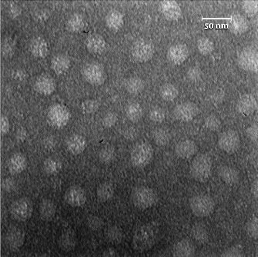
The morphology and size of the obtained exosomes were explored by TEM. The membrane integrity of exosomes was maintained during the isolation process so that their spherical structure remained intact. The particle sizes were within the reasonable range for spherical-shaped exosomes (30 - 100 nm) (Figure 1).

Characterization of exosomes released from MDA-MB-231 cells; transmission electron microscopy image of exosomes derived from MDA-MB231 cells. The sizes of exosomes were within the acceptable range of 30 to 100 nm. Their sphericity is apparent.

4.1.2. Atomic Force Microscopy

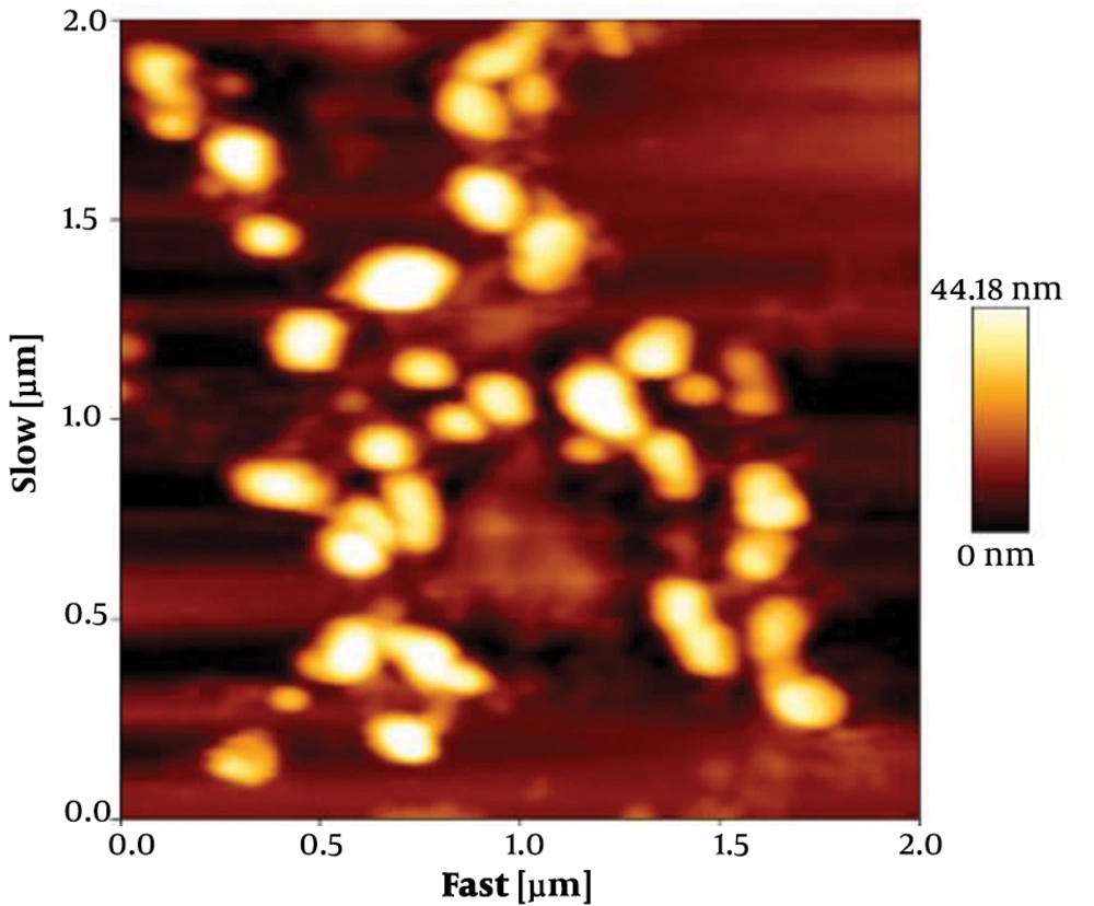
The size of exosomes was analyzed by tapping mode. Data analysis showed that the size of particles were within the acceptable range of 30 to 100 nm. The collected data showed that the membrane integrity of exosomes was maintained during the isolation (Figure 2).

Characterization of exosomes released from MDA-MB-231 cells; atomic force microscopy image of exosomes derived from MDA-MB-231 cells; the sizes of exosomes ranged from 30 to100 nm.

4.1.3. Dynamic Light Scattering

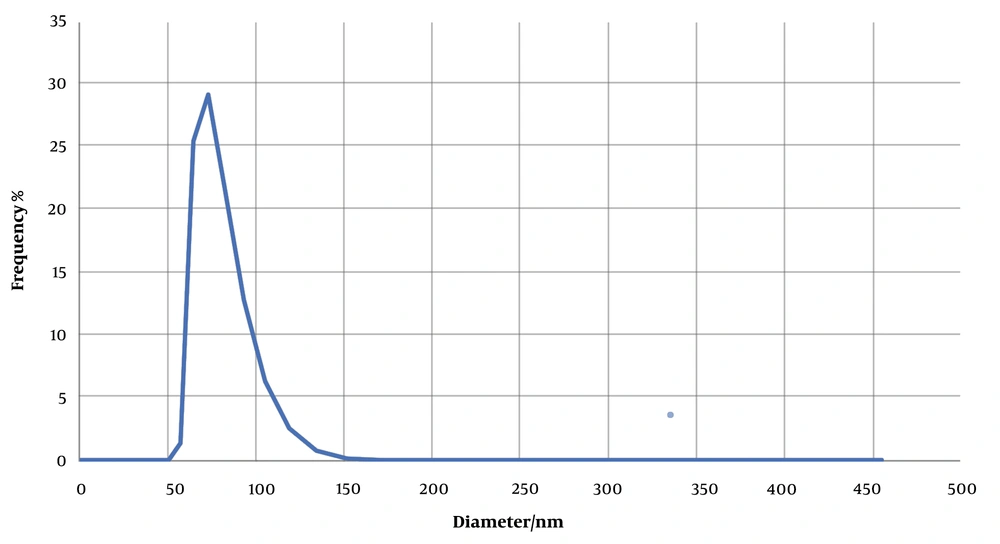
The size of exosomes isolated from MDA-MB-231cells was determined using a Zetasizer Nano and DLS technique. For analysis, 1 mL of the prepared sample was poured into a low-volume cuvette. Assessment of the distribution of sizes by DLS showed a peak of approximately 70 nm for the population of extracted exosomes (Figure 3).

Characterization of released by MDA-MB-231 cells; dynamic light scattering analysis of exosomes derived from MDA-MB-231 cells; the majority of exosomes isolated by the ultracentrifugation had an average diameter of 70 nm.

4.1.4. Scanning Electron Microscopy

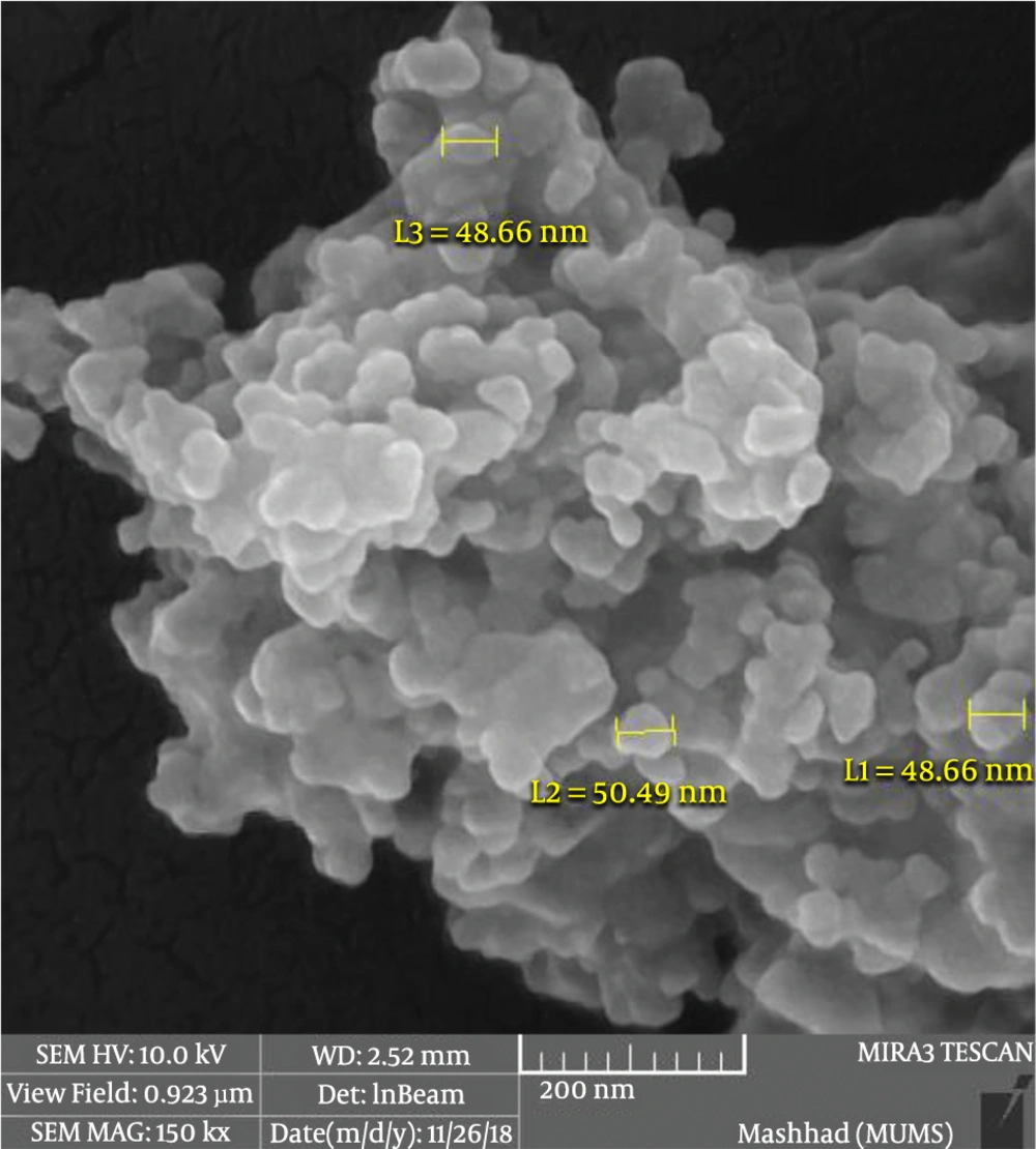
Exosomes were analyzed by SEM to confirm their morphology and size, as described in the Materials and Methods section. As shown in Figure 4, the exosomes were spherical, and their sizes ranged from 30 to 100 nm. The results confirmed the purity of exosomes isolated from the cell culture media (Figure 4).

Characterization of exosomes released from MDA-MB-231 cells; scanning electron microscopy analysis of exosomes derived from MDA-MB-231 cells; spherical morphology was apparent. Their sizes ranged from 30 to 100 nm.

The obtained results confirmed the methods employed for the isolation and characterization of exosomes.

5. Discussion

Under normal and pathological conditions, all cells release vesicles called exosomes, which attracted researchers’ attention in recent years due to their numerous roles (17). Exosomes contain genetic information and proteins, which vary across the cells they originated. Information can be transmitted through the exosome to the recipient cell, near or far from the release site, and induce functional and phenotypic changes in the target cell (18, 19). It opens up the possibility that cancer cells exert their biological effects by transmitting information to the target cells through exosomal transfer. Therefore, exosomes can act as a paracrine agent between cancer or tumor cells and normal cells (20). Exosomes derived from tumor cells are considered as agents promoting contact among nearby cells and influencing cell activities linked to the growth of cancer (7). As a result, the purification and identification of exosomes, as the first step towards human health promotion, is an important topic of discussion in basic research and clinical applications (20).

Hence, it is necessary to develop appropriate methods to isolate and characterize exosomes, which may be useful in the prognosis, prevention, and treatment of diseases. Therefore, due to the importance of the pure isolation of exosomes to understand their modes of action and application in medical sciences, the present study aimed at isolating exosomes derived fromMDA-MB-231 cells by differential centrifugation and filtration before ultracentrifugation to obtain pure exosomes and evaluate their size and appearance. The exosomes may be used as biomarkers for the early detection of breast cancer. To date, various strategies are proposed for exosome isolation. The difference in these methods is based on their efficiency (19). In standard isolation methods, the removal of excess particles and protein concentration are also considered (21). Previous researches reported ultracentrifugation as an efficient and inexpensive method, in which a considerable amount of exosomes precipitates, although it leads to exosome aggregation. Several studies suggested ultracentrifugation in sucrose gradients, which does not cause exosomal aggregation. However, the contamination of exosomes with sucrose, as a gradient reagent, may interfere with exosome functional studies (20). Thus, every method of purification has advantages and disadvantages, and it may be suitable to use a combination of the two. An effective manner due to using the differential centrifuge and filtering of the culture medium of the breast cancer cells was performed before ultracentrifugation. Then, exosomes were sonicated for homogenization and identified based on size and appearance using reliable methods. The current study findings showed that exosomes derived from MDA-MB-231 cells were spherical vesicles with 30 - 100 nm in diameter, indicating that the employed strategy was effective. The current study results were consistent with those of other research. Reimondi et al. (22) analyzed the size of myeloma-derived exosomes using the DLS method and reported that the U266- and OPM2-derived exosomes were approximately 100 and 50 nm in diameter, respectively. In another study, the size of mesenchymal stem cells isolated by ultracentrifugation, examined by DLS and TEM, were 80 and 40 - 100 nm, respectively.

In general, the technique proposed for isolation, verification, and identification of exosomes is advantageous in analyzing their roles, functions, and applications. However, there is no gold standard for exosome isolation. Further studies are required to achieve a uniform protocol for exosome isolation.

Acknowledgements

References

  • 1.

    Raposo G, Stoorvogel W. Extracellular vesicles: exosomes, microvesicles, and friends. J Cell Biol. 2013;200(4):373-83. [PubMed ID: 23420871]. [PubMed Central ID: PMC3575529]. https://doi.org/10.1083/jcb.201211138.

  • 2.

    Inder KL, Ruelcke JE, Petelin L, Moon H, Choi E, Rae J, et al. Cavin-1/PTRF alters prostate cancer cell-derived extracellular vesicle content and internalization to attenuate extracellular vesicle-mediated osteoclastogenesis and osteoblast proliferation. J Extracell Vesicles. 2014;3. [PubMed ID: 25018864]. [PubMed Central ID: PMC4072912]. https://doi.org/10.3402/jev.v3.23784.

  • 3.

    An T, Qin S, Xu Y, Tang Y, Huang Y, Situ B, et al. Exosomes serve as tumour markers for personalized diagnostics owing to their important role in cancer metastasis. J Extracell Vesicles. 2015;4:27522. [PubMed ID: 26095380]. [PubMed Central ID: PMC4475684]. https://doi.org/10.3402/jev.v4.27522.

  • 4.

    Bouyssou JM, Manier S, Huynh D, Issa S, Roccaro AM, Ghobrial IM. Regulation of microRNAs in cancer metastasis. Biochim Biophys Acta. 2014;1845(2):255-65. [PubMed ID: 24569228]. [PubMed Central ID: PMC4170185]. https://doi.org/10.1016/j.bbcan.2014.02.002.

  • 5.

    Kahlert C, Kalluri R. Exosomes in tumor microenvironment influence cancer progression and metastasis. J Mol Med (Berl). 2013;91(4):431-7. [PubMed ID: 23519402]. [PubMed Central ID: PMC4073669]. https://doi.org/10.1007/s00109-013-1020-6.

  • 6.

    Tomasetti M, Lee W, Santarelli L, Neuzil J. Exosome-derived microRNAs in cancer metabolism: possible implications in cancer diagnostics and therapy. Exp Mol Med. 2017;49(1). e285. [PubMed ID: 28104913]. [PubMed Central ID: PMC5291842]. https://doi.org/10.1038/emm.2016.153.

  • 7.

    Lim J, Choi M, Lee H, Kim YH, Han JY, Lee ES, et al. Direct isolation and characterization of circulating exosomes from biological samples using magnetic nanowires. J Nanobiotechnology. 2019;17(1):1. [PubMed ID: 30612562]. [PubMed Central ID: PMC6322342]. https://doi.org/10.1186/s12951-018-0433-3.

  • 8.

    Cvjetkovic A, Lotvall J, Lasser C. The influence of rotor type and centrifugation time on the yield and purity of extracellular vesicles. J Extracell Vesicles. 2014;3. [PubMed ID: 24678386]. [PubMed Central ID: PMC3967015]. https://doi.org/10.3402/jev.v3.23111.

  • 9.

    Minciacchi VR, Freeman MR, Di Vizio D. Extracellular vesicles in cancer: exosomes, microvesicles and the emerging role of large oncosomes. Semin Cell Dev Biol. 2015;40:41-51. [PubMed ID: 25721812]. [PubMed Central ID: PMC4747631]. https://doi.org/10.1016/j.semcdb.2015.02.010.

  • 10.

    Tauro BJ, Greening DW, Mathias RA, Ji H, Mathivanan S, Scott AM, et al. Comparison of ultracentrifugation, density gradient separation, and immunoaffinity capture methods for isolating human colon cancer cell line LIM1863-derived exosomes. Methods. 2012;56(2):293-304. [PubMed ID: 22285593]. https://doi.org/10.1016/j.ymeth.2012.01.002.

  • 11.

    Kaplan RN, Riba RD, Zacharoulis S, Bramley AH, Vincent L, Costa C, et al. VEGFR1-positive haematopoietic bone marrow progenitors initiate the pre-metastatic niche. Nature. 2005;438(7069):820-7. [PubMed ID: 16341007]. [PubMed Central ID: PMC2945882]. https://doi.org/10.1038/nature04186.

  • 12.

    Karlsson T, Lundholm M, Widmark A, Persson E. Tumor Cell-Derived Exosomes from the Prostate Cancer Cell Line TRAMP-C1 Impair Osteoclast Formation and Differentiation. PLoS One. 2016;11(11). e0166284. [PubMed ID: 27832183]. [PubMed Central ID: PMC5104397]. https://doi.org/10.1371/journal.pone.0166284.

  • 13.

    Tiedemann K, Sadvakassova G, Mikolajewicz N, Juhas M, Sabirova Z, Tabaries S, et al. Exosomal Release of L-Plastin by Breast Cancer Cells Facilitates Metastatic Bone Osteolysis. Transl Oncol. 2019;12(3):462-74. [PubMed ID: 30583289]. [PubMed Central ID: PMC6305809]. https://doi.org/10.1016/j.tranon.2018.11.014.

  • 14.

    Xiao J, Pan Y, Li XH, Yang XY, Feng YL, Tan HH, et al. Cardiac progenitor cell-derived exosomes prevent cardiomyocytes apoptosis through exosomal miR-21 by targeting PDCD4. Cell Death Dis. 2016;7(6). e2277. [PubMed ID: 27336721]. [PubMed Central ID: PMC5143405]. https://doi.org/10.1038/cddis.2016.181.

  • 15.

    Balnois E, Wilkinson KJ. Sample preparation techniques for the observation of environmental biopolymers by atomic force microscopy. Colloids Surf A. 2002;207(1-3):229-42. https://doi.org/10.1016/s0927-7757(02)00136-x.

  • 16.

    Nguyen DB, Ly TB, Wesseling MC, Hittinger M, Torge A, Devitt A, et al. Characterization of Microvesicles Released from Human Red Blood Cells. Cell Physiol Biochem. 2016;38(3):1085-99. [PubMed ID: 26938586]. https://doi.org/10.1159/000443059.

  • 17.

    Chernyshev VS, Rachamadugu R, Tseng YH, Belnap DM, Jia Y, Branch KJ, et al. Size and shape characterization of hydrated and desiccated exosomes. Anal Bioanal Chem. 2015;407(12):3285-301. [PubMed ID: 25821114]. https://doi.org/10.1007/s00216-015-8535-3.

  • 18.

    Szatanek R, Baj-Krzyworzeka M, Zimoch J, Lekka M, Siedlar M, Baran J. The Methods of Choice for Extracellular Vesicles (EVs) Characterization. Int J Mol Sci. 2017;18(6). [PubMed ID: 28555055]. [PubMed Central ID: PMC5485977]. https://doi.org/10.3390/ijms18061153.

  • 19.

    Nojehdehi S, Hashemi SM, Hesampour A. Isolation and characterization of exosomes separated from stem cells by ultra-centrifuge method. Res Med. 2017;41(4):244-50.

  • 20.

    Pakravan K, Babashah S, Javan M, Sadeghizadeh M, Mowla SJ. Isolation, Characterization and Cellular Uptake of Exosomes Derived from Bone Marrow Mesenchymal Stem Cells. Pathobiol Res. 2016;19(3):17-31.

  • 21.

    Zhang Z, Wang C, Li T, Liu Z, Li L. Comparison of ultracentrifugation and density gradient separation methods for isolating Tca8113 human tongue cancer cell line-derived exosomes. Oncol Lett. 2014;8(4):1701-6. [PubMed ID: 25202395]. [PubMed Central ID: PMC4156197]. https://doi.org/10.3892/ol.2014.2373.

  • 22.

    Raimondi L, De Luca A, Amodio N, Manno M, Raccosta S, Taverna S, et al. Involvement of multiple myeloma cell-derived exosomes in osteoclast differentiation. Oncotarget. 2015;6(15):13772-89. [PubMed ID: 25944696]. [PubMed Central ID: PMC4537049]. https://doi.org/10.18632/oncotarget.3830.