1. Background
Breast cancer has had an increased incidence in developed countries due to the lifestyle factors associated with, for example, smoking, inadequate exercise, and unhealthy food programs (1). This cancerous disease has been detected in, approximately more than 10% of womankind in the north Americas (2). Presently, cancer treatment is administered using many chemo-therapeutic agents, surgical procedures, and chemotherapy approaches. However, taking chemotherapy drugs produces side effects. Therefore, developing new anti-cancer compounds based on medicinal plants and traditional medicine has attracted considerable interest, particularly due to their proven effectiveness in improving the treatment process (3). It has long been demonstrated that traditional medicinal plants play an important role in the treatment of various cancer diseases, and the application of medicinal plants is now one of the most basic methods to treat cancer (4).
Bioactive compounds, such as Taxol, Vinblastine, Vinflunine, Vinorelbine, Camptothecin, Vincristine, and Vindesine have therapeutically significant effect on the treatment of the various cancer diseases (5). Various medicinal plants belonging to different plant families have been used in nutrition, cosmetics, and medicine for centuries. The genus Lavandula Lamiaceae are annuals, shrubs, and herbaceous plants. The antioxidant, insecticidal, anti-inflammatory, sedative, and spasmodic properties of Lavandula plants have long been recognized (6). Potential anticancer and ant-proliferative activities of the lavender essential oil distilled from Lavandula angustifoli has also been demonstrated (7). Anticancer properties of this species, however, have not received sufficient research attention.
The present study, therefore, aimed to investigate and compare the effects of some plant extract on two MCF7 and KBR3 cell lines by performing cell viability assay and gene expression analysis. Using Ephedra foeminea infusion has been widely reported to treat their ailments by cancerous patients (8).
Using E. foeminea as the primary ingredient in supplementing treatment has been documented in 68% of breast cancer patients. Moreover, E. foeminea has only become one of the most common plants utilized by the Palestinian population recently (i.e., from 0.0% in 2011 to 55.2% in 2014) (4). Analyzing genes expressed in a given cell or tissue could serve as a novel bio-markers, and overexpression of caspase genes has been sufficient to induce apoptosis in mammalian cells (9).
2. Objectives
This study aimed to evaluate the effects of some plant species such as L. angustifolia, E. major, and Scenedesmus obliquus on cell viability of two MCF7 and SK-BR3 cancer cell lines. The gene expression levels of two Bcl-2 and Caspase 3 genes involved in breast cancer were analyzed.
3. Methods
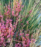
Lavander and ephedra plants investigated in this study were harvested in Yazd province of Iran and then were identified as L. angustifolia and E. major species by expert botanist (Figure 1). Dried leaves and algae mass were powdered using an electric mill and were later immersed in 80% ethanol: distill water 1: 10 (w/v). Solutions were placed on a shaker for one day at room temperature. Then, the mixture was passed through filter paper (Blue Ribbon, Grade 589, and Germany). Vacuum evaporator (Heidolphe, Germany) was used to concentrate the extracts, and lyophilizing was performed to obtain hydrous extract using freeze dryer (10).
3.1. Cell Culture and Cell Viability Assay
MCF-7 and SK-BR-3 cells created by human breast cancer were purchased from the National Cell Bank of Iran (Tehran, Iran). Gibco medium cell culture was used to culture the cells, and then fetal bovine serum (10%) was added to 96-well culture plates at 37°C in a 5%-CO2 incubator (Memert, Germany). Two penicillin and streptomycin antibiotics (Sigma Aldrich, Germany) from Passage 17 were used for performing all experiments under a humidified air atmosphere. MTT reagent was used to carry out the cell viability assay adopting the colorimetric method (11, 12). Cell treatment was performed for 24 hours using the obtained alcoholic extracts (25, 100, 500, 100 μg/μL). The cells were then subjected to MTT solution) and incubated for four hours. The formed purple formazan crystal was dissolved using 100 μL of DMSO (13). Measurement of cell viability was carried out using a plate reader at 570nm wavelength.
3.2. Molecular Manipulation
Treated and untreated cells were harvested by centrifugation, and then the cells were washed three times using PBS solution. Total RNA extraction kit (Parstos Biotechnology, Iran) was employed to perform RNA extraction. The quality and quantity of the extracted RNA were monitored using gel electrophoresis agarose and NanoDrop spectrophotometer, respectively (14). Then, 50 ng of purified RNA was applied for first-strand cDNA production using a superscript First-strand Synthesis system (Amplicon, USA).
3.3. Primers and qRT-PCR
The Primer was designed using Primer III software based on presented sequences in the bioinformatics database. All sequences of target and GAPDH as reference genes are summarized in Table 1. Real-time PCR was accomplished on a Curbet3000 Real-Time PCR System (Qiagene, Germany) employing qPCR Master Mix Green-High Rox. PCR reactions were performed on a final volume of 20 μL containing 4 μL of cDNA, 10 μL of Master Mix (2×), 2 μL of related primers (0.25 μM), and 4 μL of distilled water. The PCR program was adjusted according to the following conditions: initial denaturation step at 94°C for 5 minutes, 40 cycles of 94°C for 15 seconds, 58°C for 30 seconds, and extension at 72°C for 20 seconds.
3.4. Statistical Analysis
Statistical analysis of the obtained data was carried out using Graphpad Prism version 8.3.0 (Graphpad Software), and A 2-sided P < .05 was considered statistically significant. Obtained data were analysis adopting one-way ANOVA method, and Pearson correlation coefficient was performed where appropriate. Relative gene expressions analysis was conducted according to the ∆∆Ct method, using the following formula: 2-∆∆Ct (17).
4. Results
4.1. MCF7 Cell Viability Assay
The result of the cell viability (IC-50) showed that the extract of S. obliquus, among the tested plants, had the highest effect on both tested cells. These results also revealed that all plant extracts had significant cytotoxicity (P < 0.05) effect on MCF7 and SK-BR3 cell lines (Figures 2 and 3). The application of both extracts in the concentration of above 500 µg/mL significantly increased the death incidence. After 24 h treatment with plant extracts, MCF-7 cell line showed higher cell death compared to SK-BR3 cell line (Figure 2).
As shown in Figures 2 and 3, cell viability gradually decreased with an increase in plant extract concentration. Among plant extract concentrations, the concentration of 1000 µg/mL showed the most significant effect on cell viability. As for MCF7 cell line, the maximum effect of plant extract on SK-BR3 was also recorded at 1000 µg/mL concentrations of all three plant extracts. No significant effect of plant extract on cell viability was observed at the concentration of 25 - 100 µg/mL, while cell viability was decreased approximately to 50% with an increase of 500 µg/mL in plant extract concentration.
These results showed that an adequate concentration of plant extract was needed in order to decrease cell growing. The results from the MTT assay also demonstrated that the decrease in the viability of both MCF-7 and Sk-BR3 cancer cell lines was dose-dependent. The IC50 value corresponding to each extract are shown in Table 2.
| Variables | MCF-7 cells | SK-BR-3 cells |
|---|---|---|
| Lavandula angustifolia | 416.5 ± 2.58 | 517.9 ± 2.34 |
| Ephedra major | 644.9 ± 2.18 | 784.9 ± 2.47 |
| Scenedesmus obliquus | 402.8 ± 2.29 | 465.5 ± 2.25 |
a Data are expressed as mean ± SD.
4.2. Gene Expression Assay
The expression levels of Caspase 3 gene were statistically different between non-treated and treated SK-BR3 and MCF7 cell lines. Caspase 3 gene expression was uniformly higher in MCF7 compared to SK-BR3. The expression of the Bcl-2 gene in MCF7 cells significantly decreased only in the group treated with the S. obliquus extract. In SK-BR3 cell lines, relative expression of Caspase 3 gene was only significant in the cells treated with S. obliquus extract, and the expression level of Bcl-2 gene gradually decreased in SK-BR3 cells in all three cells treated with extracts. This expression was significant in the groups treated with L. angustifolia and S. obliquus extracts after 24 hours (Figure 4).
Relative expression of two Caspase 3 and Bcl-2 genes in treatment MCF-7 and SK-BR3 cells lines with Lavandula angustifolia, Ephedra major, and Scenedesmus obliquus extracts during 24h [bars represent the mean and standard error of four independent experiments. Control: Untreated sample (*P > 0.05, ** P < 0.05, ***P < 0.01)].
5. Discussion
The application of aqueous extracts from L. angustifolia, S. obliquus, and E. major in dealing with MCF-7 and SK-BR-3 cells showed that some groups significantly declined the cell viability in dose-dependent manner compared to control cells.
Ephedrine alkaloid has shown anti-proliferative potential against lung cancer cell lines (18). Mendelovich et al. have shown that ethanolic extracts from leaves and fruit of E. foeminea species have significant effect on anti-proliferation and pro-apoptotic potential of MDA-MB-231 breast cancer cell line in women (19). The efficacy of the leaves and seeds of ephedra in improving the breast cancer proliferation has been the subject of many ethno-pharmacological studies (20), and the results have confirmed the fact that ephedra consummation is effective in treating the cancer. The application of Lavandula oil as therapeutic and aromatic agent has been also reported in traditional medicine literature (21). This plant genus is used in cosmetic industrial due to its flavor and fragrance properties. Lavender oil mainly consists of terpenoid compounds as mono- and sesquiterpenes (22). Our MTT results revealed that treating SK-BR3 and MCF7 cancer cell lines with L. angustifolia extract, compared to untreated cells, had significant effect on cell death after 24 hours due to a direct relation with extract concentration. According to the results obtained from the examination of cytotoxic effect of L. intermedia on human cancer cell lines, a dose- and time-dependent manners were observed at 1000 μg/mL (P < 0.05); while cell viability was only dose-dependent at lower concentrations of 800, 400, and 200 μg/mL (23), which was in agreement with these results. Investigating the cytotoxic effects of L. angustifolia on cervical carcinoma cell lines demonstrated that this species may have been considered as a potential chemo-preventive agent to prevent or delay cancer development (24). Exploring the effect of Lavander extract on cell viability of MCF-7 and HeLa cell lines showed that the extract had a strong effect on the tested cell lines (25), which was in line with our study results. Scenedesmus obliquus is one of the most common micro-algae genera with easy production, harvesting, and drying process. Therefore, it has become the most popular species in microalgal biotechnology due to its antimicrobial and anticancer activities (26). According to our study findings, green alga S. obliquus was significantly capable of diminishing MCF-7 and Sk-BR3 cell viability after undergoing treatment for 24 h.
The effect of S. obliquus extract on different human cancers such as breast cancer (MCF7), hepatic (HePG2), Colon (HCT116), and human cervical adenocarcinoma (HeLa ) cancer cell lines has been investigated (27, 28) and, as a result, the strong efficacy of this algae species in treating cancer has been confirmed, which may open the way to develop new drugs using it. Relative expressions of both genes were affected differently in the cells treated with three tested extracts compared to non-treated cells; however, the maximum efficacy was observed when S. obliquus extract was applied. Therefore, algae may have been used in cancer treatment and pharmaceutical industry. In our previous study, Ephedra major extract had been shown to influence cell viability and Caspase gene expression in treated MCF7 cell line (12). The activation and high expression of Caspase 3 gene have also been detected to influence the induction of apoptosis process in cancer cell (29, 30). According to our study findings, the relative expression of Caspase 3 gene was increase in almost all extract applications, which was in line with the results from similar studies (31, 32).
Caspase 3 gene plays a key role in both death receptor pathway, initiated by the Caspase 8 gene, and the mitochondrial pathway, involving Caspase 9 (33). Expression of Bcl-2 gene may lead to the survival of cells with damaged DNA and increase the mutations, which, in turn, may promote tumor development (34). Our study results suggested that the expression of the Bcl-2 gene associated with cell proliferation and differentiation in MCF7 cells was significantly decreased only in cell line treated with the green alga S. obliquus. The related expression of the Bcl-2 gene, however, was reduced in SK-BR3 cells treated with three tested extracts.
5.1. Conclusion
It was concluded that all three extracts had the potential to gradually affect cancer cell death in experimental conditions. Therefore, these materials may have been considered as a promising anticancer reagents. The integration of medicinal plants with conventional therapy of breast cancer was found extremely important when undergoing the treatment process. The perception of edible compounds by two cancer cell lines in some plants and algae was assayed, and it was found that the effect of plant and algae extracts greatly altered the viability of MCF7 and KBR3cells. These effects depended on both concentration and time periods profile; however, a cell-line dependent dose and time was also detected. This behavior may have been attributed to the type of cell (immortalized vs. cancerous). It was recommended that further studies should be conducted to confirm the clinical effects of these extracts on breast cancer. It was also suggested that the isolation of all compounds present in plant extracts, as well as the examination of their preventive effects on cancer cell proliferation should be the subjects of further investigations.


![Cell viability of MCF-7 cell line treated with <i>Lavandula angustifolia</i> A, <i>Ephedra major</i>; B, and <i>Scenedesmus obliquus</i>; C, extracts during 24h [bars represent the mean and standard error of four independent experiments. Control: Untreated sample (*P > 0.05; ** P < 0.05, ***P < 0.01)]. Cell viability of MCF-7 cell line treated with <i>Lavandula angustifolia</i> A, <i>Ephedra major</i>; B, and <i>Scenedesmus obliquus</i>; C, extracts during 24h [bars represent the mean and standard error of four independent experiments. Control: Untreated sample (*P > 0.05; ** P < 0.05, ***P < 0.01)].](https://services.brieflands.com/cdn/serve/3170b/d92f63b5e338ea71846b4e982f462d5ee12892cc/gct-119837-i001-F2-preview.webp)
![Cell viability of SK-BR3 cell line treated with A, <i>Lavandula angustifolia</i>; B, <i>Ephedra major</i>; and C, <i>Scenedesmus obliquus</i> extracts during 24h [bars represent the mean and standard error of four independent experiments. Control: Untreated sample (*P > 0.05, ** P < 0.05, ***P < 0.01)]. Cell viability of SK-BR3 cell line treated with A, <i>Lavandula angustifolia</i>; B, <i>Ephedra major</i>; and C, <i>Scenedesmus obliquus</i> extracts during 24h [bars represent the mean and standard error of four independent experiments. Control: Untreated sample (*P > 0.05, ** P < 0.05, ***P < 0.01)].](https://services.brieflands.com/cdn/serve/3170b/e4394a0d492c183f069ec1e6b41336a280c35548/gct-119837-i002-F3-preview.webp)
![Relative expression of two <i>Caspase 3</i> and <i>Bcl-2</i> genes in treatment MCF-7 and SK-BR3 cells lines with <i>Lavandula angustifolia</i>, <i>Ephedra major</i>, and <i>Scenedesmus obliquus</i> extracts during 24h [bars represent the mean and standard error of four independent experiments. Control: Untreated sample (*P > 0.05, ** P < 0.05, ***P < 0.01)]. Relative expression of two <i>Caspase 3</i> and <i>Bcl-2</i> genes in treatment MCF-7 and SK-BR3 cells lines with <i>Lavandula angustifolia</i>, <i>Ephedra major</i>, and <i>Scenedesmus obliquus</i> extracts during 24h [bars represent the mean and standard error of four independent experiments. Control: Untreated sample (*P > 0.05, ** P < 0.05, ***P < 0.01)].](https://services.brieflands.com/cdn/serve/3170b/1d5d5610d6a153fb78c3cd53ad0faf65af28ebbf/gct-119837-i003-F4-preview.webp)