Frequency of Cytolethal Distending Toxin Genes in Escherichia coli Isolates from Bovine Mastitis in Tabriz City in 2020

authors:

avatar Saman Mahdavi ORCID 1 , * , avatar Masoumeh Salimi 1 , avatar Jafar Rahmani Kahnamoei 2

Department of Microbiology, Maragheh Branch, Islamic Azad University, Maragheh, Iran
Department of Clinical Sciences, Faculty of Veterinary, Tabriz Medical Sciences, Islamic Azad University, Tabriz, Iran

how to cite: Mahdavi S, Salimi M, Rahmani Kahnamoei J. Frequency of Cytolethal Distending Toxin Genes in Escherichia coli Isolates from Bovine Mastitis in Tabriz City in 2020. Gene Cell Tissue. 2023;10(2):e132228. https://doi.org/10.5812/gct-132228.

Abstract

Background:

Cytolethal distending toxin (CDT) gene is one of the important virulence factors in Escherichia coli isolated from different human and animal sources strains.

Objectives:

The aim of this study was to investigate the frequency of CDT genes in E. coli isolates from bovine mastitis in Tabriz City (East Azarbaijan province) in 2020.

Methods:

In this cross-sectional descriptive study, after isolation and biochemical and molecular identification of 100 E. coli from bovine mastitis, the frequency of cdt-I, cdt-III, and cdt-IV genes was assessed by PCR method.

Results:

Of a total of 100 tested E. coli samples, 94 isolates were confirmed as E. coli by PCR test. Of the 96 E. coli isolates, none had the cdt-I, cdt-III, and cdt-IV genes.

Conclusions:

The results of this study showed that the frequency of the cdt gene in E. coli isolates from cow mastitis is very low, like the other animal sources in past studies.

1. Background

A pathological change in glandular tissue and abnormalities in milk is caused by bovine mastitis, an inflammation of the udder tissue parenchyma. As a result, the dairy industry around the world suffers significant economic losses (1). In the United States, mastitis-related economic losses are estimated to be USD 2 billion annually (2), with each mastitis case costing producers USD 326 (3). Today, one of the important causes of economic losses in the animal husbandry industry is mastitis caused by coliforms (4). Environmental or coliform mastitis in cattle, the factors involved in which include Escherichia coli, Klebsiella pneumoniae, and Enterobacter aerogenes, often occurs in acute form and is the most common fatal form of mastitis, especially in the case of those dairy cows that are kept in a closed space during the cold season (5). In addition to its effects on dairy cattle, mastitis threatens human health due to the risk of transmitting zoonotic pathogens through ingesting contaminated milk or direct contact with infected cattle (6, 7). The presence of virulent factors such as the cytolethal distending toxin (CDT) gene is related to the pathogenicity of E. coli in the digestive tract and outside it. This gene was isolated for the first time in 1987 from patients with diarrhea caused by E. coli, and its presence has been proven in different pathotypes, such as enteropathogenic E. coli (8). The complete toxin is a heterotrimer consisting of three protein subunits, A, B, and C, coded by three adjacent genes that sometimes overlap each other to a small extent. The expression of all three genes is necessary to produce an active toxin. The cdtB subunit is the active subunit of this toxin, which has deoxyribonuclease activity (9). Subunits A and C are involved in the transfer of subunit B to the target cell, and after entering the target cell, cdtB causes DNA damage. Following the breakdown of DNA connections, irreversible cell cycle arrest occurs in the M/G2 phase, which leads to cell death. So far, five different types of this toxin have been identified in E. coli (CDT I - V). Type 3 is mostly found in animal isolates, and types 1 and 2 are common in human isolates (9, 10).

2. Objectives

The aim of this study was to determine the frequency of CDT genes in E. coli isolates from bovine mastitis in Tabriz city.

3. Methods

3.1. Samples

The present study was performed in the microbiology laboratory at the Islamic Azad university, Maragheh branch, from March to October 2020. In this study, we isolated 100 E. coli strains from 100 cows with acute mastitis in Tabriz city (Northwest of Iran), which had already been identified with biochemical experiments (e.g., Mac Conkey agar, Simmons citrate agar, SIM medium, Triple sugar iron agar (TSI), Methyl red and Voges Proskauer tests (MR-VP), Urea agar, oxidase test, catalase test, and Gram staining).

3.2. DNA Extraction

Escherichia coli DNA was extracted in brain-heart infusion (BHI) agar medium (Merck company, Germany) by boiling at 35°C for 24 hours (11). An Eppendorf tube containing 200 l of sterile TE buffer was filled with 3 - 5 colonies of each sample and thoroughly mixed using a shaker. A boiling ban Marry (100°C) was used to boil the vials for 10 minutes, resulting in boiling water covering two-thirds of the vials. The vials were centrifuged at 9000 g for 5 - 10 minutes for the final step. PCR testing was performed on the supernatant of the DNA vials in sterile Eppendorf tubes. The quality and quantity of DNA extracted were checked using electrophoresis on a 1.5% agarose gel and nanodrop device (Thermo scientific company, Germany).

3.3. PCR Test to Detect Escherichia coli and Intended Genes

In order to confirm the phenotypic diagnosis of E. coli bacteria isolated from bovine mastitis, genotypic diagnosis of the isolates was performed using the genus-specific primer by PCR (polymerase chain reaction) method (Table 1). To detect the tested genes, the PCR method was done in 25 µL, including 12.5 µL of Master mix PCR, 0.5 µL of each specific primer (25 nanomoles) (Table 1), 1 µL (50 ng) of DNA template, and 10.5 µL of double distilled water. The timetable and thermal schedule for each gene are presented in Table 2. The amplified products were run on 1% agarose gel and stained with ethidium bromide (0.5 mg/mL) in a dark room. This study used pathotype STEC O113: NM (Shiga toxin-producing E. coli) as a positive control, and double distilled deionized water was used as the negative control. The PCR product was electrophoresed on 1.5% agarose gel for 1 h.

Table 1.

Sequence of Specific Primers Related to the Genes Under Investigation

GenePrimer Sequence (5′→3′)Amplicon Size (bp)References
16srRNAF- AGAGTTTGATCMTGGCTCAG919(12)
R- CCGTCAATTCATTTGAGTTT
cdt-IBF-GATTTTGCCGGGTATTTCT700(13)
R- CCCTCAACAGAGGAAGAA
cdt-IIIBF- TGTGCAGGAGGCAGGT490(13)
R- TTGTGTCGGTGCAGCA
cdt-IVBF-TACCATCTTCAGCTACAC298(13)
R- GCTCCAGAATCTATACCT
Table 2.

Conditions for Performing PCR of Escherichia coli Isolates to Amplify the Desired Genes

StageNumber of CyclesTested Gene TimeTemperature (°C)
16s rRNA/cdt-I/cdt-III/cdt-IV
Primary denaturation14′/4′/4′/4′94/94/94/94
Denaturation3530′′/30′′/30′′/30′′94
Annealing60′′/30′′/30′′/30′′55
Extension60′′/60′′/60′′/60′′72
Terminal extension15′/5′/5′/5′72

4. Results

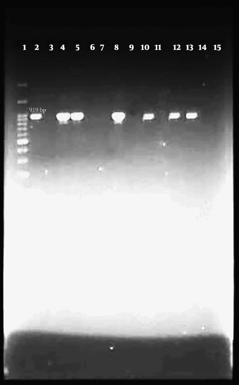
Out of 100 isolates of E. coli that were isolated from bovine mastitis samples by biochemical method and identified by PCR test with specific 16s rRNA primer, 94 isolates were confirmed as E. coli (Figure 1).

Lane 1 is a marker (100 bp). Lane 2 is positive control (Escherichia coli PTCC 1163) that bands within 919 bp. Lane 3 is negative control (double distilled water). Lanes 4, 5, 8, 10, 12, and 13 are positive samples. Lanes 6, 7, 9, 11, 14, and 15 are negative samples.

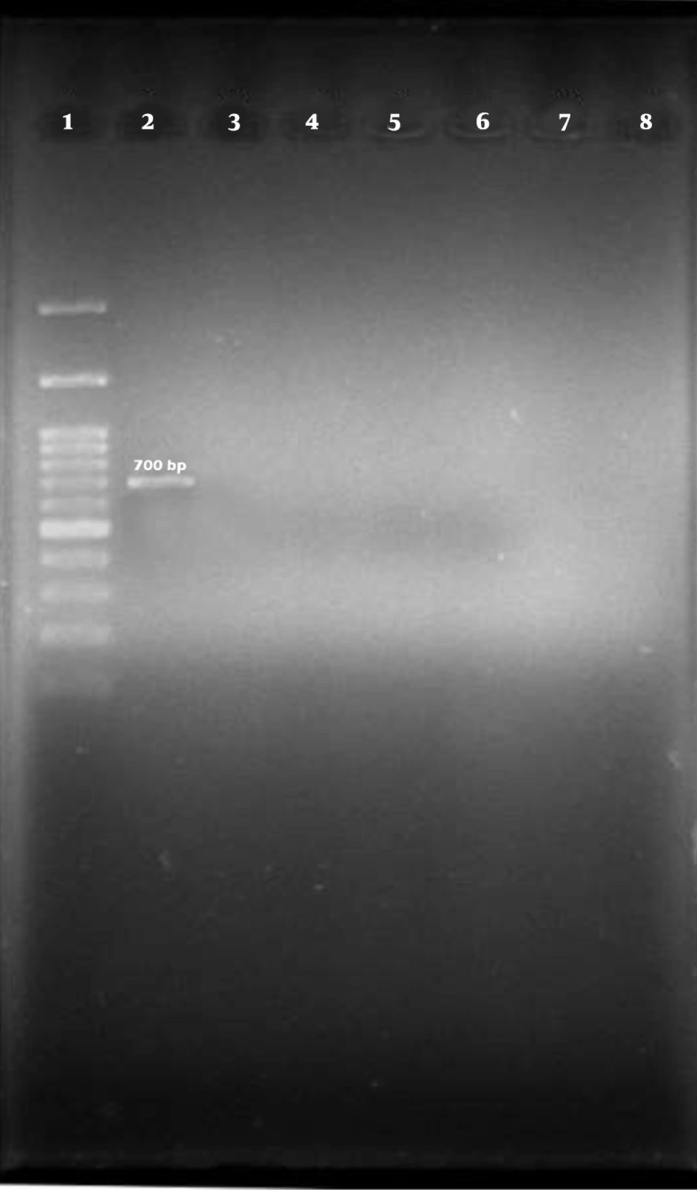
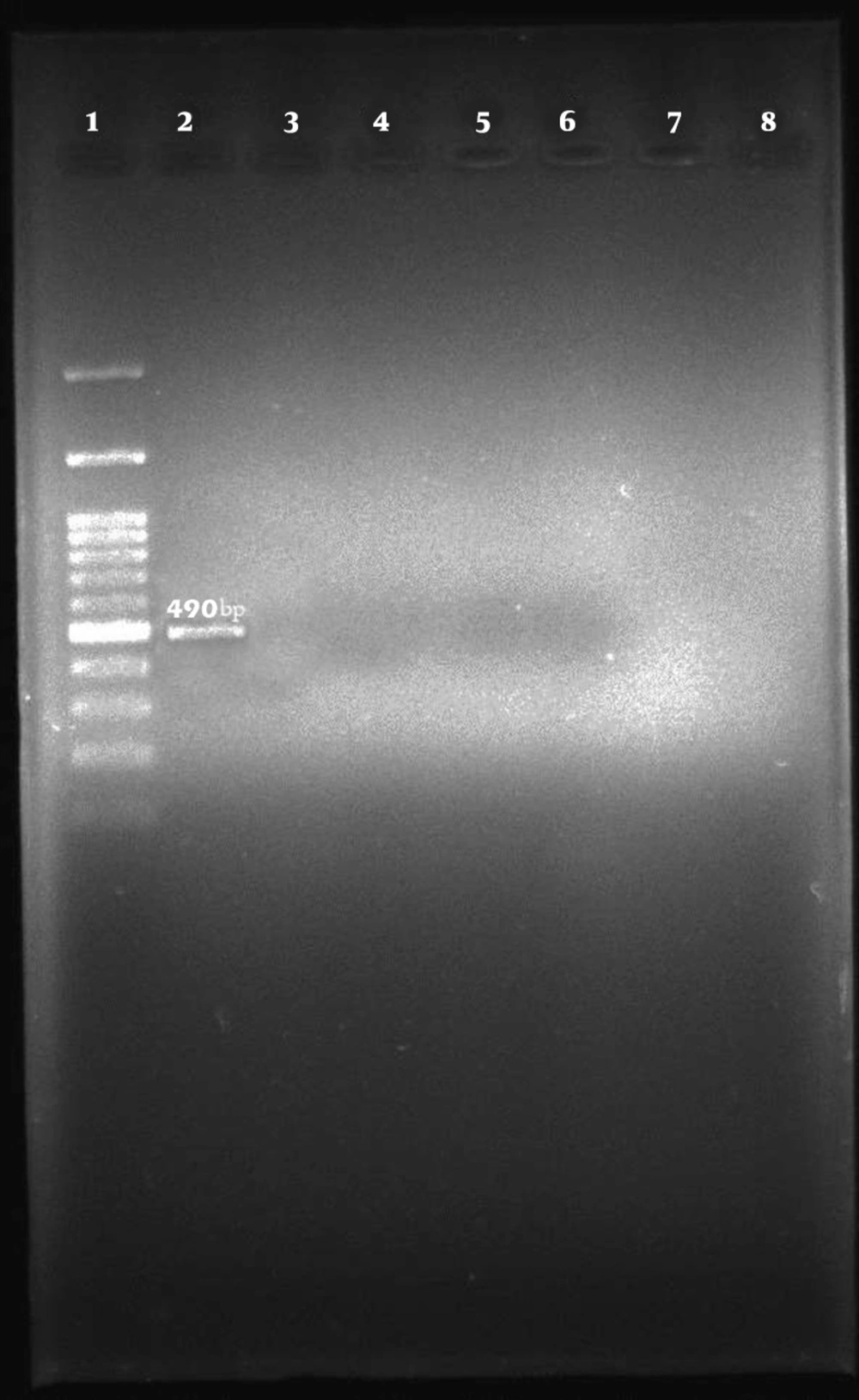
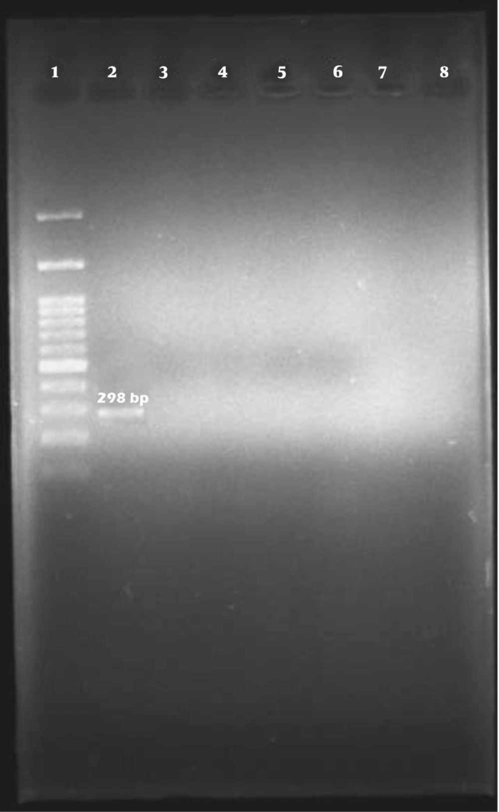
Among the 96 E. coli isolates examined, none of the samples harbored cdt-I, cdt-III, and cdt-IV genes (Figures 2, 3, and 4).

Lane 1 is a marker (100 bp). Lane 2 is positive control (Escherichia coli O113:NM) that bands within 700 bp. Lane 3 is negative control (double distilled water). Lanes 4 - 8 are negative samples.
Lane 1 is a marker (100 bp). Lane 2 is positive control (Escherichia coli O113:NM) that bands within 490 bp. Lane 3 is negative control (double distilled water). Lanes 4 - 8 are negative samples.
Lane 1 is a marker (100 bp). Lane 2 is positive control (Escherichia coli O113:NM) that bands within 298 bp. Lane 3 is negative control (double distilled water). Lanes 4 - 8 are negative samples.

5. Discussion

Environmental health, stress, and livestock immune system are three important factors in coliform mastitis (14), and clinical mastitis occurs most frequently on stressful days after calving (1). Escherichia coli is the most common causative species of coliform mastitis, followed by Klebsiella spp. responsible for most cases of clinical coliform mastitis (15). The importance of CDT toxin-producing E. coli in causing human gastrointestinal and extra-gastrointestinal infections is still unknown. Isolation of CTEC (Cytolethal distending toxin-producing E. coli) strains from patients suffering from septicemia, diarrhea, and urinary tract infections strengthen the hypothesis that these strains are related to human disease (16). Due to the connection of different alleles of this toxin with mobile genetic elements and prophages, its horizontal transfer between different strains is very likely. cdt-III gene is located on a large plasmid containing virulence genes (17), and cdt-I and cdt-IV genes are encoded by lambda prophage (18). Based on the results of this research, out of 100 E. coli isolates identified by biochemical method, 94 samples were identified as E. coli by PCR method and with a specific 16s rRNA primer. This indicates that molecular methods have higher diagnostic sensitivity and accuracy than biochemical phenotypic methods. In the recent study, cdt-I, cdt-III, and cdt-IV genes were not observed in any tested samples. Bouzari et al. reported that the frequency of cdt-I, cdt-III, and cdt-IV genes in patients with diarrhea in Tehran was 45.3%, 52%, and 2.6%, respectively (9). Kafshdouzan and Zahraei Salehi showed that 1.62% of poultry suffering from colibacillosis disease in Tehran had the cdt gene (19). Gomes et al. reported that the frequency of the cdt gene in patients with gastroenteritis, livestock, and food sources in Qatar is 3%, 17%, and 8%, respectively (20). Mainil et al. showed that the frequency of the cdtB-III gene in bovine type 2 necrotoxigenic E. coli (NTEC2) was 83%. Furthermore, they reported that the frequency of cdtB-I and cdtB-IV genes in bovine type 1 necrotoxigenic E. coli (NTEC1) were 3.81% and 4.96%, respectively (21).

5.1. Conclusions

In this study, the frequency of cdt-I, cdt-III, and cdt-IV genes in E. coli isolated from cow mastitis is much lower than in other studies, which indicates the difference in the distribution of the mentioned virulence genes among the different strains. This is probably due to geographical differences and differences in the ecological origin of the isolated strains (food, human, and livestock).
References

  • 1.

    Radostitis O, Gay C, Hinchcliff K, Constable P. Veterinary medicine: A textbook of the diseases of Cattle, Sheep, Pigs, Goats and Horses. Philadelphia: Saunders; 2007.

  • 2.

    Rollin E, Dhuyvetter KC, Overton MW. The cost of clinical mastitis in the first 30 days of lactation: An economic modeling tool. Prev Vet Med. 2015;122(3):257-64. [PubMed ID: 26596651]. https://doi.org/10.1016/j.prevetmed.2015.11.006.

  • 3.

    Liang D, Arnold LM, Stowe CJ, Harmon RJ, Bewley JM. Estimating US dairy clinical disease costs with a stochastic simulation model. J Dairy Sci. 2017;100(2):1472-86. [PubMed ID: 28012631]. https://doi.org/10.3168/jds.2016-11565.

  • 4.

    Seegers H, Fourichon C, Beaudeau F. Production effects related to mastitis and mastitis economics in dairy cattle herds. Vet Res. 2003;34(5):475-91. [PubMed ID: 14556691]. https://doi.org/10.1051/vetres:2003027.

  • 5.

    Tabatabayi AH, Firouzi R. Diseases of animals due to bacteria. 1st ed. Tehran University Press; 2001.

  • 6.

    Maity S, Ambatipudi K. Mammary microbial dysbiosis leads to the zoonosis of bovine mastitis: a One-Health perspective. FEMS Microbiol Ecol. 2020;97(1). [PubMed ID: 33242081]. https://doi.org/10.1093/femsec/fiaa241.

  • 7.

    Sugrue I, Tobin C, Ross RP, Stanton C, Hill C. Foodborne pathogens and zoonotic diseases. Raw milk - Balance between hazards and benefits. London, UK: Academic Press; 2019. p. 259-72. https://doi.org/10.1016/b978-0-12-810530-6.00012-2.

  • 8.

    Sahilah AM, Audrey LYY, Ong SL, Wan Sakeenah WN, Safiyyah S, Norrakiah AS, et al. DNA profiling among egg and beef meat isolates of Escherichia coli by enterobacterial repetitive intergenic consensus-PCR (ERIC-PCR) and random amplified polymorphic DNA-PCR (RAPD-PCR). Int Food Res J. 2010;17:853-66.

  • 9.

    Bouzari S, Oloomi M, Oswald E. Detection of the cytolethal distending toxin locus cdtB among diarrheagenic Escherichia coli isolates from humans in Iran. Res Microbiol. 2005;156(2):137-44. [PubMed ID: 15748977]. https://doi.org/10.1016/j.resmic.2004.09.011.

  • 10.

    Svab D, Horvath B, Maroti G, Dobrindt U, Toth I. Sequence variability of P2-like prophage genomes carrying the cytolethal distending toxin V operon in Escherichia coli O157. Appl Environ Microbiol. 2013;79(16):4958-64. [PubMed ID: 23770900]. [PubMed Central ID: PMC3754688]. https://doi.org/10.1128/AEM.01134-13.

  • 11.

    Chen J, Su Z, Liu Y, Wang S, Dai X, Li Y, et al. Identification and characterization of class 1 integrons among Pseudomonas aeruginosa isolates from patients in Zhenjiang, China. Int J Infect Dis. 2009;13(6):717-21. [PubMed ID: 19208492]. https://doi.org/10.1016/j.ijid.2008.11.014.

  • 12.

    Goudarzi V, Mirzaee M, Ranjbar R. O-serogrouping of Escherichia coli strains isolated from patients with Urinary tract infection by using PCR method. Iran J Med Microbiol. 2017;10(6):1-8. Persian.

  • 13.

    Wu Y, Hinenoya A, Taguchi T, Nagita A, Shima K, Tsukamoto T, et al. Distribution of virulence genes related to adhesins and toxins in shiga toxin-producing Escherichia coli strains isolated from healthy cattle and diarrheal patients in Japan. J Vet Med Sci. 2010;72(5):589-97. [PubMed ID: 20103992]. https://doi.org/10.1292/jvms.09-0557.

  • 14.

    Radostitis OM, Blood DC. Verterinary medicine. London: W.B. Saunders co; 2000.

  • 15.

    Mohammad Sadegh M, Askari Badouei M, Gorjidooz M, Daneshvar M, Koochakzadeh A. A study on the clinical coliform mastitis of Holstein cows on Garmsar suburban dairy farms. J Vet Microbiol. 2012;8(2):137-49.

  • 16.

    Hinenoya A, Shima K, Asakura M, Nishimura K, Tsukamoto T, Ooka T, et al. Molecular characterization of cytolethal distending toxin gene-positive Escherichia coli from healthy cattle and swine in Nara, Japan. BMC Microbiol. 2014;14:97. [PubMed ID: 24742173]. [PubMed Central ID: PMC4001111]. https://doi.org/10.1186/1471-2180-14-97.

  • 17.

    Peres SY, Marches O, Daigle F, Nougayrede JP, Herault F, Tasca C, et al. A new cytolethal distending toxin (CDT) from Escherichia coli producing CNF2 blocks HeLa cell division in G2/M phase. Mol Microbiol. 1997;24(5):1095-107. [PubMed ID: 9220015]. https://doi.org/10.1046/j.1365-2958.1997.4181785.x.

  • 18.

    Toth I, Nougayrede JP, Dobrindt U, Ledger TN, Boury M, Morabito S, et al. Cytolethal distending toxin type I and type IV genes are framed with lambdoid prophage genes in extraintestinal pathogenic Escherichia coli. Infect Immun. 2009;77(1):492-500. [PubMed ID: 18981247]. [PubMed Central ID: PMC2612248]. https://doi.org/10.1128/IAI.00962-08.

  • 19.

    Kafshdouzan KH, Zahraee Salehi T. Detection of cytolethal distending toxin (cdt) genes in Escherichia coli isolates from poultry colibacillosis. J Vet Microbiol. 2016;12(1):61-8. Persian.

  • 20.

    Gómez AM, Valenzuela NN, Peters KE, Salem A, Sultan A, Doiphode S, et al. Detection of cytolethal distending toxin and other virulent factors in Escherichia coli samples from animal livestock, retail foods and gastroenteritis human cases in Qatar. J Food Saf Hyg. 2020. https://doi.org/10.18502/jfsh.v5i2.3949.

  • 21.

    Mainil JG, Jacquemin E, Oswald E. Prevalence and identity of cdt-related sequences in necrotoxigenic Escherichia coli. Vet Microbiol. 2003;94(2):159-65. [PubMed ID: 12781483]. https://doi.org/10.1016/s0378-1135(03)00102-0.