Abstract
Background:
The attention of the scientific community has been recently focused on the role of tumor-infiltrating lymphocytes in non-small-cell cancer and there is a convergence of results, supporting a direct proportionality between lymphocytic infiltrate and good prognosis.Objectives:
The aim of our research was to investigate the immune response around and inside the small-cell lung neoplastic tissue and its related neo-lymphangiogenesis in a group of 20 patients.Materials and Methods:
We have examined 20 bioptic samples of small cell lung cancer, obtained from 18 men and two women, aged 56-74 years old. Besides hematoxylin-eosin staining, immunohistochemistry for neuroendocrine markers (CD56, chromogranin) and D2-40 was performed following the standard protocols.Results:
Our histological analysis, in all cases, has emerged the absence of an immune response supported by an adequate neo-lymphangiogenesis inside the neoplasia.Conclusions:
The absence of an immune response is interpreted as a sign of tissue tolerance and host acceptance towards the cancer, in favor of the cancer metastatic spread. The development of a paraneoplastic syndrome can be considered a biochemical proof of this tissue compatibility.Keywords
Small Cell Lung Carcinoma Antigens CD56 Chromogranins Monoclonal Antibody D2-40 Paraneoplastic Syndrome
1. Background
The immune response to different types of cancer has been the subject of many previous researches. Over years, several aspects of this topic have been considered, including the relative lymphocytosis in the peripheral blood and the histological presence of immunoreactive cells around and inside the neoplastic tissue or in the metastatic regional lymph nodes. These studies have been addressed also to lung neoplasia, mainly focused on non-small-cell lung cancer (NSCLC), demonstrating that a local infiltration by immunocompetent cells, primarily lymphocytes, correlates to a better prognosis (1-6).
2. Objectives
We investigated 20 patients’ immune response to small-cell lung cancer (SCLC), in order to evaluate the state around and inside the neoplastic tissue. For this reason, particular attention was paid to observe the presence of tumor-infiltrating lymphocytes and to recognize neo-lymphangiogenesis. The presence of a connective stromal network was also carefully evaluated.
3. Materials and Methods
Nowadays, surgical resection specimens of SCLC are absolutely rare, therefore our investigation was performed on bronchopulmonary biopsies. In our series we included 20 cases with classic SCLC, not combined with other histological types of lung cancer. None of the patients had received treatments prior to their surgeries. There were 18 male and two female patients, aged 56-74 years in the study group, without signs of immunological diseases and with no previous or concomitant steroid therapy. All bioptic samples with an evident of necrotic background were excluded. The diagnosis was based on the typical SCLC morphology, with the standard hematoxylin-eosin staining and all ascertained by immunohistochemistry for CD56 and chromogranin. The implementation of a new immunostaining method with the D2-40 antibody, specifically against podoplanin (7), permitted the evaluation of both lymphatic capillaries and the neo-lymphangiogenesis.
4. Results
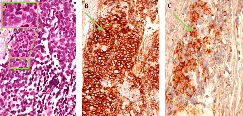
Our results can be outlined as follows: first of all, there is no leucocyte infiltration within the tumor or at its borders. Particularly an immune reaction, supported by activated monocytes or lymphocytes, is absent.(Figure 1). Secondly, a lymphatic vascular network was not particularly textured inside the neoplastic tissue or in its proximity and there was no evidence of neo-lymphangiogenesis. Finally, the stromal network of the neoplastic tissue was scantly represented and no desmoplastic reaction was observed (Figure 1).
Core Needle Biopsy of SCLC
5. Discussion
Our histological findings showed biological and clinical behaviors of SCLC, profoundly different from any other lung cancer histotype (8). The absence of immunological competent cells can be interpreted as a sign of tissue tolerance and of host acceptance towards SCLC, causing expanded subsequent diffusion. Same feature is found with carcinoid tumors, histo-genetically correlated to SCLC (9), for their common origin from neuroendocrine cells, belonging to the Amine Precursor Uptake and Decarboxylation (APUD) system. The APUD system consists of chromaffin secreting cells, with a coordination function between nervous and endocrine systems, widely diffused along all the respiratory and digestive tracts, with higher incidence of carcinoids and small-cell cancers.
Development of the paraneoplastic syndrome, which is common in patients affected by SCLC, can be considered a biochemical proof of its tissue compatibility. Secretion of the immunosuppressive factors, instead of pro-inflammatory molecules, may be suspected.
Lymphatic vessel invasion is considered a negative prognostic factor in lung cancers (10, 11). The same conclusion cannot be drawn for SCLC, where a complete absence of neo-lymphangiogenesis is emerged. The lack of an evident stromal network can be correlated with the speediness of this neoplasia cellular replication and its particular local aggressiveness. On the contrary, the lymphocytic reaction to NSCLC, develops in parallel, through its stromal architecture (12-15). The absence of tumor-infiltrating lymphocytes, not supported by a textured stromal network, can be considered a histological marker of SCLC, correlating with a poor prognosis.
Acknowledgements
References
-
1.
Al-Shibli KI, Donnem T, Al-Saad S, Persson M, Bremnes RM, Busund LT. Prognostic effect of epithelial and stromal lymphocyte infiltration in non-small cell lung cancer. Clin Cancer Res. 2008;14(16):5220-7. [PubMed ID: 18698040]. https://doi.org/10.1158/1078-0432.CCR-08-0133.
-
2.
Shigematsu Y, Hanagiri T, Shiota H, Kuroda K, Baba T, Ichiki Y, et al. Immunosuppressive effect of regulatory T lymphocytes in lung cancer, with special reference to their effects on the induction of autologous tumor-specific cytotoxic T lymphocytes. Oncol Lett. 2012;4(4):625-30. [PubMed ID: 23205074]. https://doi.org/10.3892/ol.2012.815.
-
3.
Karagoz B, Bilgi O, Gumus M, Erikci AA, Sayan O, Turken O, et al. CD8+CD28- cells and CD4+CD25+ regulatory T cells in the peripheral blood of advanced stage lung cancer patients. Med Oncol. 2010;27(1):29-33. [PubMed ID: 19148592]. https://doi.org/10.1007/s12032-008-9165-9.
-
4.
Hiraoka K, Miyamoto M, Cho Y, Suzuoki M, Oshikiri T, Nakakubo Y, et al. Concurrent infiltration by CD8+ T cells and CD4+ T cells is a favourable prognostic factor in non-small-cell lung carcinoma. Br J Cancer. 2006;94(2):275-80. [PubMed ID: 16421594]. https://doi.org/10.1038/sj.bjc.6602934.
-
5.
Ruffini E, Asioli S, Filosso PL, Lyberis P, Bruna MC, Macri L, et al. Clinical significance of tumor-infiltrating lymphocytes in lung neoplasms. Ann Thorac Surg. 2009;87(2):365-71. discussion 371-2. [PubMed ID: 19161739]. https://doi.org/10.1016/j.athoracsur.2008.10.067.
-
6.
Black CC, Turk MJ, Dragnev K, Rigas JR. Adenocarcinoma contains more immune tolerance regulatory t-cell lymphocytes (versus squamous carcinoma) in non-small-cell lung cancer. Lung. 2013;191(3):265-70. [PubMed ID: 23494387]. https://doi.org/10.1007/s00408-013-9455-7.
-
7.
Roncati L, Manenti A, Sighinolfi P. Immunohistochemical improvement in the analysis of the lymphatic metastases from lung carcinoma. Ann Thorac Surg. 2014;97(1):380-1. [PubMed ID: 24384208]. https://doi.org/10.1016/j.athoracsur.2013.06.072.
-
8.
Dingemans KP, Mooi WJ. Ultrastructure of tumour invasion and desmoplastic response of bronchogenic squamous cell carcinoma. Virchows Arch A Pathol Anat Histopathol. 1987;411(3):283-91. [PubMed ID: 3113068].
-
9.
Filosso PL, Ruffini E, Di Gangi S, Guerrera F, Bora G, Ciccone G, et al. Prognostic factors in neuroendocrine tumours of the lung: a single-centre experience. Eur J Cardiothorac Surg. 2014;45(3):521-6. discussion 526. [PubMed ID: 24092506]. https://doi.org/10.1093/ejcts/ezt442.
-
10.
Wang J, Wang B, Zhao W, Guo Y, Chen H, Chu H, et al. Clinical significance and role of lymphatic vessel invasion as a major prognostic implication in non-small cell lung cancer: a meta-analysis. PLoS One. 2012;7(12). ee52704. [PubMed ID: 23285161]. https://doi.org/10.1371/journal.pone.0052704.
-
11.
Strano S, Lupo A, Lococo F, Schussler O, Loi M, Younes M, et al. Prognostic significance of vascular and lymphatic emboli in resected pulmonary adenocarcinoma. Ann Thorac Surg. 2013;95(4):1204-10. [PubMed ID: 23415237]. https://doi.org/10.1016/j.athoracsur.2012.12.024.
-
12.
Schwartz AM, Rezaei MK. Diagnostic surgical pathology in lung cancer: Diagnosis and management of lung cancer, 3rd ed: American College of Chest Physicians evidence-based clinical practice guidelines. Chest. 2013;143(5 Suppl):e251S-62S. [PubMed ID: 23649441]. https://doi.org/10.1378/chest.12-2356.
-
13.
De Wever O, Mareel M. Role of tissue stroma in cancer cell invasion. J Pathol. 2003;200(4):429-47. [PubMed ID: 12845611]. https://doi.org/10.1002/path.1398.
-
14.
Bremnes RM, Donnem T, Al-Saad S, Al-Shibli K, Andersen S, Sirera R, et al. The role of tumor stroma in cancer progression and prognosis: emphasis on carcinoma-associated fibroblasts and non-small cell lung cancer. J Thorac Oncol. 2011;6(1):209-17. [PubMed ID: 21107292]. https://doi.org/10.1097/JTO.0b013e3181f8a1bd.
-
15.
Wald O, Izhar U, Amir G, Kirshberg S, Shlomai Z, Zamir G, et al. Interaction between neoplastic cells and cancer-associated fibroblasts through the CXCL12/CXCR4 axis: role in non-small cell lung cancer tumor proliferation. J Thorac Cardiovasc Surg. 2011;141(6):1503-12. [PubMed ID: 21463876]. https://doi.org/10.1016/j.jtcvs.2010.11.056.