
Dear Editor,
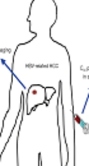
Hepatocellular carcinoma (HCC) is a common primary liver malignancy worldwide with a poor prognosis (1) and is the third leading cause of cancer - related deaths (2).
Chronic hepatitis B (HBV) infection, which can cause liver inflammation, has been seen in many individuals with HCC. In addition, fibrosis and cirrhosis are inevitable complications of the infection, which leads to the death of a patient (3, 4).
Early diagnosis of this can prevent the death of a patient and is a crucial factor in the treatment process (5).
It seems that urea cycle enzymes are reliable marker for hepatocytes. For example, arginase - 1 (Arg - 1), which mainly concentrates in periportal hepatocytes (6), is one of the urea cycle enzymes that is expressed in a healthy liver (7).
Chrzanowska et al., assessed Arg - 1 expression by advanced PCR methods and found decreased expression of Arg - 1 in cirrhotic nodules and HCCs (8).
Recently, we found that the expressions of Arg - 1 significantly reduced in patients with HBV - related HCC compared to the patients with only HBV, which can probably manage the diagnosis process of HCC in a way that is time - consuming. The specificity of Arg - 1, among all groups we tested, was 88.4%. Our data emphasized that Arg - 1 can be one of the best markers for HCC on fine - needle aspiration specimens. The results of this study will be released. We also reported Arg - 1 as a very sensitive marker for hepatocytes, which supports Benjamin et al., conclusion as well (9).
Benjamin et al., (9) found the overall sensitivity to be about 96.0% for Arg - 1 in hepatocytes and hepatocellular neoplasms. In addition, all HCCs in their study were reactive for Arg - 1. Moreover, the study of nonhepatocellular tumors revealed that only 2 non - HCC tumors were reactive for Arg - 1.
Furthermore, proper combination of Arg - 1, with some reliable biomarkers such as HepPar - 1 could increase the specificity, accuracy, and precision of natural history of HBV - related HCC diagnosis. We proposed an algorithm to study the natural history of HCC using 2 markers of malignancy (HepPar - 1 and Arg - 1) and found that the respective specificity of Arg - 1 + HepPar - 1 is 100% when we examined HCC cases that were infected previously with HBV.
In another study, Timek et al., (10) performed a triplet algorithm including Arg - 1, hepatocyte paraffin - 1 (HepPar1), and glypican - 3 on liver specimens. They showed that none of the nonhepatic tumor cases were positive for Arg - 1. A total of 19 HCCs samples responded to all three markers, 9 samples responded with only 1 or 2 markers and only 1 case was negative for all 3 markers. Timek suggested the use of 3 markers in differentiating between HCC and metastatic carcinoma.
In conclusion, most of the Arg - 1 studies demonstrated that Arg - 1 has a high sensitivity and specificity in diagnosis of HBV - related HCC and it seems that Arg - 1 can probably be used in detecting HBV infected patients, which are susceptible to HCC. Arg - 1 can develop as an identifier biomarker in pathology of HCC. In addition, the proper combination of Arg - 1 and some well - known and reliable biomarkers is more effective in the study of natural history of the liver malignancy in different stages.
