1. Background
Hepatitis E virus (HEV) is an important cause of acute clinical hepatitis in endemic countries and can lead to detrimental prognostics, such as severe acute hepatitis, liver failure, chronicity in immunocompromised patients, and death in pregnant women (1, 2). Four HEV genotypes have been reported to infect humans. Genotypes 1 and 2 are strictly human, transmitted by fecal–oral route, and responsible for water-borne HEV outbreaks in Asia, Africa, and Central America. HEV genotypes 3 and 4 are found in swine and other animal species in many countries where direct and indirect zoonotic transmission in industrialized countries have been reported (3-6).
The disease is considered emerging in many parts of the world because of the increased awareness and availability of effective diagnostics. The HEV seroprevalence has been found to be elevated even in areas classified non-endemic (7). North Africa is a region classified as endemic for hepatitis E. For instance, half of the population aged above five years in Egypt is serologically positive for HEV (8), and high HEV seroprevalence was reported in Tunisia, Libya, and Morocco (9). Furthermore, two HEV outbreaks occurred in Algeria. The first one occurred in Mostaganem (Northwest Algeria) in 1980 and the second in Tanefdour (Northeast Algeria) in 1986 - 1987 (10, 11). Both epidemics were traced back to contaminated water sources, and the causative pathogen was HEV genotype 1. Another outbreak of hepatitis non-A and non-B occurred in Medea (Northern Algeria) from October 1980 to January 1981, and it affected 788 people, mostly young adults, with a mortality rate of 100% in pregnant women (12). Although the causative pathogen in this latter outbreak was not identified, it was likely to be HEV given the circumstances of the occurrence and the development of the disease.
2. Objectives
To date, no accurate estimates of the prevalence of HEV in Algeria are available. Moreover, no studies on HEV or anti-HEV antibodies have been conducted, and HEV infection is not investigated in the hospitals when assessing acute hepatitis. Therefore, this study aimed to investigate the presence of anti-HEV antibodies in Northern Algeria, determine the HEV genotype(s) of the circulating strains, and elucidate the contamination routes (zoonotic and/or water-borne).
3. Methods
3.1. Study Population
Sample size was calculated to be 517 based on an anticipated anti-HEV IgG rate of 15%, a margin of error of 3%, a confidence level of 95%, and a population size of approximately 10,000 (information from the commune chiefs on the total population depending on the three health facilities). Therefore, the intended sample size was determined to be 590. The blood samples were collected from three hospitals in central Algeria: 379 samples from blood donors and 211 samples from outpatients. Sera were separated and stored at -20°C until further analysis. At their convenience, the subjects were enrolled in the study from January to May 2014. The demographic characteristics of the study population are presented in Table 1. This study was conducted in accordance with the national ethics regulation and was approved by the research ethics committee of Southeast University in Nanjing.
3.2. Preparation of HEV P166 Antigens
The truncated p166 capsid protein was generated from the amino acid position 452–617 of the open reading frame 2 of the following HEV strains: W01 (genotype 1, JX857689), Mexico-14 (genotype 2, M74506), US-1 (genotype 3, AF060668), and China-9829 (genotype 4, AY789225) strains, and expressed in Escherichia coli (13). Briefly, the polymerase chain reaction fragment encoding aa 452 - 617 of the HEV strains was inserted into the pET28a vectors (Novagen, Darmstadt, Germany). Then, the plasmids were used to transform competent E. coli BL21 (DE3) cells (Promega, Madison, USA). After the confirmation of the sequence of aa 452 - 617 in the plasmids by DNA sequencing, the gene expression was induced. The cells were pelleted and lysed after an incubation period and constant shaking. The suspension was clarified by centrifugation, and then the supernatant was loaded onto a column containing Ni-NTA super flow affinity resin. The column was washed, and the fusion proteins were eluted as described previously (14). The four p166 proteins were designated as p166W01, p166Mex, p166US, and p166Chn, and a mixture (p166mix) containing equal concentrations of each of the four p166 proteins was prepared.
3.3. Detection of Anti-HEV Total Antibodies
Sera were screened for the presence of anti-HEV antibodies with a high performance assay, namely, the in-house sandwich enzyme immunoassay, according to Dong et al. (14). A double-antigen sandwich assay using the p166 proteins was adopted. Briefly, microplate wells were coated with His-p166 mix and incubated at room temperature overnight. Unbound antigens were washed with 10 mM phosphate-buffered saline containing 0.05% Tween 20 (PBS-T). Then, undiluted test serum was added, and the plates were incubated at 37°C for 1 hour. After a washing step with PBS-T, the horseradish peroxidase (HRP)-conjugated p166 mix was added, and the plates were incubated at 37°C for 1 hour. After washing, tetramethylbenzidine was added as substrate, and the plates were read using a kinetic microplate reader at a wavelength of 450 nm. All sera were tested in duplicate, and a signal/cutoff (s/co) value of ≥ 1 was considered a positive reaction.
3.4. Detection of Anti-HEV IgM Antibodies
The presence of anti-HEV IgM antibodies was also assessed as previously described (15). Briefly, the purified p166 proteins were used as antigens to coat microplate wells. After an incubation period of 2 h at 37°C, followed by three washings with PBS containing 0.05% Tween 20, test and control were distributed into wells and incubated for 1 h at 37°C. After three washings, the HRP-conjugated goat anti-human IgM (KPL) was added to each well and incubated at 37°C for 1 hour. After a final washing, the colorimetric reactions were developed using tetramethylbenzidine substrate (Sigma) for 15 minutes at room temperature and stopped with 2 M H2SO4. The plates were read using a kinetic microplate reader at 450 nm wavelength.
3.5. HEV Genotype Prediction by Assessment of Anti-HEV IgG in the Positive Sera
An indirect ELISA was adopted to detect IgG antibodies in serially diluted positive sera. The p166 proteins generated from the four genotypes were used as antigens, and each p166 protein was used in a separate analysis as previously described (16). Briefly, the purified His-p166 proteins were used as antigens to coat microplate wells. After an incubation period of 2 hours at 37°C, followed by three washings with PBS containing 0.05% Tween 20, test and control sera serial dilutions (1: 200, 1: 400, 1: 800, 1: 1600, 1: 3200, and 1: 6400) were distributed into wells and incubated for 1 h at 37°C. After three washings, the HRP-conjugated goat anti-human IgG (KPL) was added to each well and incubated at 37°C for 1 hour. After a final washing, the colorimetric reactions were developed using tetramethylbenzidine substrate (Sigma) for 15 minutes at room temperature and stopped with 2 M H2SO4. The plates were read using a kinetic micro-plate reader at a wavelength of 450 nm.
3.6. Statistical Analysis
Seropositivity rates were calculated and compared according to age group and gender. Differences were evaluated using logistic regression analysis and the chi-square test. P < 0.05 was considered statistically significant. For the cross-genotype neutralization assay, two-way ANOVA with Bonferroni posttest was performed using GraphPad Prism version 5.00 for Windows (GraphPad Software, San Diego, CA, USA).
4. Results
4.1. Detection of Anti-HEV Antibodies
The serological screening detected an overall seropositivity of 20.17% as shown in Table 1. Although a slight difference was found in the prevalence rates between males and females (Table 1), statistically no significant correlation was noted between HEV seroprevalence and subjects’ gender (P = 0.5).
Table 1 represents the number of positive cases by age group. Initially, the patients were grouped by an interval of 10 years. The results showed that majority of the positive cases were aged between 21 and 60 years old and that no positive case was found from 0 to 10 years old. However, this result must be taken with caution given the small number of sample in this group (only four). However, compared with the group of ≥ 70 years, which also had a small number of sample (n = 11), four positive cases were detected. To better appreciate these results, they are represented as percentages compared with the total number of sample in each age group. The groups of 21 - 30, 31 - 40, and 41 - 50 years almost had equal rates at 18.82%, 18.50%, and 22.37%, respectively, but these rates were significantly higher than that of the first group. The group of patients aged less than 20 years had the lowest rate (6.25%).
| A. Anti-HEV Antibodies Prevalence by Gender | ||||
|---|---|---|---|---|
| Samples, No. (%) | Age, Mean (Range) | Positive, No. (%) | Negative, No. (%) | |
| Female | 289 (48.98) | 38.87 (6 - 78) | 55 (19.03) | 234 (80.97) |
| Male | 301 (51.02) | 39.86 (9 - 83) | 64 (21.26) | 237 (78.74) |
| Overall | 590 (100) | 39.365 (6 - 83) | 119 (20.17) | 471 (79.83) |
| B. Anti-HEV Antibodies prevalence by Age Group | ||||
| Age group, y | Total | Positive, No. (%) | Negative, No. (%) | |
| 0 - 10 | 4 | 0 (0.00) | 4 (100.00) | |
| 11 - 20 | 60 | 4 (6.67) | 56 (93.33) | |
| 21 - 30 | 85 | 16 (18.82) | 69 (81.18) | |
| 31 - 40 | 173 | 32 (18.50) | 141 (81.50) | |
| 41 - 50 | 152 | 34 (22.37) | 118 (77.63) | |
| 51 - 60 | 84 | 25 (29.76) | 59 (70.24) | |
| 61 - 70 | 21 | 4 (19.05) | 17 (80.95) | |
| ≥ 71 | 11 | 4 (36.36) | 7 (63.64) | |
| Overall | 590 | 119 (20.17) | 471 (79.83) | |
| C. Logistic Regression Analysis | ||||
| Odds Ratio (95% CI) | P Value | Chi Square test P Value | ||
| Gender | 0.926 (0.619 - 1.388) | 0.711 | 0.500 | |
| Age | 1.025 (1.010 - 1.040) | 0.001 | 0.033 | |
Prevalence of HEV Antibodies in Relation to Gender (A) and Age (B) of Subjects and the Logistic Regression Analysis Results (C)
The positivity rate continued to increase to reach a maximum of 29.76% in group 51 - 60 years to finally decrease to 25% in the last group (≥ 61 years). Logistic regression analysis showed a significant correlation between age of the patients and presence of anti-HEV antibodies (P = 0.001) as shown in Table 1. Processing the results of the blood donors alone was important. Note that among the 379 blood donors (mean age 38.73, age range 18 - 65, 55.67% are men), 83 (21.9%) were diagnosed positive for anti-HEV antibodies, 37 of whom were women.
Testing the presence of anti-HEV IgM antibodies in all samples revealed only two weakly positive cases who were both blood donors (a 48-year-old woman and a 32-year-old man).
4.2. Presence of Anti-HEV Antibodies in the Different Age Groups
In a second step, the subjects were grouped into three age groups according to whether they were born before or after the 1987 - 1988 and 1979 - 1980 outbreaks. The results showed that 24.29% of the subjects aged over 36 years (born before the first outbreak of 1978) were positive for the presence of anti-HEV antibodies. This rate decreased to 17.4% in the subjects aged between 25 and 35 years (born after the 1978 outbreak and before the 1987 outbreak). The anti-HEV antibody prevalence rate was only 9.9% for the subjects aged less than 25 years (born after the last outbreak). Statistically, a significant correlation was found between age and presence of anti-HEV antibodies (P = 0.033).
4.3. HEV Genotype Prediction by Assessing Anti-HEV IgG in Positive Sera
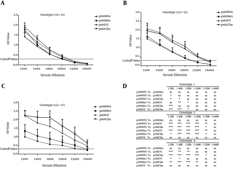
We previously assessed the immunoreactivity of anti-HEV antibodies present in the serum sample collected from patients infected with different HEV genotypes (genotype 1: n = 15, genotype 3: n = 12, and genotype: 4 n = 17) using p166 antigens generated from the four HEV genotypes (16). The sera were serially diluted, and the anti-HEV antibodies were detected by an indirect ELISA. The four p166 proteins were used as antigens, and each p166 antigen was used in a separate experiment. The results revealed that the immunoreactivity of anti-HEV antibodies raised against genotype 1 strains was stronger than that against the p166 antigens generated from genotypes 1 and 2 (p166W01 and p166Mex) and that against the p166 antigens generated from genotypes 3 and 4 (p166US and p166Chn). By contrast, the reaction of anti-HEV antibodies raised against the zoonotic genotypes 3 and 4 was more significant than that against the p166 antigens generated from genotypes 3 and 4 (p166US and p166Chn) as shown in Figure 1.
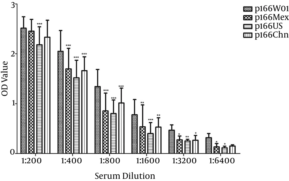
Moreover, we exploited this immunoreactivity difference for the prediction of the HEV genotypes in this serum panel collected in Algeria. As expected, the detection of anti-HEV IgG antibodies in the positive sera using the different p166 antigens generated from the different HEV genotypes revealed a variation of immunoreactivity (Figure 2). The IgG antibodies reacted strongly against p166W01 (generated from HEV genotype 1) at all dilution titers. These results showed the obvious effects of the antigen origin on the IgG-binding ability and suggested that the IgGs were more likely to be raised against an HEV genotype 1 strain (Figure 2).
A, Sera of patients infected by HEV genotype 1 strains; B, Sera of patients infected by HEV genotype 3 strains; C, Sera of patients infected by HEV genotype 4 strains; D, Bonferroni multiple comparison test results. Each point represents mean ± SD; *P < 0.05; **P < 0.01 and ***P < 0.001(16).
5. Discussion
To our knowledge, this study is the first to determine the presence and immunoreactivity of anti-HEV antibodies in Northern Algeria and revealed a positivity rate of 20.17%. This prevalence is higher than those of the countries on the northern side of the Mediterranean Sea, such as France and Italy (17, 18), but is relatively lower than those of neighboring countries on the southern side. In Egypt, the prevalence of anti-HEV antibodies reached 84.3% in pregnant women, 67.6% in rural areas, 56.4% in semi-urban areas, and 45.3% in blood donors (19-21). In Morocco, the prevalence is 8.5% among blood donors (22). In Tunisia, the seroprevalence of HEV is 46% in healthy people, 22% in blood donors, and 12% in pregnant women (23, 24). However, these results should be taken with caution because of the small number of subjects included in these studies.
In the present study, no significant correlation was found between gender and presence of anti-HEV antibodies, whereas a significant difference was found in seroprevalence among the different age groups. These results are similar to those previously reported in other studies conducted in various countries (25-27). This similarity is probably due to the comparable exposure of both sexes to the virus sources. However, exposure time is long in the elderly, and this long exposure increases the chances of contracting the virus, thus explaining the difference in HEV prevalence among the different age groups. The presence of anti-HEV antibodies in people under 25 years (9.9%) and the two cases that were weakly positive for anti-HEV IgM contradicted the exposure to virus during the last outbreaks (1979 - 1980 and 1987 - 1988). These circumstances explain the results and indicate clearly that HEV infection is still present in Algeria.
Several strains of genotype 3 were isolated from humans and animals across different continents, where they cause sporadic cases mainly after the consumption of undercooked swine products. Several studies reported the isolation of HEV from several other animal species (28-32). Except in swine, deer, rabbits, and mongooses, viral RNA has not been detected in other animal species. The distribution of genotype 3 and its dispersion throughout the world (33) raises the question of its presence in Algeria. However, Algeria, which is a Muslim country, has no swine consumption and breeding, thus making the presence of genotype 3 unlikely. In this study, only two cases were weakly positive for anti-HEV IgM antibodies. According to (34), viral RNA is no longer detectable at such a low rate of IgM antibodies. To predict the genotype of the causative strain that infected the subjects, we exploited the immunoreactivity difference among the p166 proteins generated from the four genotypes as reported previously (16). We showed that the IgG-binding ability is significantly stronger in the presence of antigens generated from the same genotype than from the genotype they were raised against. Using the same approach in this study, when the IgG-positive sera were assessed by different p166 proteins, the antibodies showed a stronger immunoreactivity against p166W01, which was generated from a genotype 1 strain. Moreover, for the HEV outbreaks that occurred in Algeria (1979 - 1980 and 1987 - 1988), the isolated virus belonged to HEV genotype 1, which contaminated the water sources after a period of intense rain (10, 11). Therefore, given the present results and the available history of HEV in Algeria, the presence of genotype 1 HEV is clearly the most likely reason, and this genotype 1 strain(s) still causes sporadic cases.
Recently, research on hepatitis E has been directed to investigate the risk of HEV transmission via blood transfusions. Therefore, several studies on the seroprevalence of hepatitis E in blood donors were conducted (35-37). Although the positivity rates for anti-HEV IgM antibodies were relatively low, several cases of post-transfusion infection were reported (38-40). In this context, our study reveals a relatively high seroprevalence of anti-HEV antibodies (21.9%) among blood donors, and only two cases weakly positive for anti-HEV IgM antibodies as discussed above were found. The accumulated data on this topic demonstrate a potential transfusion-associated risk. Given the high mortality rates in pregnant women and immune-compromised patients, detrimental effects will occur if these patients receive HEV-contaminated blood products. However, making the screening of donated blood for the presence of HEV as a mandatory test is still early, and more detailed investigations are required especially in endemic areas.
In conclusion, we presented a new approach for the prediction of the genotype of HEV strains circulating in a given region in seroprevalence studies using different antigens generated from the four genotypes. This pilot study on the field application of this method revealed that the sera positive for anti-HEV antibodies presence reacted strongly against the antigens derived from HEV genotype 1. This finding indicates that hepatitis E in Northern Algeria is most likely caused by genotype 1 strains. Moreover, this study also revealed a relatively high seroprevalence of anti-HEV antibodies within the targeted population in Northern Algeria. Therefore, to prevent future outbreaks, the management strategy of Algerian clinicians in assessing acute hepatitis requires an urgent re-evaluation. Finally, this study raises several issues that require further investigation: assess the prevalence and incidence of HEV infection throughout the Algerian territory, identify the risk factors other than age (e.g., socioeconomic condition, working in animal breeding, working in the health sector, and co-infection with other pathogens), and evaluate the risk of transmission via blood donation.

