1. Background
The liver carries out several complex and important functions, and liver diseases are considered potential threats to human life (1). Liver cancer is a complicated disease resulting from a several-phase process that includes the deregulation of a number of various signaling cascades (2). Hepatocellular carcinoma (HCC) is the most common liver cancer worldwide, the most common early cancer of hepatocytes, and the fifth most common deadly malignant tumor worldwide (3, 4). The important risk factors for HCC are environmental agents (such as hepatitis C virus, hepatitis B virus, and chemical carcinogen exposure). Several other risk factors, including food additives, non-alcoholic fatty liver disease, obesity, industrial and environmental toxic chemicals, and water and air pollutants are also involved in the etiology of HCC (5, 6). HCC is rarely detected at the primary phase, and once detected, there is a poor prognosis in most cases (7). HCC treatment methods, including chemotherapy, liver transplantation, and resection, show poor tolerance, low efficacy, and poor subsequent survival with a high recurrence rate (8). The diet contains several biologically active substances, nutrients, and non-nutritive compounds that may be changed into metabolites and isomers with various abilities, resulting in different bioavailability profiles (2). Natural products have made important contributions to therapies for various cancers (for example, breast cancer). In the past 40 years, more than 300 patents have been issued for potential anticancer candidates from the sea, and at least 10 compounds are in various phases of clinical trials (9).
Sea cucumbers, belonging to the Holothuroids (Holothuroidea), are marine invertebrates that exist in benthic areas and deep seas. They are one of several marine animals that are underutilized as food, particularly among Asian populations (for example, China, Korea, and Taiwan). Sea cucumbers are well known to exert useful effects on human health, and are used for medicinal purposes (10, 11). In developed countries, such as the United States and Canada, sea cucumber tissues are dried, pulverized and encapsulated as nutraceuticals for over-the-counter dietary health supplements that are primarily directed at inflammatory conditions in humans and companion animals (9).
Natural marine products (especially sponges) have attracted the notice of chemists and biologists around the world over the last five decades, as the ocean is considered to be a source of potential drugs (12). The marine sponge genus Haliclona (of the Chalinidae family) has been extensively examined, and about 190 metabolites that exhibit cytotoxic, antifungal, antimalarial, antimicrobial, and anti-fouling activities have been isolated (13). Haliclona oculata, a marine demospongiae belonging to order Haplosclerida, family Chalinidae, is a soft rosy-brown to yellow-brown branching sponge with small mouth-like openings that cigar-shaped oxeas, mammiform elevations, stylote or strongylote forms, and laterally compressed branches (14). H. oculata reportedly shows pharmacological activity against several diseases, such as cancer, fungal and microbial infections, neurodegeneration, and type 2 diabetes (14-17).
2. Objectives
Despite several worldwide studies that have revealed the efficacy of some sea cucumber and sponge species as potential sources of cytotoxic compounds, there is still a shortage of information about levels of this activity, especially in Persian Gulf species, including Holothuria parva and H. oculata. Hence, we aimed to investigate the selective toxicity of Persian Gulf sea cucumber (H. parva) and sponge (H. oculata) extracts on liver mitochondria isolated from an animal model of hepatocellular carcinoma as part of a national project that hopes to identify novel potential anticancer candidates among Iranian Persian Gulf flora and fauna.
3. Materials and Methods
3.1. Sea Cucumber Samples
Sea cucumber samples, including H. parva (10 specimens), were collected during low tide from the Bandar-e Lengeh coast in southern Iran. They were kept in iced boxes and transported to the laboratory, where they were washed with cold water, weighed, and measured.
3.2. Extraction of Samples and Isolation of H. parva
Bioactive compounds were extracted based on their polarity, using water and organic solvents according to the method described by Sarhadizadeh et al. (18). The samples from the gonad (G), respiration tree (RT), Cuvierian organ (CO), and body wall (BW) were defrosted before use. The recovered body wall was cut into small pieces and the samples were homogenized with a blender, then suspended. This was followed by successive extractions with methanol (50%) by percolation (72 h for each solvent) at room temperature. After filtration and centrifugation (15 minutes, 30,000 ×g, 4°C), the extracts were evaporated under a vacuum at 45°C with a rotary evaporator. The powdered extracts of each sample were obtained with a freeze dryer and stored at -20°C.
3.3. Sponge Samples
H. oculata was collected from tidal and subtidal habitats via scuba diving at depths between 0 - 20 m, near Larak Island in the mouth of the Strait of Hormuz of the Persian Gulf. The samples were cleaned and washed with distilled water, then immediately frozen and maintained at -20°C prior to extraction. They were transferred to the laboratory as soon as possible.
3.4. Extraction, Fractionation, and Isolation Procedure of H. oculata
Freshly collected H. oculata (2.0 kg) was cut into small pieces and extracted with methanol (4 × 4 L) at room temperature. The combined extract was filtered, then concentrated into a viscous mass (45.0 g) under reduced pressure, below 45°C, in a Rotavapor®. The animal residue was further extracted with 50% methanol-chloroform (4 × 4 L) and the combined extract was filtered and concentrated under reduced pressure as described above, into a green viscous mass (35.0 g). The remaining residue was rejected. The dried residue was stored at -20°C, to be used in anticancer assays.
For standardization of methanolic extracts, the total phenolic (TP) determination was performed as follows: 2.5 g of the oil samples were diluted with 2.5 mL of n-hexane, and extracted three times by 5 minutes of centrifugation (5000 rpm) with CH3OH/H2O (80:20 v/v) extract. The extract was added to 2.5 mL of Folin-Ciocalteu reagent and 5 mL of Na2CO3 (7.5%) in a 50 mL volume flask, reaching the final volume with deionized water. The samples were stored overnight and the spectrophotometric analysis was performed at λ = 765 nm. The methanolic extracts of H. parva and H. oculata consisted of 1045 ± 73 mg/g and 785 ± 42 mg/g of TPs, respectively.
3.5. Animals
Male Sprague-Dawley rats (120 - 130 g), fed a standard chow diet and given water ad libitum, were used in all experiments. They were purchased from Institute Pasteur (Tehran, Iran) and were kept in individual cages under controlled room temperature (20 - 25°C) and humidity (50% - 60%), and exposed to 12 hours light/dark cycles. All experiments were conducted according to the ethical standards and protocols approved by the Committee of Animal Experimentation of Shahid Beheshti University of Medical Sciences in Tehran, Iran. All efforts were made to minimize the number of animals used, and their suffering.
3.6. Experimental Design
The rats were divided into two groups of ten animals each. Group A, was untreated and served as the normal control. Hepatocarcinogenesis was induced in each rat of Group B, by a single intraperitoneal (i.p.) injection of DEN dissolved in corn oil, at a dose of 200 mg/kg body wt. Two weeks after DEN administration, cancer development was promoted with dietary 2-AAF (0.02%, w/w) for two weeks (19).
3.7. Serum Alpha-Fetoprotein
Serum alpha-fetoprotein (AFP) concentrations were determined using the ADVIA Centaur AFP bioassay (Siemens, Germany) (19).
3.8. Liver Function Tests
Serum alanine transaminase (ALT), aspartate transaminase (AST), and alkaline phosphatase (ALP) determinations were done spectrophotometrically using the Hitachi-912 Chemistry Analyser (Mannheim, Germany) and standard diagnostic kits (Roche Diagnostics) (19).
3.9. Histopathological Evaluation
Pieces of liver tissue were formalin-fixed and paraffin-embedded (FFPE), then stained with hematoxylin and eosin (H and E) according to the standard method. They were then examined for lesions under light microscopy (19).
3.10. Isolation of Mitochondria From rat Hepatocytes
Preparation of isolated rat liver cells is usually performed using the two-step collagenase liver perfusion technique (20, 21). In order to evaluate cellular integrity (or viability), the trypan blue exclusion test was performed (22, 23). The mitochondria were prepared from hepatocytes (30 × 106 cells); 1 × 10⁶ cells/mL were resuspended in Krebs-Henseleit medium (pH 7.4), supplemented with 5 mM of glucose, and incubated under an atmosphere of 95% O2/5% CO2 in a shaking bath at 37°C for 2 hours (24). The cells were then pelleted (300 g for 3 minutes) and resuspended in 10 mL of Solution A (0.25 M of sucrose, 0.01 M of tricine, 1 mM of EDTA, 10 mM of NaH2PO4, and 2 mM of MgCl2; pH = 8). Next, they were supplemented with 0.4% BSA and frozen at -80°C for 10 minutes to break the plasma membrane, then centrifuged at 760 g for 5 minutes. The supernatant was kept while the pellet was homogenized, using Ultra-Turrax® homogenizer for 10 minutes, followed by centrifugation at 760 g for 5 minutes. The supernatants from the previous two steps were combined and centrifuged for 20 minutes at 8,000 g. With the exception of the mitochondria used to assess ROS production, MMP, and swelling, the final mitochondrial pellets were suspended in Tris buffer ( 0.05 M of Tris-HCl, 0.25 M of sucrose, 20 mM of KCl, 2.0 mM of MgCl2, and 1.0 mM of Na2HPO4; pH = 7.4) at 4°C. The mitochondria used to assess ROS production, MMP, and swelling were suspended in respiration buffer (0.32 mM of sucrose, 10 mM of Tris, 20 mM of Mops, 50 μM of EGTA, 0.5 mM of MgCl2, 0.1 mM of KH2PO4, and 5 mM of sodium succinate), MMP assay buffer (220 mM of sucrose, 68 mM of D-mannitol, 10 mM of KCl, 5 mM of KH2PO4, 2 mM of MgCl2, 50 μM of EGTA, 5 mM of sodium succinate, 10 mM of HEPES, and 2 μM of rotenone), and swelling buffer (70 mM of sucrose, 230 mM of mannitol, 3 mM of HEPES, 2 mM of Tris-phosphate, 5 mM of succinate, and 1 μM of rotenone). Protein concentrations were determined with the Coomassie blue protein-binding method as explained by Bradford (25). The isolation of mitochondria was confirmed by the measurement of mitochondrial complex II (succinate dehydrogenase) activity (25).
3.11. Complex II Activity Assay Using the MTT Test
The activity of mitochondrial complex II (succinate dehydrogenase) was assayed by measuring the reduction of MTT (3-[4, 5- dimethylthiazol-2-yl]-2, 5-diphenyltetrazolium bromide). Briefly, 100 μL of mitochondrial suspension was incubated with different concentrations of H. parva (0 - 1000 μg/mL) at 37°C for 60 minutes, then 0.4% of MTT was added to the medium and incubated at 37°C for 30 minutes. The product of formazan crystals was dissolved in 100 μL DMSO and the absorbance at 570 nm was measured with an ELISA reader (Tecan, Rainbow Thermo, Austria) (26).
3.12. Determination of Mitochondrial ROS Levels
Mitochondrial ROS measurements were performed using the fluorescent probe dichlorodihydrofluorescein-diacetate (DCFH-DA). Briefly, isolated mitochondria from hepatocytes were placed in respiration buffer, then DCFH-DA was added (final concentration, 10 μM) to the mitochondria, which were then incubated for 10 minutes at 37°C. For the next step, the fluorescence intensity of dichlorofluorescein (DCF) was measured using the Shimadzu RF-5000 U fluorescence spectrophotometer at an excitation wavelength of 488 nm and an emission wavelength of 527 nm (21).
3.13. Determination of MMP
Rhodamine 123 (Rh 123) (10 μM) was added to the mitochondrial suspensions (1000 µg mitochondrial protein/mL) in MMP assay buffer. The cytosolic Rh 123 fluorescence intensity, which represents the redistribution of the dye from the mitochondria into the cytosol, was determined using the Shimadzu RF-5000U fluorescence spectrophotometer at an excitation wavelength of 490 nm and an emission wavelength of 535 nm (21).
3.14. Determination of Mitochondrial Swelling
The isolated mitochondria from the hepatocytes were suspended in swelling buffer and incubated at 30°C with 250, 500, and 1000 µg/mL of H. parva and 200, 400, and 800 µg/mL of H. oculata. The absorbance was then measured at 549 nm at 10-min intervals with an ELISA reader (Tecan, Rainbow Thermo, Austria). A decrease in absorbance indicated an increase in mitochondrial swelling (21).
3.15. Measurement of Cytochrome c Expulsion
The cytochrome c expulsion was assayed with a Quantikine Rat/Mouse Cytochrome c Immunoassay kit (R&D Systems, Inc., Minneapolis, MN, USA).
3.16. Determination of Caspase-3 Activity
Caspase-3 activity was determined in the cell lysates of hepatocytes from different groups with a caspase-3 assay kit (CASP-3-C; Sigma-Aldrich, Taufkirchen, Germany). In brief, this colorimetric assay is based on the hydrolysis of substrate peptide by caspase-3. The released moiety (p-nitroaniline) had a high absorbance at 405 nm. The concentration of the p-nitroaniline released from the substrate was calculated from the absorbance values at 405 nm, and a calibration curve was prepared with defined p-nitroaniline solutions.
3.17. Statistical Analysis
Results are presented as mean ± SD. All statistical analyses were performed using SPSS software, version 20. The assays were performed five times, and the mean was used for statistical analysis. Statistical significance was determined using the one-way ANOVA test, followed by the post hoc Tukey test. In some experiments, the two-way ANOVA test, followed by the post hoc Bonferroni test, was also performed. Statistical significance was set at P < 0.05.
4. Results
4.1. Effect of DEN/2-AAF on Body Weight and Liver Weight
There was difference in food and water consumption between the two groups of rats during the entire period of this study. In general, the HCC-induced rats ate and drank less than those of the normal group. Table 1 shows the final body weights and liver weights of the control and HCC-induced rats. According to this table, the average weight of the control rats was 282.33 ± 13.65 g, which was significantly higher than that of the HCC rats (215.33 ± 4.72 g; P < 0.05). Moreover, in the HCC rats, the average liver weight was significantly increased compared to the controls (13.58 ± 0.62 g versus 9.66 ± 1.04 g, respectively; P < 0.05).
| Group | Initial Body Weight, g | Final Body Weight, g | Liver Weight, g |
|---|---|---|---|
| Normal group (A) | 128.33 ± 5.85 | 282.33 ± 13.65 | 9.66 ± 1.04 |
| DEN/2-AAF (B) | 128.66 ± 5.55 | 215.33 ± 4.72 c | 13.58 ± 0.62 c |
aValues are presented as mean ± SD of data determined from five separate rats in each group.
bRats were administered a single i.p. injection of DEN (200 mg/kg) and dietary AAF (0.02%) for two weeks.
cP < 0.05 compared with group A.
4.2. Effect of DEN/2-AAF on Serum Markers of Liver Damage and Hepatocarcinogenesis
Rats treated with DEN/2-AAF to develop the induced hepatocellular carcinoma showed a significant (P < 0.05) increase in serum ALT, AST, and ALP, as well as in serum AFP concentrations (Table 2).
Table 2.
| Group | ALT, IU/L | AST, IU/L | ALP, IU/L | AFP, IU/L |
|---|---|---|---|---|
| Normal group (A) | 97 ± 15 | 86 ± 17 | 628 ± 10 | 0.46 ± 0.05 |
| DEN/2-AAF (B) c | 789 ± 54 | 680 ± 67 | 772 ± 38 | 2.86 ± 0.32 |
aValues are presented as mean ± SD of data determined from five separate rats in each group.
bThe HCC rats were administered a single i.p. injection of DEN (200 mg/kg) and were given dietary AAF (0.02%) for 2 weeks.
cP < 0.05 compared with group A.
4.3. Histopathology
The histopathological findings in the liver sections from both groups of animals are illustrated in Figure 1, the hepatic sections from the untreated control animals showed normal liver parenchyma, with the typical architecture characterized by granulated cytoplasm, a central vein, and small uniform nuclei (Figure 1A). The hepatic sections from the HCC rats following DEN/2AAF treatment showed abnormal architecture, with irregular-shaped cytoplasm and enlarged, hyperchromatic nuclei. A large number of abnormal hepatocytes containing irregular lipid droplets and with significant variations in nuclear size, even including binucleation, were also observed (Figure 1B and C)
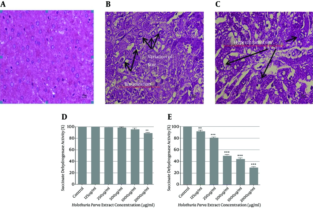
Figure 1.
A, liver section from the control group shows normal cellular architecture (H and E; 40 × magnification); B and C, liver sections from the HCC group show areas of aberrant hepatocellular phenotype with variation in nuclear size, hyperchromatism, binucleation, and irregular sinusoids (H and E, 40 × magnification). The effect of H. parva concentrations on complex II (succinate dehydrogenase) activity in the liver mitochondria obtained from hepatocytes of untreated control D, and HCC groups E, values are represented as mean ± SD (n = 3). ** and *** indicate significant differences in comparison with the corresponding control mitochondria (P < 0.01 and P < 0.001, respectively).
4.4. Effect of H. parva Extract Concentrations on Mitochondrial Complex II
The effects of different concentrations of H. parva extract (0 - 2000 μg/mL) on the collapse of mitochondrial succinate dehydrogenase activity (determined as % of enzyme activity) after 1 hour of incubation were measured with the MTT assay, using mitochondria isolated from hepatocytes of both the untreated control group and the HCC group. Statistical analysis showed a significant concentration-dependent decrease in the mitochondrial metabolism of MTT to formazan in the HCC group, but not in the untreated control group (P < 0.05) (Figure 1E). Only the highest concentration of H. parva extract (2000 μg/mL) induced a significant decrease in the mitochondrial metabolism of MTT to formazan in the control group (P < 0.05) (Figure 1D).
4.5. Effects of H. oculata Extract Concentrations on Mitochondrial Complex II Activity
The effect of H. oculata (0, 50, 100, 200, 400, and 1600 μg/mL) on the collapse of mitochondrial succinate dehydrogenase activity (determined as % of enzyme activity) after 1 h of incubation was measured with the MTT assay, using mitochondria isolated from liver hepatocytes of groups A and B. Statistical analysis showed a significant concentration-dependent decrease in the succinate dehydrogenase activity (P < 0.05) (Figure 2B) in the mitochondria isolated from hepatocytes of the HCC rats, but not from those of the control group (Figure 2A).
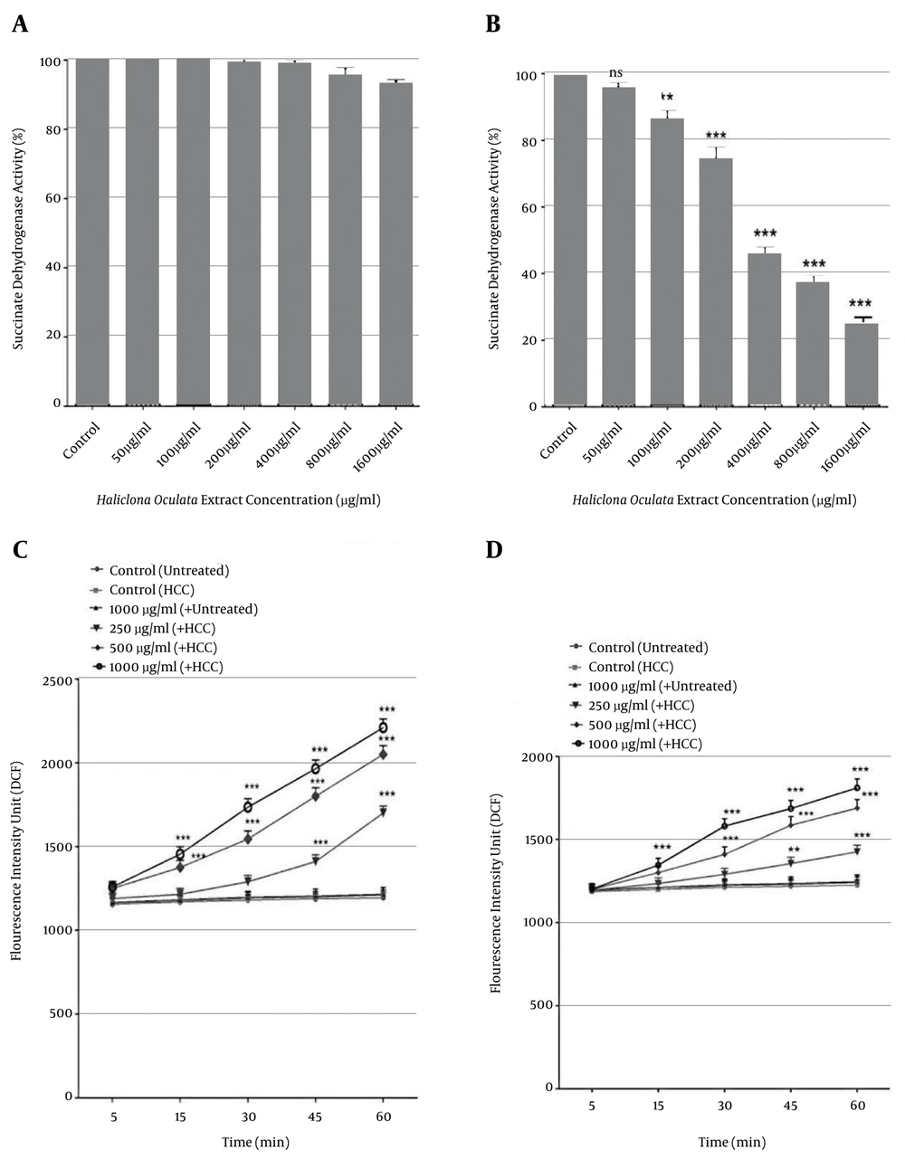
Figure 2.
The effect of H. oculata concentrations on complex II (succinate dehydrogenase) activity in the liver mitochondria obtained from hepatocytes of both the untreated control A, and HCC B, groups. Values are represented as mean ± SD (n = 3). ** and *** indicate a significant difference in comparison with the corresponding control mitochondria (P < 0.01 and P < 0.001, respectively). Measurement of mitochondrial ROS formation showing increases after addition of various concentrations of C, H. parva (250, 500, and 1000 µg/mL) and D, H. oculata (200, 400, and 800 µg/mL) extracts at different time intervals within 60 min of incubation, in the mitochondria obtained from hepatocytes of the HCC group but not the control group. Values are presented as mean ± SD (n = 3). *, **, *** and **** indicate significant differences between the control and HCC groups (P < 0.05, P < 0.01, P < 0.001, and P < 0.0001, respectively).
4.6. Effects of H. parva and H. oculata Extract Concentrations on Mitochondrial ROS Production
As shown in Figure 2C and D, different concentrations of H. parva (250, 500, and 1000 µg/mL) and H. oculata (200, 400, and 800 µg/mL) extracts induced significant (P < 0.05) H2O2 formation, demonstrated as fluorescence intensity units emitted from highly fluorescent DCF, in the mitochondria obtained from hepatocytes of the HCC group but not of the control group. This activity occurred in a concentration- and time-dependent manner.
4.7. Effects of H. parva and H. oculata Extract Concentrations on Mitochondrial Membrane Potential (MMP)
As shown in Figure 3A, H. parva concentrations of 250, 500, and 1000 μg/mL significantly (P < 0.05) decreased the MMP (demonstrated as fluorescence intensity units emitted from Rh 123, redistributed from damaged mitochondria into the cytosol) in a time- and concentration-dependent manner in the mitochondria obtained from hepatocytes of the HCC rats compared to the controls. As shown in Figure 3B, H. oculata extract concentrations (400 and 800 µg/mL) significantly decreased the MMP in a time- and concentration-dependent manner (P < 0.05) in the mitochondria obtained from hepatocytes of the HCC rats, but not of the untreated control rats. A low concentration of H. oculata extract (200 µg/mL) did not significantly decrease MMP within 60 min of incubation in the above-referenced HCC mitochondria. On the other hand, all of the applied concentrations of both H. parva and H. oculata did not induce significantly decreased MMP (P < 0.05) within 60 min of incubation in the mitochondria isolated from hepatocytes of the control rats (group A).
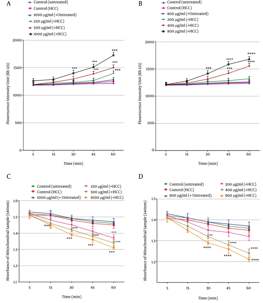
Figure 3.
Determination of the collapse of mitochondrial membrane potential (MMP). Decreased MMP after the addition of various concentrations of A, H. parva (250, 500, and 1000 µg/mL) and B, H. oculata (200, 400, and 800 µg/mL) extracts at different time intervals within 60 min of incubation in the mitochondria obtained from hepatocytes of the HCC group but not the control group. Values are presented as mean ± SD (n = 3). *, **, *** and **** indicate significant differences in the comparison with the control group (P < 0.05, P < 0.01, P < 0.001 and P < 0.0001, respectively). Determination of mitochondrial swelling showed an increase after the addition of various concentrations of C, H. parva (250, 500, and 1000 µg/mL) and D, H. oculata (200, 400, and 800 µg/mL) extracts at different time intervals within 60 min of incubation in the mitochondria obtained from hepatocytes of the HCC group but not of the control group. Values are presented as mean ± SD (n = 3). ** and **** indicate significant differences in the comparison with the control group (P < 0.01 and P < 0.0001).
4.8. Effect of H. parva and H. oculata on Mitochondrial Swelling
We measured the decrease of absorbance in the mitochondrial samples at 540 nm in order to assay mitochondrial swelling, another indicator of the mitochondrial permeability transition (MPT). The addition of different concentrations of H. parva extract (250, 500, and 1000 µg/mL) to mitochondrial suspensions obtained from liver hepatocytes of the HCC group led to significant mitochondrial swelling in a time-dependent manner (P < 0.05) (Figure 3C). The results in Figure 3D also show that H. oculata extract (400 and 800 µg/mL) induced significant swelling in a time- and concentration-dependent manner (P < 0.05) in the mitochondria obtained from hepatocytes of the HCC group. Only a low concentration of H. oculata extract (200 µg/mL) did not significantly increase mitochondrial swelling within 60 min of incubation. The addition of the same concentrations of H. parva and H. oculata to the mitochondria obtained from liver hepatocytes of the control group did not induce any mitochondrial swelling.
4.9. Effects of H. parva and H. oculata extract Concentrations on Cytochrome c Release
Our previous results showed that H. parva and H. oculata extracts significantly (P < 0.05) decreased MMP and induced mitochondrial swelling. Consequently, it is expected that H. parva and H. oculata extracts induce the release of cytochrome c from the mitochondria into the cytosolic fraction. As shown in Figure 4A and B, H. parva (500 µg/mL) and H. oculata (400 µg/mL) extracts induced a significant (P < 0.05) release of cytochrome c (ng/mg mitochondrial protein) in the mitochondria isolated from liver hepatocytes of the HCC group, but not from the untreated control group. Pretreatment of both H. parva (500 µg/mL) and H. oculata (400 µg/mL)-treated mitochondria with MPT inhibitors, such as cyclosporine A (CsA), and antioxidants, such as butylated hydroxyl toluene (BHT), caused inhibition of cytochrome c release (P < 0.05). These results confirm the direct role of oxidative stress and MPT pore-opening in cytochrome c release resulting from exposure to H. parva and H. oculata extract.
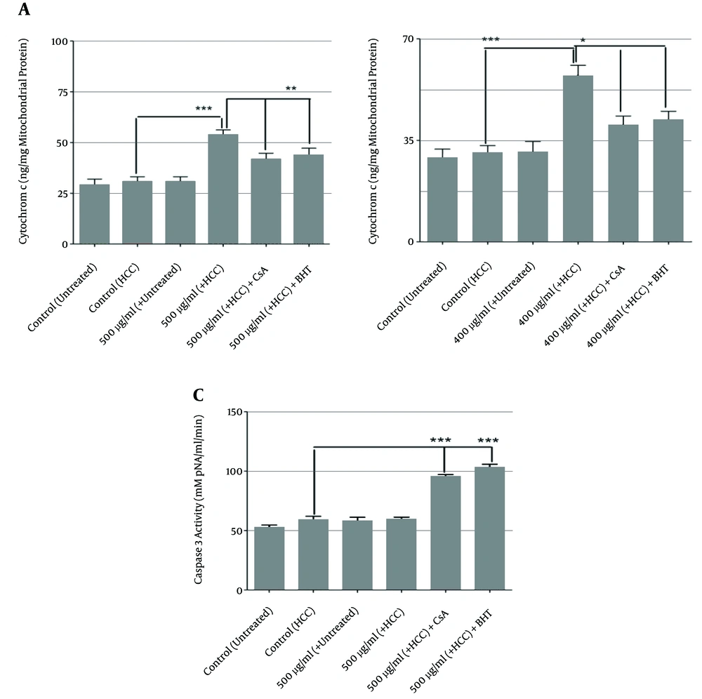
Figure 4.
Measurement of cytochrome c expulsion. Increased cytochrome c release after addition of A, H. parva (500 µg/mL) and B, H. oculata (400 µg/mL) to the mitochondria obtained from hepatocytes of the HCC group but not from the control group. Pretreatment with BHT or CsA significantly inhibited cytochrome c release in the HCC liver mitochondria. The amount of expelled cytochrome c from the mitochondrial fraction into the suspension buffer was determined using a rat/mouse cytochrome c ELISA kit. Values are presented as mean ± SD (n = 3). *** indicates significant difference in comparison with the untreated group (P < 0.001). * and ** indicate significant differences in comparison with H. oculata (400 µg/mL) and H. parva (500 µg/mL)-treated HCC group (P < 0.05). C, Determination of caspase-3 activity. Caspase-3 activation was measured in the HCC and control hepatocytes following exposure to H. parva (500 µg/mL) and H. oculata (400 µg/mL) extracts, using a Sigma-Aldrich kit. The kit measures pNA released from the interaction between caspase-3 and AC-DEVD-pNA (peptide substrate). Values are expressed as mean ± SD from three separate experiments (n = 3). *** indicates a significant difference in comparison with the untreated HCC group (P < 0.001).
4.10. Effects of H. parva and H. oculata Extract Concentrations on Caspase-3 Activation
The caspases are the most important effector molecules in the execution of apoptosis, and the progression of the caspase activation cascade ends in the activation of caspase-3, the final mediator of apoptosis. In this study, caspase-3 activity (mM pNA/mL/min) was significantly (P < 0.05) increased in hepatocytes obtained from only the HCC rats (group B) when incubated with H. parva (500 µg/mL) and H. oculata extracts (400 µg/mL), but not in hepatocytes from the control rats (group A) (Figure 4C).
5. Discussion
Cancer is a disease with tremendous negative implications at the personal, health care, social, and economic levels. The alarming increase in the worldwide death toll from cancer combined with alternative approaches to cancer therapy have fueled the search for novel effective anti-tumor drugs through biological testing of both marine and terrestrial organisms (11, 27). Natural products are a main source of new, complex chemicals, many of which show potent cytotoxic activity and are currently being used in cancer therapies (28). There is increasing evidence suggesting that the marine environment contains various classes of biologically active compounds with strong anticancer properties, in particular marine sponges, from which multiple potent cytotoxic compounds containing alkaloids, steroids, terpenes, peptides, macrolides, and polyketides have been isolated (29).
Sea cucumbers are one of the marine animals. The therapeutic properties of these animals are related to the presence of functional components with numerous anticipated biological activities (11, 18). Multiple unique pharmacological and biological activities, including anticancer, anti-angiogenic, antitumor, anticoagulant, anti-inflammatory, and anti-hypertensive effects, are related to chemical compounds extracted from various sea cucumber species. These therapeutic benefits and health applications may be attributed to the presence of numerous arrays of bioactive compounds, including triterpene glycosides (saponins), phenolics, cerebrosides, sulfated polysaccharides, chondroitin sulfate, glycoprotein, glycosaminoglycan, sterols, peptides, and lectins (11, 18, 30).
The results of the present study seem to provide support for the effects of DEN/2-AAF on induced liver cancer in rats. There was a significant reduction in body weight and an increase in liver weight in the HCC group compared to the control group. An in vivo study also confirmed this fact and reported that rats treated with DEN/2-AAF showed marked loss of body weight and increased liver weight (31, 32).
In comparison with the control rats, serum markers such as ALT, AST, and ALP showed significant increases in the cancer group (P < 0.05). These serum enzymes are indicators of hepatic function and their increased levels in the blood indicate liver damage. AFP is a cancer marker that can be produced by regenerating hepatic tumors. Increased serum AFP in the HCC-induced rats in our study may have resulted from DEN/AAF intoxication, which caused genetic alterations in the hepatocytes (33).
The histopathological findings in our study were supported by biochemical results obtained in experimental animals. The histopathological observations of the livers of DEN-treated rats revealed well-differentiated HCC hepatocytes with disorganized hepatic lobular architecture and obvious cellular damage.
Mitochondria play important roles in cellular metabolism and apoptosis pathways. Multiple significant differences in the function and structure of mitochondria between cancerous and normal cells have been reported. For example, there are alterations in the size, number, and shape of the mitochondria in cancerous liver cells compared to corresponding normal cells. In addition to structural and functional changes, genomic mitochondrial alterations have also been correlated with cancer. It has been reported that mitochondria in liver cancer cells are more fragile than normal liver mitochondria (21, 34, 35).
H. parva and H. oculata at concentrations of 0 - 2000 µg/mL and 0 - 1600 µg/mL, respectively, significantly reduced the activity of complex II (succinate dehydrogenase) in the mitochondria isolated from the HCC rats, but not in the untreated control-rat hepatocytes.
In this report, H. parva and H. oculata extracts significantly increased ROS production in a time- and concentration-dependent manner in mitochondria obtained from the HCC group compared to the untreated normal group. ROS are intracellular second messengers that affect numerous cellular processes, including metabolism, differentiation, and cell proliferation and death by regulating critical signaling pathways. It has been recognized that ROS bring about complex and irreversible damage to the cellular constituents that impair cellular homoeostasis, and elevated levels of ROS can influence central cellular processes, including apoptosis and proliferation (36).
Our results showed that all of the applied concentrations of H. parva and H. oculata extracts significantly induced decreased levels of ΔΨm in mitochondria isolated from cancerous, but not normal, hepatocytes.
Alteration of mitochondrial swelling as an indicator of MPT was also monitored in our study. H. parva and H. oculata extracts induced significant mitochondrial swelling in the mitochondria obtained from cancerous, but not normal, hepatocytes.
MMP is a necessary factor in the regulation of mitochondrial activity, and MMP collapse is the major stimuli for apoptosis and necrosis. Briefly, mitochondrial membrane damage results in MPT pore-opening and the release of cytochrome c into the cytosol. Once released into the cytosol, cytochrome c, along with apoptotic protease activating factor 1 (Apaf-1) protein and procaspase-9, forms the apoptosome. In the presence of ATP, caspase-9 is activated, leading to activation of the downstream effector caspase-3, which ultimately leads to the degradation of cell components and the final steps of apoptosis (37, 38).
Our results showed that the applied concentrations of H. parva and H. oculata extracts induced significant dismissal of cytochrome c from the mitochondria. Moreover, pretreatment with both CsA (the MPT pore-sealing agent) and BHT (an ROS scavenger) completely blocked the H. parva- and H. oculata-induced release of cytochrome c from the mitochondria, which supports our hypothesis that apoptosis induction via H. parva and H. oculata is due to oxidative stress and depends on the opening of the MPT pore.
It was reported that frondoside A (from Cucumaria frondosa) induced significant morphological changes consistent with apoptosis. The results indicated that frondoside A induced apoptosis of AsPC-1 human pancreatic cancer cells via the mitochondrial pathway and activation of the caspase cascade (39).
Another study also showed that stichoposide C (isolated from the holothurian Thelenota anax) caused apoptosis in A549, HCT-116, and MCF-7 cells in a dose-dependent manner due to the activation of Fas and caspase-8, cleavage of Bid, mitochondrial damage, and caspase-3 activation (40).
As suggested by many other investigators, the regulation of apoptosis involves, in particular, the decreased expression of proteins such as Bcl-2 and Mcl-1, increased Bax expression, and enhanced mitochondrial cytochrome c release, which ends in the induction of apoptosis (41-43).
We obtained consistent and relevant mitochondrial data that could be used as supporting evidence for the initiation of apoptosis signaling in cancerous hepatocytes through mitochondrial dysfunction. Following the addition of two natural extracts, from H. parva and H. oculata, to determine whether we could selectively induce apoptosis in the cancerous hepatocytes, we decided to measure caspase-3 activity. Our results showed that H. parva (500 µg/mL) and H. oculata (400 µg/mL) extracts induced significant caspase-3 activation in hepatocytes obtained from cancerous, but not normal, rats. It has been reported that the apoptosis-inducing lead compounds isolated from marine sponges, divided by putative biogenetic origin, include alkaloids, terpenoids, lipids, and macrolides. Because of the pressing need to develop non-cytotoxic anticancer treatments, novel apoptosis-inducing drug candidates with the potential to be developed into effective targeted cancer therapies are of interest to the cancer research community. In this regard, marine sponge-derived bioactive metabolites will continue to be some of the most promising sources of new drug leads (44).
Gupta et al. reported that H. oculata is a main source of alkaloids, steroids, terpenoids, unsaturated fatty acids, and cyclic peptides. Some of these compounds have been reported to possess diverse biological activities (14).
In conclusion, as an outcome of this comprehensive investigation, we can recommend H. parva and H. oculata as new anti-HCC drug candidates. This study provides evidence that mitochondrial targeting is the critical mechanism by which H. parva and H. oculata could potentially and selectively induce apoptosis in HCC hepatocytes, and could inhibit tumor growth.