1. Introduction
Giant cell tumors are mesenchymal neoplasms primarily seen in bone and soft tissues; but they have also been rarely reported from other organs such as thyroid, gall bladder, and pancreas (1). Occurrence of this type of tumor in the liver is an extremely rare event and to the best of our knowledge, only 5 cases have been reported in the English literature (2-5). Herein, we report our experience with an aggressive giant cell rich non-epithelial mesenchymal tumor in the liver (osteoclastoma-like giant cell tumor) in a 64-year-old female as the 6th case in the English literature.
2. Case Report
A 64-year-old woman presented with abdominal pain. Her past medical history was unremarkable. Her physical examination was also unremarkable. The heart and the lung were normal. There was no hepatomegaly or splenomegaly in abdominal examination. The head and neck examinations were also normal.
Laboratory examination showed WBC = 9000/mL, Hb = 13 g/dL, and platelet = 250,000/mL. ALT, AST, GGT, and alkaline phosphates were all unremarkable. Tumor markers of CEA, CA125, and CA19-9 were all in normal range.
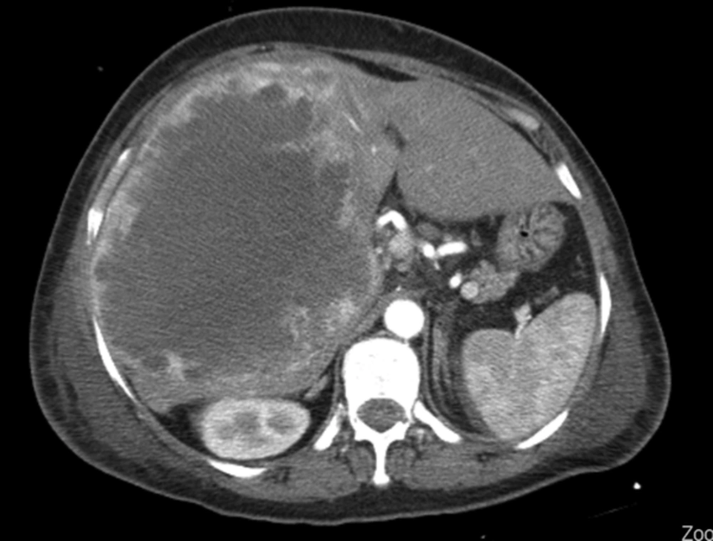
Abdominal sonography showed a liver mass. CT scan showed an enlarged liver with a large mass in the right lobe of the liver with irregular borders and central necrosis, measuring 20 cm in the greatest diameter. A few smaller lesions were also present. Portal vein thrombosis was also identified. Non-tumoral liver was unremarkable, meaning that there was no underlying disease in the non-tumoral liver parenchyma (Figure 1).
The patient was scheduled for surgery with the preoperative imaging diagnosis of angiosarcoma. In the operation, the mass was reported to be hypervascular by the surgeon and the surgeon’s diagnosis was angiosarcoma. Incisional biopsy was performed. The received specimen in the pathology lab was fragmented, brown, and full of blood, measuring totally 4X3X1 cm3.
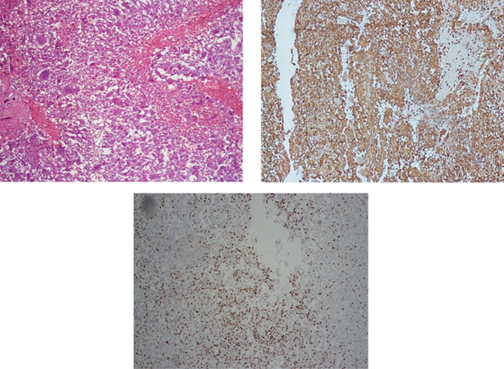
Microscopic sections showed bloody background with many osteoclast-like giant cells intermingled with mononuclear cells (Figure 2A), which were reactive with CD68 and vimentin (Figure 2B) but nonreactive with all the epithelial markers. The histopathologic findings were very similar to giant cell tumor of the bone. Many mitotic figures were present in the atypical pleomorphic mononuclear cells, with high Ki67 (Figure 2C).
After surgery, the patient had an uneventful postoperative period; however, in less than 2 months she died of cardiorespiratory arrest.
3. Discussion
Osteoclastoma-like giant cell tumor of the liver is an extremely rare tumor in the liver so that only 5 cases have been reported in the English literature since 1980. Table 1 shows the characteristics of the above-mentioned 5 cases. The first case was reported by Munoz in 1980 (1). All of the reported patients including ours were older than 50 years. It does not seem to have any sex preferences, and it is equally distributed in females and males. All of the previously reported cases, except two, have been in the liver with no underlying disease. The first reported case in 1980 by Munoz was observed in a patient with alcoholic cirrhosis who presented symptoms of portal hypertension (1). The other case autopsied and reported by Zhang was also a cirrhotic patient with no definite cause (5).
| Author | Year Reported | Age, y | Sex | Clinical Presentation | Size, cm | Outcome | Underlying liver disease |
|---|---|---|---|---|---|---|---|
| Munoz 1 | 1980 | 87 | M | Portal Hypertension | 11 | Died 32 days after operation | Alcoholic Cirrhosis |
| Horie 2 | 1987 | 66 | F | Hematoemesis; Nausea; Abdominal pain | 5 | Died in 42 days after operation | - |
| Rudloff3 | 2005 | 61 | F | Right upper quadrant pain | 8 | Died after cardiorespiratory failure | - |
| Bauditz4 | 2006 | 54 | M | Incidental | 7 | Died after 15 months | - |
| Zhang5 | 2006 | 65 | M | Right upper quadrant pain | 13 | Died after 5 days Of hypovolemic shock | Cirrhosis |
| Current Case | 2017 | 64 | F | Abdominal pain | 20 | Died after cardiorespiratory arrest | - |
The most common presenting symptom has been abdominal pain; however, as the table shows, one case was incidentally detected (4).
In all non-cirrhotic cases, liver function tests and tumor markers have been in normal range and therefore, seem to have no diagnostic value (1-5).
All of the reported tumors have been hemorrhagic, which has been misdiagnosed as angiosarcoma before pathologic diagnosis (2).
Microscopic examination has been the same in all the 6 cases including osteoclast-like giant cells with abundant eosinophilic cytoplasm and a variable number of nuclei, mixed with malignant pleomorphic mononuclear cells and hemorrhagic background. The most important point in histopathology of this tumor is the differential diagnosis of hepatocellular carcinoma rich in osteoclast like giant cells, which shows positive epithelial markers instead of mesenchymal (6). These tumors, which have most commonly been reported in pancreas, sometimes have typical areas of carcinoma (7).
All of the previously reported cases of osteoclastoma-like giant cell tumor of the liver expired shortly after surgery, indicating that the disease has a very poor prognosis.

