1. Context
Hepatitis B virus (HBV), a member of the Hepadnaviridae family, is a small enveloped and partially double-stranded circular deoxyribonucleic acid (DNA) virus. Its genome is made up of four overlapping reading frames, which are Pol (P), Core (C), Surface (S), and X, encoding HBV proteins and promoter regions. In this regard, P encodes viral polymerase enzyme that is required for DNA synthesis, C encodes Hepatitis B envelope antigen (HBeAg), a precore/core antigen protein, which is a marker for an active infection and replication, S encodes surface proteins that are essential for viral entry and encapsulation of the core during replication, and X encodes X protein that is a key regulator of replication (1, 2). Formation of covalently closed circular DNA (cccDNA) during replication differentiates HBV from other retroviruses. This is the stable form of viral DNA that allows its persistence in the hepatocytes with significant clinical impacts, including chronicity, carcinogenesis, and anti-viral drug inefficiency. The high mutagenesis rate of HBV that is caused due to a lack of proof-reading of reverse transcriptase has resulted in the formation of ten HBV genotypes (A - J) and their several corresponding sub-genotypes with a well-characterized geographical distribution (3).
Hepatitis E virus (HEV), which is an icosahedral single-stranded RNA genome virus, has been classified as a single member of the genus Hepevirus in the family Hepeviridae. It has three overlapping open reading frames (ORFs). ORF1, ORF2, and ORF3 which respectively encode Ribonucleic acid (RNA)-dependent RNA polymerase (RdRp) protein, a capsid protein, and a small protein which induces immune suppression in HEV-infected patients (4, 5). To date, only one serotype and five genotypes (HEV-1, HEV-2, HEV-3, HEV-4, and HEV-7) are known to exist (6). Based on the new labeling approach established by the International Committee on the Taxonomy of Viruses (7), a total of 24 sub-genotypes of HEV have been identified. These include five, two, ten, and seven subtypes in HEV genotypes 1, 2, 3, and 4, respectively.
Both HBV and HEV infections affect a large number of people globally. Therefore, an interaction between the two infections in endemic areas is likely to occur, and when it does, a synergetic impact is usually observed. This review summarizes recent information in the epidemiology of both infections, HEV superinfection, pertaining to clinical interactions and prevention.
2. Evidence Acquisition
In order to achieve a comprehensive approach to find relevant articles, an extensive search was done using relevant keywords in Google Scholar, PubMed, and Scopus. The new papers were considered first and appropriate combination of the following keywords were used: Hepatitis B, Hepatitis E, coinfection, superinfection, epidemiology, liver disease, liver failure, liver cirrhosis, and hepatocellular carcinoma.
3. Results
3.1. Epidemiology
Chronic hepatitis B (CHB) infection, which embraces a large spectrum of the disease, remains a serious public health problem in the world with 257 million people affected and causes around 900000 deaths annually. The African and Western-Pacific World Health Organization’s regions harbor 68% of all infections globally (8). Most of the countries of these regions are of higher-intermediate endemicity (prevalence 5% - 7.9%) or highly endemic (prevalence ≥ 8%) for HBV. The European region is of lower-intermediate endemicity (prevalence 2% - 4.9%) and most of the countries in the American region have low endemicity (prevalence < 2%) (9). Globally, it is estimated that HEV affects a total of 20 million cases, 70000 deaths, and 3000 stillbirths (10). The HEV is endemic in many countries of Asia, Central America, and Africa. South-East Asia harbors most of the infections (60%) and deaths (65%) (11). Epidemiology of HEV infection varies greatly in each geographical location with a well-characterized viral genotype distribution.
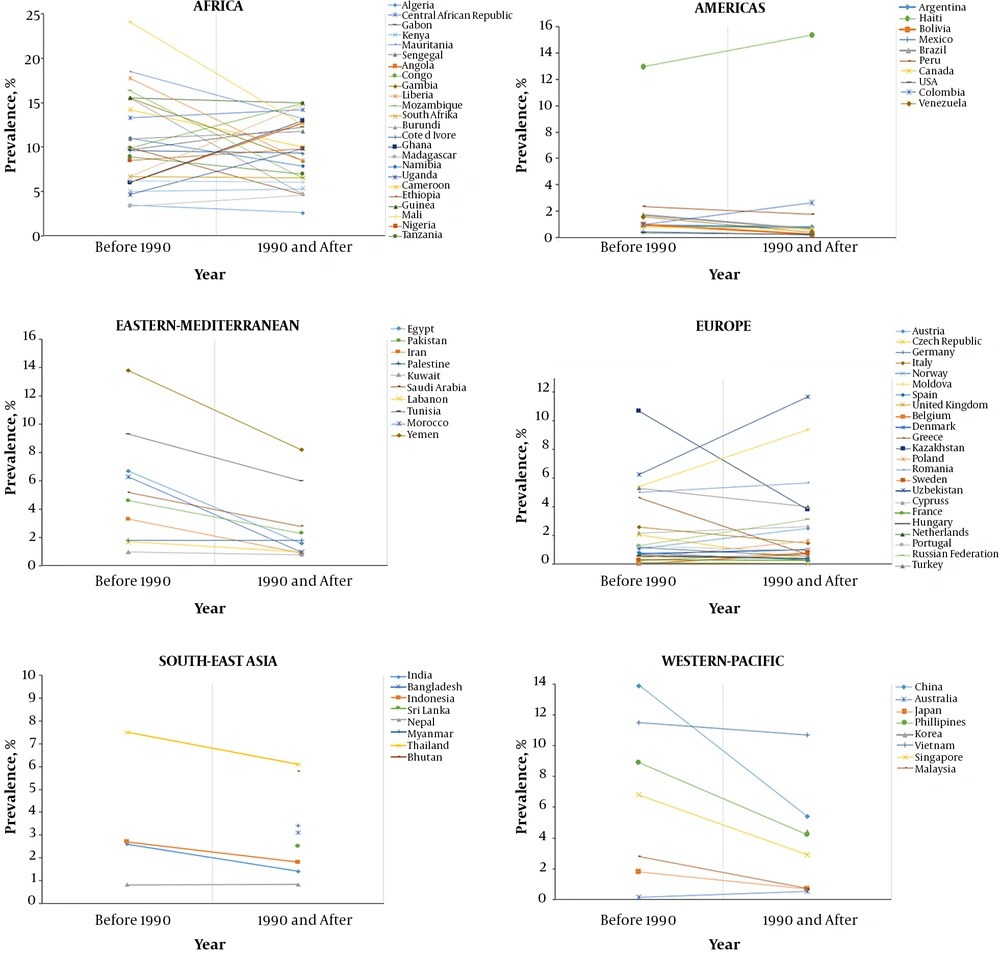
While there is a decline in the prevalence of HBV in many regions, the prevalence of HEV is generally increasing in most countries. A recent systemic review of the studies published between 1965 and 2013 by Schweitzer et al. (8), has shown a clear decline in HBV infection rates in many countries in several WHO regions, including Western-Pacific, South-East Asia, America, Europe and Eastern-Mediterranean during the periods 1965 - 1989 and 1990 - 2013. However, this trend was not seen in the African region throughout the periods considered. Another national-wide review from China, which is one of the most affected countries by HBV infection, also showed a significant decrease of HBV seroprevalence in the general population from 7.1% in 2006 to 6.1% in 2013 (12). This pattern clearly deciphers an expanded adaptation of universal HBV immunization recommendations in newborns from the year 1992 in several countries (13), with a poor response by many African countries due to economic constraints. These data are summarized in Figure 1.
HBV seroprevalence in different countries in two-time periods (before and after the year 1990) (9). Graphs show the studies describing HBV seroprevalence (HBsAg) in countries from six WHO regions. The correlation was shown between studies that were published before the year 1990 and those published in 1990 and afterward. Apart from Africa and few countries in Europe and Americas, general declination of HBV seroprevalence is evident between two time periods.
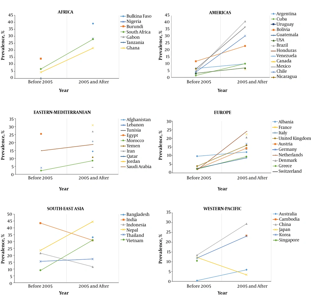
On the other hand, gathered evidence suggests that the rate of HEV infection is clearly escalating in many countries (14-17). In Europe for instance, the number of autochronous symptomatic cases has steadily increased from 124 cases in 2003 to 1243 cases in 2016 in England (18) and 31 cases in 2001 to 1991 cases in 2016 in Germany (19). A large multicenter study in Scotland has recently reported an increase in HEV detection among blood donors from 4.5% in 2004 - 2005 to 9.3% in 2014 - 2015 (20). Furthermore, in China, it was reported in the Third National Viral Hepatitis Prevalence Survey of 2005 - 2006 (21) that HEV seroprevalence rate was 23.5% in the country, which was higher than the previously reported seroprevalence of 17.2% in 1997 (22). These data are illustrated in Figure 2 and Appendix 1 in Supplementary File. The increasing detection rate of HEV might be a result of increased awareness and hence increased testing for the virus and/or a true increase in numbers of new infections. Moreover, the new serological tests, particularly rapid Wantai, have shown to have a significantly higher sensitivity than most of the older tests (23) and hence, it might possibly contribute to the variability.
HEV seroprevalence in different countries in two-time periods (before and after year 2005). Graphs showing the studies describing HEV seroprevalence (anti-HEV IgG) in countries from six WHO regions. The correlation was shown between studies that were published before the year 2005 and those published in 2005 and afterward. A clear inclination of HEV seroprevalence was observed between two time periods in most countries.
Several meta-analysis studies have revealed that older subjects and males are more susceptible to HEV infection than younger people and females, respectively (14, 24-26). In most countries where HEV is prevalent, infection in children is relatively rare. However, in Egypt where the prevalence of HEV in the general population is among the highest in the world, a very high rate (56.7%) of HEV infection was seen in children among patients with acute viral hepatitis (27). These observations were at least partly attributed to possible parenteral transmission as up to 20% of these children pre-received blood transfusion (BT) that was not screened for HEV. Indeed, BT has previously correlated with the possibility of HEV transmission (28). Late presentation of HEV infection in life might be due to cumulative viral exposure in a lifetime and probably aging of the immune system. However, the reasons as to why males are more susceptible than females are yet to be clarified.
HBV affects different age groups depending on the time of infection. In countries with a high prevalence of HBV (prevalence > 2%), infection is usually acquired perinatally or during early childhood and the majority of the patients live with the infection to its chronic phase, while in countries with low prevalence (prevalence < 2%), HBV infection is usually acquired during adulthood, mostly through intravenous drug uses (IDU) or sexual intercourse (29). To date, there is no evidence of any sex predilection of HBV infection, but male sex is known to be an independent risk factor for increased progression to cirrhosis and HCC (30).
In endemic areas, HEV typically superimposes CHB in most cases since the former occurs during adulthood, while the latter occurs in early life in these areas. Thus older and male subjects comprise a higher risk group for HBV-HEV superinfection (31-33). The rates of HBV-HEV superinfection vary considerably. The prevalence of 9% - 38% has been reported in previous studies in China (34-38). In Southeast Asia, the HBV-HEV superinfection rate was reported to be 45%, 14% and 9.5% in Vietnam, India and Bangladesh, respectively (39-41). A clear territory-wise distribution of HBV-HEV superinfection has been observed in Africa where a higher prevalence of 56.7% was reported in Egypt in the northern part of the continent, while insignificant rates of 1% and 0% were reported in Kenya, the Eastern part of Africa (27, 42, 43) (Table 1).
| Country | Reference | Cohort | Prevalence (%) | Sample Size | Year of Sampling | |||
|---|---|---|---|---|---|---|---|---|
| HEV Diagnostic Method | Agea | |||||||
| IgG | IgM | RNA | ||||||
| China | (44) | Pregnant women | 10.7 | 0 | 26.4 ± 4.1 | 391 | 2016 | |
| Turkey | (45) | Positive HBV DNA | 13.7 | 8.4 | 14.7 | 42.2 ± 9.1 | 190 | 2004 |
| China | (37) | General | 38.1 | 1.57 | 52.1 ± 13.1 | 1022 | 2014 | |
| India | (46) | Cirrhotics | 6.3 | 46 ± 14.6 | 192 | 2004 | ||
| Vietnam | (39) | CHB | 41 | 9 | 41 (9 - 84) | 744 | 2013 | |
| Hongkong | (32) | Acute HEV | 19 | 51 (22 - 85) | 161 | 2000 - 2012 | ||
| Spain | (47) | HIV-infected | 13.3 | 0 | 0 | 46.3 (21 - 80) | 448 | 2013 |
| Nepal | (48) | HIV-infected | 14.3 | 36.2 ± 10.3 | 459 | 2015 | ||
| Italy | (49) | HIV-infected | 5.9 | 41.7 ± 6.7 | 34 | 2013 | ||
| Kenya | (42) | Acute hepatitis | 1 | 38.9 (19.83) | 100 | 2012 | ||
| Bangladesh | (50) | Pregnant with acute viral hepatitis | 9.4 | 32 (8 - 82) | 31 | 2004 - 2006 | ||
| Ghana | (51) | Jaundiced | 18.7 | NS | 155 | 2016 | ||
| Central African Republic | (52) | Fever and jaundice | 4.9 | NS | 198 | 2008 - 2010 | ||
| India | (40) | Children with acute viral hepatitis | 0.7 | < 16 | 149 | 1998 - 2002 | ||
| Egypt | (27) | Children with sporadic acute viral hepatitis | 56.7 | NS | 162 | 2008 | ||
Abbreviations: IQR, interquartile range; NS, not specified; SD, standard deviation.
aValues are expressed as mean ± SD or mean (IQR).
3.2. Interaction Between HBV and HEV Infections
Endemic areas for both HBV and HEV infections provide an important intersection between two diseases. Following HEV superinfection, the ordinary clinical evolution of CHB infection is usually distorted with a deterioration of most of the manifestations, with the subsequent poor patient outcomes.
3.2.1. Clinical Features
Following HEV infection, the majority of immunocompetent subjects clear an infection spontaneously with only mild and unspecific symptoms, while up to 60% of immunosuppressed individuals progress to the chronic course of the disease (53, 54) that occurs exclusively in genotype 3 (55). In rare cases, however, HEV can cause acute severe liver injury, and/or acute liver failure and acute-on-chronic liver failure (ACLF) (56). Pregnant women and individuals with chronic liver disease (CLD) are at increased risk of developing ALF.
Although the clinical progression of HBV infection is complex and usually based on the state of balance between the host’s immune response and viral replication, HBV patient may develop acute (self-limiting) infection, acute fulminant infection or chronic state that may facilitate increased risks of developing liver cirrhosis or hepatocellular carcinoma (HCC) (3). According to a new nomenclature system, the natural history of CHB has been re-categorized into 5 phases that are not necessarily sequential. These are phase 1 which was previously referred to as immune tolerant and is more frequent and prolonged in perinatally-infected people. It is characterized by minimum or no liver necroinflammation or fibrosis. Phase 2 usually occurs after several years of the first phase and is more frequently and/or rapidly reached in subjects infected during adulthood. There is a moderate or severe liver necroinflammation with a rapid progression of fibrosis. Phase 3 which was previously known as an inactive carrier and results in minimum necroinflammation activity and fibrosis. If remained in this phase, the affected patients have a low risk of progression to cirrhosis or HCC. Phase 4 is associated with a significant necroinflammation and fibrosis, and a low rate of spontaneous disease remission. Phase 5 which is also known as occult HBV infection, is associated with minimal risk of cirrhosis, decompensation, and HCC if HBsAg loss occurred before the occurrence of cirrhosis (57).
HEV superinfection has been shown to adversely modify the natural clinical progression of CHB in various aspects. Several clinical symptoms were found to be more frequently reported among HBV-HEV superinfected patients. In one study, for instance, fever was found to be significantly common among HEV + HBV group (37.7%) compared to HBV monoinfection group (1.4%). Other symptoms, including inappetence, fatigue, nausea, vomiting, and epigastric discomfort also followed the same trend (35). These findings suggest that CHB patients who develop a fever and other non-specific symptoms should be thoroughly screened for HEV. Other features such as accelerated rates of liver injury, a progression of HBV-related CLD to more severe forms and rapid decompensation to liver failure have also correlated with HEV superinfection in CHB patients. In a recent large Vietnamese survey by Hoan et al. (39), HEV seropositivity was found to be associated with the presence of liver cirrhosis and HCC among CHB patients. The poor prognosis of cirrhotic patients based on the Child-Pugh score also correlated with HEV superinfection in this study. Another study from India reported that CHB patients with liver cirrhosis that were superinfected with HEV developed decompensation to liver failure more rapidly than those without HEV. The HEV was also found to be an independent risk factor for 12-month mortality as 64.3% in HEV-infected vs 3% in HEV-uninfected (46). Similarly, a recent country-wide survey in Hong Kong that analyzed data from the year 2000 - 2016 reported that underlying CHB infection was an independent risk factor for 30-day liver-related mortality among patients with acute HEV infection (58). Similar findings have also been reported elsewhere (36, 59, 60).
3.2.2. Treatment
To date, there is no curative therapy for HBV infection; hence, the supportive treatment that is available is only recommended for patients who are likely to benefit from it (i.e. that are at higher risk for liver cirrhosis and HCC). Based on this fact, several criteria have been recommended for the initiation of therapy (61-66) that mainly consists of Nucleotide analogs and/or interferons. Regarding HEV, evidence on the effectiveness of its treatment is very limited. The currently available data from case reports and small observations are limited to chronic HEV infection among organ transplant recipients. The new European Clinical Practice Guidelines on HEV Infection (67) recommend Ribavirin, a potent antivirus, to be considered for the treatment of severe acute HEV infections, acute-on-chronic liver failure as well as chronic cases. Pegylated interferon-α should be added to ribavirin non-responder subjects. To date, there is no recommended specific treatment regime for HBV-HEV superinfected cases, especially if the patient is already on anti-HBV therapy. It is also unclear if the early initiation of anti-HEV medications will delay the development of liver dysfunction and/or liver failure in these dually infected patients. Extensive clinical studies in this area are, therefore, desirable in order to develop better management strategies for these patients as to preclude the associated complications and consequently, improve their outcomes.
3.2.3. Prevention
The HEV vaccine which has been shown to be vastly effective has so far been licensed in China only. The vaccine is recommended for individuals who are above 16 years old and have a high risk of HEV infection. These include people who are engaged in animal husbandry and catering, students, women of childbearing age, and travelers to endemic areas (68). There is limited data on the efficacy of HEV vaccine in persons with CLD, the population that is likely to be affected by the superinfection. Therefore, safety and effectiveness studies of HEV vaccines among CLD patients are essential for the potential prevention of this risk group.
Provided that the majority of HEV infections (mainly genotype 3) are acquired by eating undercooked meat, avoiding of consumption of these products or thorough cooking of the same should be advised and encouraged. In a cell culture model study, it was found that heating at 70°C for 2 minutes or more effectively eliminated an infective HEV (69). However, it is unclear whether this in vitro finding can be feasibly transformed into meat preparation. Concomitantly, the majority of immunocompetent individuals are clearly known to harbor HEV infection uneventfully, thus it might be irrational to recommend that undercooked meat should be avoided by the general population to prevent HEV. It is also unclear whether this approach can be beneficial to CLD patients. Further explorative studies are, therefore, needed to confirm the preventive benefit of boiling meat and to identify the targeted population.
Despite increasing evidence of the existence of HEV contamination in donated blood from qualified blood donors (70-72), vigorous pre-transfusion screening for HEV has not received much attention in many areas probably due to the uncertainty of HEV transmission risks. While some studies did not show any HEV transmission to blood recipients (73), others showed clear evidence of transmission of the virus (74). Thus some countries including UK, Ireland, Germany, and the Netherlands have developed a universal donor screening for HEV infection, while countries such as France and Switzerland performs selective screening of blood that has been intended for use in high-risk patients only (immunocompromised and solid organ recipients) (75, 76). The CLD patients have not been embraced as a high-risk group in spite of clear evidence of their increased risk of HEV infection. In China, where most of important intersection between HEV and CHB probably occurs due to a high prevalence of both infections, screening of HEV is not routinely done in pre-transfused blood (77).
With regard to HEV genotypes 1 and 2 that are transmitted by the fecal-oral route, improvement of general hygienic measures such as hand washing and provision of clean drinking water remains a vital preventive measure. Regular and sustainable provision of health education to the risk groups such as pregnant women and those with CLD is an important preventive measure and should be encouraged and facilitated.
4. Conclusions
The HEV infection rates are alarming. In spite of unclear mechanisms of interaction between two conditions, HEV superinfection clearly alters the course of CHB disease into a detrimental pattern with patients’ poor outcomes. Prevention of HEV infection, as well as its aggressive treatment in coinfected cases, might be an important strategy for reducing related morbidity and mortality, but the paucity of comprehensive clinical evidence hinders this approach. Thus it is highly desirable to address and fill the existing important research gaps in HBV-HEV interaction in all clinical aspects.


