1. Background
Diabetes refers to a heterogeneous group of metabolic disorders associated with a major increase in blood glucose levels as a result of a failure in insulin secretion (1). Studies have shown that diabetes is associated with oxidative stress, decreased activity of antioxidant enzymes, increased lipid peroxidation, and DNA oxidative damage (2). In addition, this disease can cause cell death by disrupting oxidative phosphorylation and reducing energy production (3). Hyperglycemia and insulin resistance seem to increase the production of free radicals and oxidative stress (2). Evidence shows that the expression of antioxidant enzymes decreases in patients with chronic diabetes. In a normal state, free radicals can increase the activity of antioxidant enzymes, while in diabetes, the balance between oxidant and antioxidant agents is disturbed due to the weakening of the antioxidant defense system (4).
Research suggests a direct association between diabetes and systemic inflammation. Serum levels of inflammatory markers, such as interleukins (IL), tumor necrosis factor-α (TNF-α), and C-reactive protein (CRP), significantly increase in diabetes (5). The increased level of TNF-α can induce insulin resistance by decreasing the activity of insulin receptor tyrosine kinase and stimulating adipocyte lipolysis. On the other hand, an increase in the level of IL-6, as a proinflammatory cytokine, results in insulin resistance in liver cells. Increased inflammatory cytokines also have inhibitory effects on the glucose transporter type 4 (GLUT-4) gene, leading to increased insulin resistance (6).
Apoptosis is a natural cellular event, regulating the growth and proliferation of body cells. During this process, the immune system shows many anti-antigenic activities. Generally, there are different pathways for apoptosis activation. If the message is internal, the first activated organ is the mitochondria. On the other hand, if the message is transmitted through surface receptors, the transmission is facilitated by adapter molecules to the caspase cascade (7). The ratio of apoptosis inducers to inhibitors plays an important role in the fate of cells. The inducible BCL2-associated X protein (Bax) and B-cell lymphoma-2 (Bcl-2) protein have been identified as the inhibitors of apoptosis (8).
Oxidative stress induced by hyperglycemia plays an important role in inducing apoptosis. High glucose concentrations seem to induce apoptosis by weakening the antioxidant defense system. Consequently, an imbalance between the production of free radicals and antioxidant agents can lead to apoptosis. This finding has been confirmed by examining the extent of DNA fragmentation and increasing the ratio of Bax to Bcl-2 (9). In diabetes, there is a significant increase in lipid peroxidation; therefore, the tissue level of Malondialdehyde (MDA) may increase. It has also been shown that the oxidation of free fatty acids in liver tissue increases in diabetes, which, together with inflammation, leads to structural and functional changes in hepatocytes (10).
Avicennia marina (Forssk.) Vierh is a species of a mangrove tree, containing a high percentage of flavonoids (11). Research has indicated the significant antioxidant properties of flavonoids. The consumption of plants rich in flavonoid compounds can increase the level of antioxidants in the blood, leading to the prevention of free radical formation (12). Evidence suggests that the administration of A. marina to diabetic rats reduces the serum glucose levels (13). Moreover, the protective effects of A. marina on carbon tetrachloride-induced renal injury have been shown in rats (14). Flavonoids and A. marina tannins show anti-nociceptive properties, and they can reduce pain sensation in the formalin-induced inflammatory phase (15). In addition, A. marina exhibits anti-inflammatory properties to improve arthritis in rats (15).
2. Objectives
With this background in mind, this study aimed to evaluate the effects of hydroalcoholic leaf extract of A. marina on apoptosis, inflammation, oxidative stress, lipid peroxidation, and tissue changes in the liver of diabetic rats.
3. Methods
3.1. Ethical Considerations
This experimental research was conducted in the Research Center of Baqiyatallah University of Medical Sciences, Tehran, Iran, in 2018. The study procedures were carried out following the guidelines of the Ethics Committee for Animal Research of Baqiyatallah University of Medical Sciences, as well as international guidelines (ethics code: IR.BMSU.REC.1396.230).
3.2. Animals
In this laboratory experiment, a total of 40 male Wistar rats, with a mean age of 155 ± 4 days and a mean weight of 168 ± 5 g, were selected. The animals were maintained in standard transparent polycarbonate cages at a temperature of 24 ± 3°C with a relative humidity of 35 ± 4% in a 12/12 h light/dark cycle. The rats had access to sufficient water and compressed food with a standard formula. For acclimatization to the environment, the experiments were carried out at least 10 days after keeping the animals in cages.
3.3. Experimental Design
Rats were randomly divided into five groups (eight rats per group) including the control group, type 1 diabetes mellitus (DM) group, diabetic rats treated with 50 mg/kg of hydroalcoholic extract of A. marina, diabetic rats treated with 100 mg/kg of hydroalcoholic extract of A. marina, and diabetic rats treated with 200 mg/kg of hydroalcoholic extract of A. marina. The control and DM groups were intraperitoneally administered with 0.5 mL of saline solution as the solvent for 30 days. The experimental groups were intraperitoneally administered with 0.5 mL of hydroalcoholic extract of A. marina at doses of 50, 100, or 200 mg/kg for 30 days.
3.4. Extraction
We collected A. marina leaves (by Seyed Damoon Sadoughi) from the northern coast of Qeshm Island, Iran, during spring (May 2017). Then, A. marina species was identified and confirmed at the Herbarium Department of the Payam Noor University of Mashhad, Iran (herbarium code: 12/3254). Next, A. marina leaves were ground by a grinder after drying in the shade at 36 ± 3°C. To prepare the extract, 200 g of dried A. marina powder was poured into a percolator. Then, 80% ethyl alcohol and distilled water (1:1 ratio) were added, and the powder was completely mixed in the solution. It was placed in the laboratory environment for 72 hours and filtered using a filter paper. It was stored at a temperature of 45°C for 48 hours to dry. After removal of the solvent, the extracts were prepared at concentrations of 50, 100, and 200 mg/kg. The extraction yield was estimated at 6.28%, based on the following formula:
3.5. Induction of Experimental Diabetes
The empirical model of type 1 DM was induced in rats by an intraperitoneal injection of alloxan monohydrate (120 mg/kg; Sigma-Aldrich, Germany). In addition, citrate buffer (pH = 5.4) was used as the alloxan solvent (16). Alloxan was administered to the DM group and experimental groups receiving 50, 100, and 200 mg/kg of A. marina. Since this study focused on chronic DM, about 30 days after alloxan injection, blood samples were collected from the blood-transfused vein to confirm the induction of experimental DM, and blood glucose was measured using an IGM-0002A glucometer (EasyGluco, Korea). A blood glucose level of above 300 mg/dL was considered as an indicator of DM on day 0 of the test.
3.6. Histomorphometric Analysis
Under sterile conditions, the liver was removed and placed in physiological serum. The liver tissue was then isolated and fixed in 10% formalin for histological evaluations. After tissue passage, 10 tissue sections (5 µm thickness) were prepared randomly and stained with hematoxylin and eosin. Subsequently, images were acquired at 400× magnification using an optical microscope (CX21FS1, Olympus, Japan), equipped with a camera (XC30, Olympus, Japan) and CellSens Dimension V1.6 software (Olympus, Japan).
3.7. Biochemical Assay
At the end of treatments, rats were terminally anesthetized by diethyl ether, and blood samples were collected from the left ventricle of the heart. Blood samples were placed in an incubator (INB400, Memmert, Germany) at 37°C for 15 minutes. After coagulation, they were placed in a centrifuge (EBA280, Hettich, Germany) at 5,000 rpm for 12 minutes. Next, blood serum was isolated to measure the serum levels of TNF-α, IL-1β, and IL-6, using ELISA kits (FineTest, China). The liver tissue was homogenized with Tris buffer at 5,000 rpm for two minutes using a homogenizer (T25 Ultra Turrax- IKA, Germany). The cellular cytoplasm was then isolated from the homogenized tissue via centrifugation (Z366, Hermle, Germany) and used for further assessments. To prevent the degradation of enzymes and proteins, all steps were carried out at 4°C, and a solution of 0.5 mM phenylmethylsulfonyl fluoride (Sigma-Aldrich, Germany) was used as the inhibitor of cell proteases. The levels of Bax, Bcl-2, caspase-9, Superoxide dismutase (SOD), catalase (CAT), glutathione peroxidase (GPx), MDA, and 8-hydroxy-2’-deoxyguanosine (HOdG-8) were also measured in liver tissues using ELISA kits (FineTest, China).
3.8. Statistical Analysis
Data analysis was performed in SPSS version 22 using the Kolmogorov-Smirnov test (for assessing the normal distribution of data), one-way ANOVA, and Tukey’s post hoc test. The results were presented as mean ± SD, and the P values of less than 0.05 were considered statistically significant.
4. Results
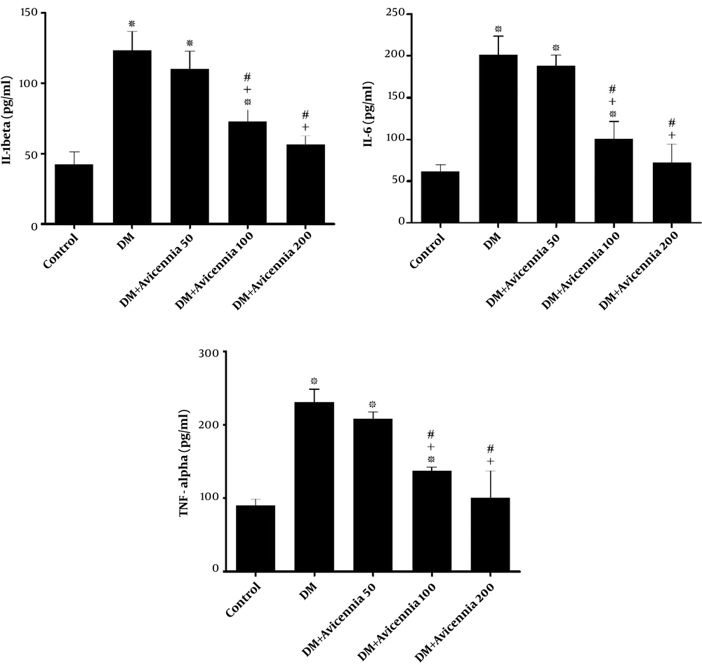
The results showed that the serum levels of IL-1β, IL-6, and TNF-α were significantly lower in diabetic groups receiving 100 and 200 mg/kg of A. marina than in the DM group, depending on the dose of injection (P < 0.05). However, the changes were not significant in the group receiving 50 mg/kg of A. marina (P > 0.05) (Figure 1).
Comparison of mean serum levels of IL-1β, IL-6, and TNF-α in groups. Values are mean ± SD of eight animals in each group. ∗Significant when compared to the control group (P < 0.01); +, significant when compared to the DM group (P < 0.01); #, significant when compared to the DM + Avicennia 50 group (P < 0.01).
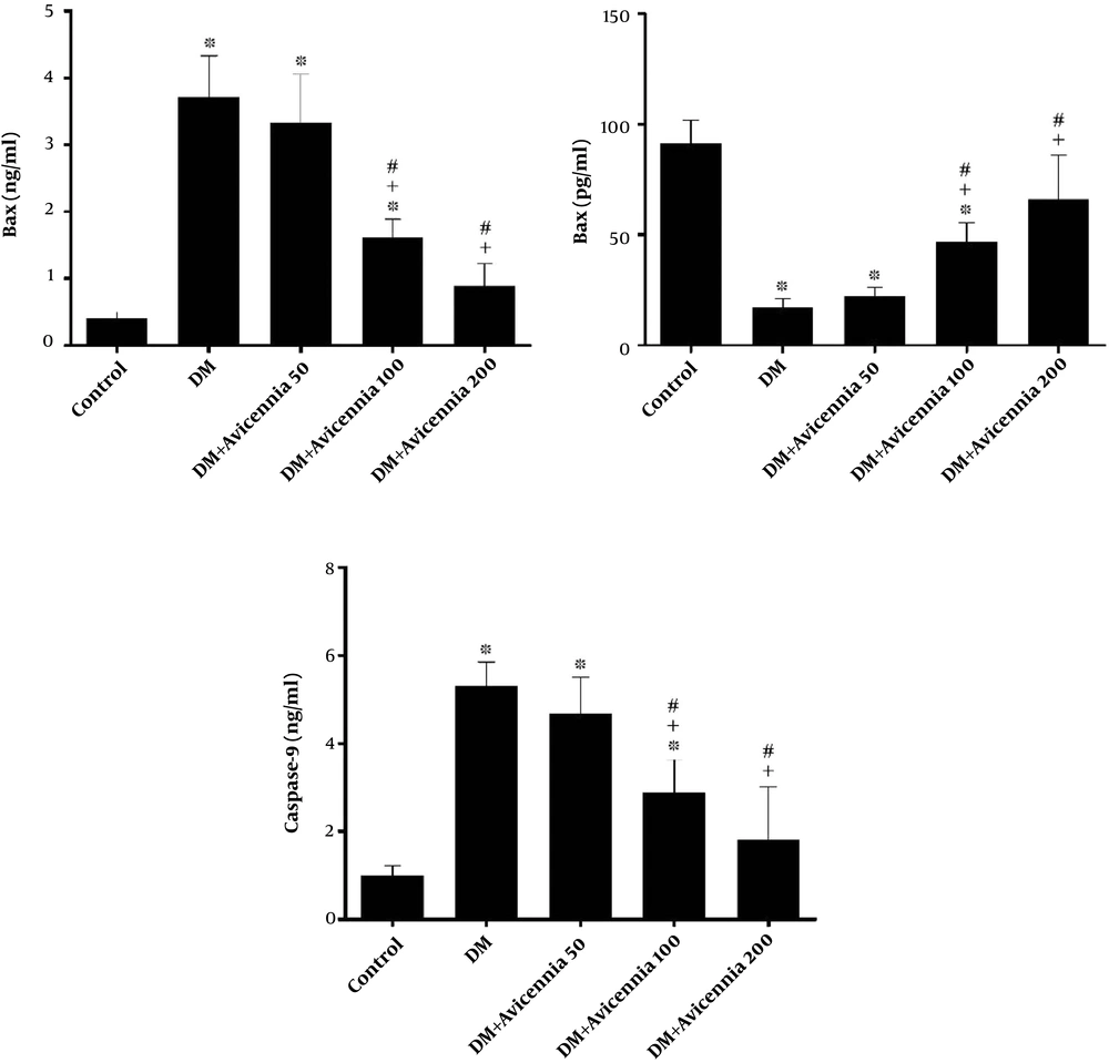
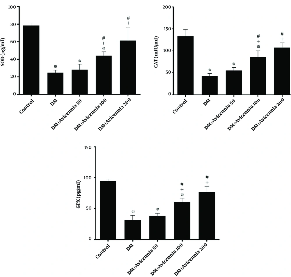
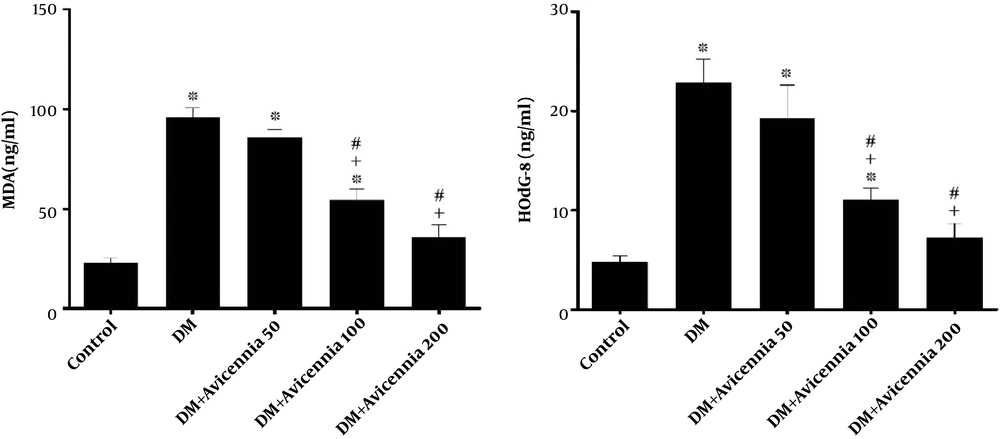
The level of Bcl-2 was significantly higher and the levels of Bax and caspase-9 were significantly lower in the groups receiving 100 and 200 mg/kg of A. marina than in the DM group (P < 0.05). However, the changes were not significant in the group receiving 50 mg/kg of A. marina (P > 0.05) (Figure 2). The levels of SOD, CAT, and GPx enzymes were significantly higher while the MDA and HOdG-8 concentrations were significantly lower in diabetic groups receiving 100 and 200 mg/kg of A. marina than in the DM group, depending on the doses of injection (P < 0.05). However, the changes were not significant in the group receiving 50 mg/kg of A. marina (P > 0.05) (Figures 3 and 4).
Comparison of mean tissue levels of Bax, Bcl-2, and caspase-9 in groups. Values are mean ± SD of eight animals in each group. ∗, significant when compared to the control group (P < 0.01); +, significant when compared to the DM group (P < 0.01); #, significant when compared to the DM + Avicennia 50 group (P < 0.01).
Comparison of mean tissue levels of SOD, CAT, and GPX in groups. Values are mean ± SD of eight animals in each group. ∗, Significant when compared to the control group (P < 0.01); +, significant when compared to the DM group (P < 0.01); #, significant when compared to the DM + Avicennia 50 group (P < 0.01).
Comparison of mean tissue levels of MDA and HOdG-8 in groups. Values are mean ± SD of eight animals in each group. ∗, Significant when compared to the control group (P < 0.01); +, significant when compared to the DM group (P < 0.01); #, significant when compared to the DM + Avicennia 50 group (P < 0.01).
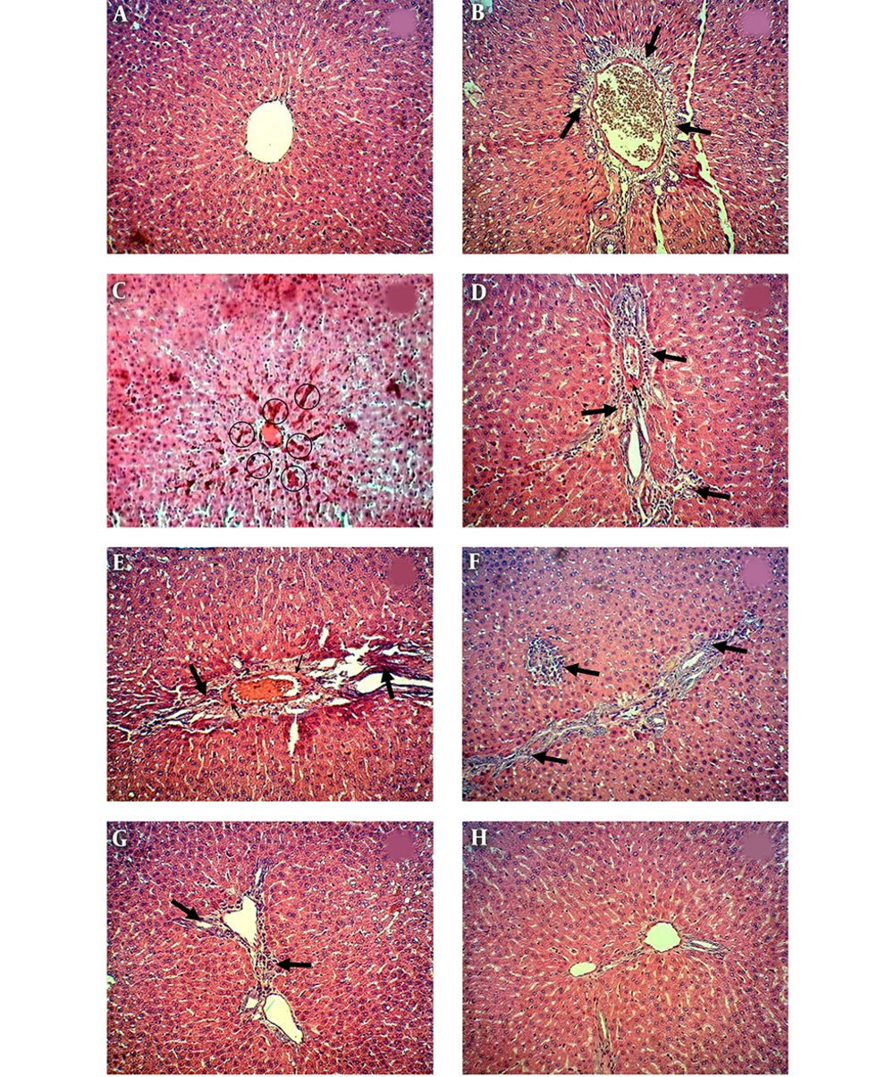
The histological examination revealed that the structures of the hepatic portal vein and liver sinusoid were normal in the control group, with no pathological changes (Figure 5A). In the DM group, inflammatory cells entered the lobule from the portal space, and diffuse inflammation was visible around the portal space. Blood accumulation was observed at the intervals of the sinusoid, and cell necrosis was found around the portal space and in different parts of liver lobules. There was also abnormal port deformity and irregularities in the liver sinusoid (Figure 5B-E).
A, Photomicrographs showing liver sections of control; B-E, DM; F, DM + Avicennia 50; G, DM + Avicennia 100; H, and DM + Avicennia 200 groups. (H & e staining, magnification 400×). In the images, the large arrow indicates necrotic tissue, the small arrow indicates inflammation, and the accumulation of inflammatory cells around the port, and the circles indicate blood accumulation in the sinusoidal duct.
Cell necrosis, inflammation, and abnormal deformity of the portal space were still visible in tissue samples of the diabetic group receiving 50 mg/kg of A. marina (Figure 5F). In the DM group receiving 100 mg/kg of A. marina, cell necrosis and inflammation were lower than in the DM group, and no blood was observed in the sinusoid space. Small deformation of the portal space was also visible (Figure 5G). In the diabetic group receiving 200 mg/kg of A. marina, we observed significantly reduced cell necrosis around the portal space, reduced local inflammation, and irregularly decreased liver sinusoid, when compared to tissue samples of the DM group. In this group, the tissue structure of the liver was approximately normal (Figure 5H).
5. Discussion
This study aimed to evaluate the effect of hydroalcoholic leaf extract of A. marina on apoptosis, inflammation, oxidative stress, lipid peroxidation, and tissue changes of the liver in diabetic rats. The results revealed that the level of Bcl-2 in liver tissues was significantly lower in the DM group than in the control group, while Bax and caspase-9 levels were significantly higher. Previous studies showed that high concentrations of glucose could induce apoptosis in PC12 cells. This finding was reported by examining the extent of DNA fragmentation and increased expression of apoptosis-inducing proteins (9). Overall, exposure to high concentrations of glucose in cardiac muscle cells seems to increase the level of Bax protein (17). Previous studies showed that a high glucose concentration was associated with increased Bax expression (18).
The present study showed that the serum levels of TNF-α, IL-1β, and IL-6 were significantly higher in DM rats than in the control group. Researchers showed that the serum level of TNF-α was significantly higher in patients with type 1 DM than in healthy controls. The increased level of TNF-α has also been implicated in the destruction of pancreatic cells by inducing the apoptotic process, leading to the development of type 1 DM (19). Moreover, studies have shown that type 1 DM increases the levels of IL-1β and TNF-α in vascular smooth muscles of rats (20).
In the present study, it was found that the levels of SOD, CAT, and GPx were significantly lower in liver tissues of rats in the DM group than in the control group. According to previous studies, the activity of antioxidant enzymes decreases in the testicular tissue of diabetic rats (21). Similar studies have indicated a significant decrease in the antioxidant enzyme activity of red blood cells (22), liver (23), and sperms (24) in diabetic rats. Moreover, based on the results of the present study, the levels of HOdG-8 and MDA were significantly lower in the liver tissue of rats in the DM group than in the control group.
Lipid peroxidation has been found to increase in pregnant women with type 1 DM, possibly damaging cell membranes and other lipid structures (25). Another study demonstrated that lipid peroxidation increases in pancreatic cells of diabetic rats (26). Evidence suggests that the accumulation of products increases due to oxidative DNA damage in diabetic retinopathy (27). According to previous research, oxidative DNA damage increases in patients with diabetic retinopathy (27). Another study found that urinary levels of HOdG-8 increased in diabetic patients with chronic hyperglycemia, and urinary levels of HOdG-8 had a direct relationship with the severity of retinopathy and diabetic neuropathy (28). Researchers also showed that the levels of 8-hydroxydeoxyguanosine and 8-hydroxyguanine, as the markers of DNA oxidative damage, increased in the urine and liver tissue of all streptozotocin-induced diabetic rats (29). Diabetes may lead to morphological changes and hepatocyte damage. Researchers have reported that the binding of glucose units to some intracellular molecules, false hypoxia induced by hyperglycemia, and imbalance in oxidation-resuscitation reactions can cause damage to hepatocytes (30). Liver tissue damage is manifested by the increased leakage of hepatic enzymes from the cytosol into the bloodstream. It also increases in diabetic patients with necrosis and hepatic steatosis (30).
The findings of the present study revealed that the treatment of diabetic rats with the hydroalcoholic extract of A. marina (100 and 200 mg/kg) could reduce apoptosis, inflammation, oxidative stress, and lipid peroxidation in a dose-dependent manner. Furthermore, this extract could improve liver damage induced by diabetes. It was found that A. marina could induce hypoglycemic effects in diabetic rats. In other words, the constituents of A. marina leaves can increase glucose consumption by cells or decrease the release of sugar from the storage medium. It is also likely that the constituents of mangrove plants can cause hypertrophy of beta cells in the remnant pancreas, leading to increased insulin secretion in type 1 DM rats (31). Studies have shown that chalcones, especially methyl hydroxy chalcone, are one of the flavonoid compounds of mangrove plants. Methyl hydroxy chalcone prevents the formation of oxygen free radicals. Therefore, due to its antioxidant properties, it is possibly effective in reducing the complications of diabetes (32, 33). On the other hand, researchers found that methyl hydroxy chalcone has insulin-like properties and that it increases glycogen formation by activating the glycogen synthase enzyme (34). Another study showed that ellagic acid, as one of the biologically active constituents of mangrove leaves, has anti-diabetic properties (35).
Previous studies showed that phenolic compounds, such as flavonoids and isoflavones, in mangrove plants have the potential to inhibit free radicals and enhance the cellular antioxidant defense system (36). A study found that mangrove leaf extract had hypoglycemic and protective effects against diabetes-induced neuronal destruction due to increased antioxidant enzyme activity. Another study showed that polyphenols, such as gallic acid, quercetin, and coumarin, derived from mangrove plants, could reduce oxidative stress and lipid peroxidation in liver tissues by increasing the plasma antioxidant capacity and endogenous antioxidant enzymes (37).
One of the complications of hyperglycemia and associated oxidative stress is the induction of apoptosis in hepatocytes. Flavonoids have protective effects on hepatocytes in high glucose-induced apoptosis. The effects of flavonoids on the improvement of hepatic apoptosis are attributed to their potential to inhibit caspase activity (38). Previous studies have also shown that flavonoids in mangrove plants can reduce the level of caspase-9 and Bax/Bcl-2 ratio by decreasing the Bax protein and increasing Bcl-2 expression (39).
Flavonoids in mangrove plants not only can reduce inflammation in damaged tissues and prevent the secretion of cytokines and inflammatory compounds induced by diabetes, but also can reduce apoptosis by decreasing inflammation at the site of injury (40). Moreover, phenolic compounds can decrease the number of inflammatory cells in the liver tissue damaged by carbon tetrachloride and inhibit the inflammatory process in the liver by inhibiting the secretion of inflammatory cytokines. Therefore, the reduction of inflammation reverses the loss of liver function and regenerates damaged cells (41). Based on the findings, the constituents of mangrove leaf may have insulin-like functions or may increase insulin secretion. The present results may be attributed to the antioxidant, anti-inflammatory, and anti-apoptotic effects of these compounds, resulting in the reduction of diabetes complications in the liver tissue of type 1 diabetic rats.
One of the most important limitations of the present study was that we did not extract the effective constituents of the hydroalcoholic extract of A. marina leaves. We also had insufficient information about the amount and type of flavonoid components.
5.1. Conclusions
It can be concluded that the dose-dependent administration of hydroalcoholic extract of A. marina leaf could reduce apoptosis, inflammation, oxidative stress, and lipid peroxidation in the liver tissue of rats with type 1 DM. Therefore, this extract can be used to improve liver tissue damage and reduce diabetes-associated liver complications.

