Fulltext
Full-text is available in PDF file.
Authors
Introduction: Recognition of risk factors in different high-risk groups such as smokers in comparison with non-high risk groups would help to develop good preventive strategies for pulmonary thromboemboli (PTE). The purpose of this study was to investigate and compare clinical findings and risk factors in smoker and nonsmoker patients with pulmonary thromboembolism and assessing anatomical variant in pulmonary computerized tomography angiographies.
Methods: In this descriptive study 260 consecutive patients suspected to have PTE underwent pulmonary computerized tomography angiographies in a training hospital since 2015 to 2018. Patient with documented PTE were enrolled. Clinical finding and risk factors were determined and compared between them.
Results: From 260 patients 172 subjects (66.15%) had PTE and enrolled in the study. Fifty-six (32%) were smoker and 116 (68%) non-smoker. The smoker group was younger and male gender was more predominant. Oxygen saturation and inspired oxygen partial pressure differed between smokers and non-smokers (P < 0.05). The predisposing factors of thromboembolism and anatomic distribution of emboli were the same in smokers and non-smokers.
Conclusion: Regarding different factors responsible for PTE in smokers and non-smokers, clinical presentation and anatomic distribution of PTE are comparable.
© 2019, International Journal of Cardiovascular Practice. This open-access article is available under the Creative Commons Attribution-NonCommercial 4.0 (CC BY-NC 4.0) International License (https://creativecommons.org/licenses/by-nc/4.0/), which allows for the copying and redistribution of the material only for noncommercial purposes, provided that the original work is properly cited.
Bozorgmehr R, Pishgahi M, Mohaghegh P, Saneitaheri M, Bayat M, et al. Prevalence of Non-Thromboembolic Incidental Findings on Computed Tomography-Pulmonary Angiography for Pulmonary. Int J Cardiovasc Pract. 2019;4(4):e130567. doi: https://doi.org/10.29252/ijcp-26716
Molaee S, Ghanaati H, Safavi E, Foroumandi M, Peiman S. Computed Tomography Pulmonary Angiography for Evaluation of Patients With Suspected Pulmonary Embolism: Use or Overuse. I J Radiol. 2015;12(3):e22383. doi: https://doi.org/10.5812/iranjradiol.12(2)2015.22383
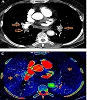
Bayraktaroglu S, Cinkooglu A, Savas R, Ceylan N, Bozdağ M, et al. Dual Energy CT Evaluation of the Severity of Pulmonary Thromboembolism: Correlation of Pulmonary Perfusion Score with CT. I J Radiol. 2020;17(3):e92990. doi: https://doi.org/10.5812/iranjradiol.92990
Faghihi Langhroudi T, Borji Esfahani M, Khaheshi I, Naderian M, Zahedi Tajrishi F, et al. Correlation of Hematologic Indices with CT-pulmonary Arterial Obstruction Index in Patients with Acute Pulmonary Emboli. Int J Cardiovasc Pract. 2019;4(3):e130555. doi: https://doi.org/10.29252/ijcp-27078
Raoufi M, Faghihi Langroudi T, Arjmand Shabestari A, Sanei Taheri M. Quantification of Pulmonary Thromboembolism in CT Angiography Reports and Predicting Right Ventricular Dysfunction and Patient Mortality. I J Radiol. 2014;11(30th Iranian Congress of Radiology):e21266. doi: https://doi.org/10.5812/iranjradiol.21266
Last Update: 3 days ago
Last Update: 3 days ago
Last Update: 3 days ago
Last Update: 1 day ago