Abstract
Keywords
Atrial Fibrillation Oxidative Stress Cell-Free Circulating Mitochondrial DNA Arrhythmia
1. Context
As we know, atrial fibrillation (AF) is the most prevalent sustained cardiac arrhythmia (1). It is an irregular and occasionally rapid arrhythmia originating from multiple ectopic foci in the atria (2). AF is known to be an important risk factor for dementia, ischemic stroke, heart failure, and decreased quality of life (3-7). The estimated risk for the incidence of this arrhythmia is 25% in people aged over 40 years (8, 9). AF affects more than 33 million people worldwide, and its overall incidence is 5 million new cases annually (10). Therefore, AF is an important public health concern. Some risk factors, including diabetes mellitus, hypertension, smoking, increased body weight, alcohol consumption, and stress, are proposed to be associated with AF (11).
Mitochondria are cellular organelles with a double-layer membrane that convert various forms of molecules into adenosine triphosphate (ATP). They do this process via ATPase enzyme and in the presence of electrons that have high energy in the internal mitochondrial membrane in a process named oxidative phosphorylation (12, 13). Mitochondria have a double-stranded, small, circular, independent DNA (known as mtDNA) that encodes 2 ribosomal RNAs (rRNAs), 22 transfer RNAs (tRNAs), and 13 proteins (14). This small molecule has specific characteristics, making it a helpful tool in evolutionary studies (15). The mt-DNA is increased in tissue injury, for example, in sepsis (16-22), trauma (23-26), and malignancy (27-36), indicating severe cellular stress that results in severe cellular death as an essential factor in the release of mtDNA (37). Cell-free circulating mitochondrial DNA (Cfc-mtDNA) has previously been used as a predictive factor for situations associated with the dysfunction of mitochondria, such as cardiac arrest survival (38), dengue severity (39), cancer progression (40, 41), and diabetes mellitus (42). Mutations and replication errors may defect mtDNA. Sufficient mtDNA damage can result in cellular and mitochondrial dysfunction, leading to accelerated aging or impaired health (43).
In this article, we present the association between AF and mtDNA levels and answer whether using mtDNA as a predictive factor for AF is possible.
2. Shared Molecular Basis of Atrial Fibrillation and Mitochondria
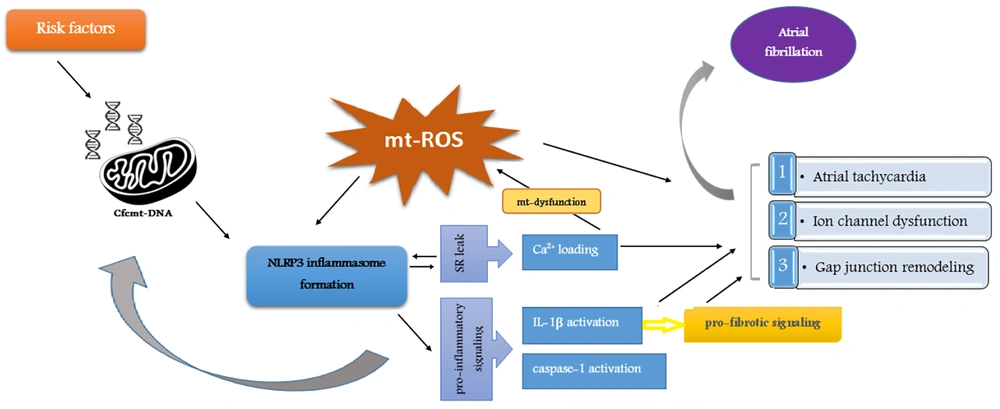
A mitochondrial molecular pathway, including Cfc-mtDNA release, has a significant role in AF pathogenesis (Figure 1). As a damage-associated molecular pattern (DAMP), Cfc-mtDNA is released into the extracellular space during significant cell stress and injury. This phenomenon acts as a signal for other organelles (44). Studies have shown that certain AF risk factors, such as diabetes mellitus and high blood pressure, can lead to mitochondrial dysfunction and a rise in DAMPs (45-50). NLRP3 inflammasome, an innate immune system receptor, regulates the inflammatory response by activating IL-1β. Recent studies have considered NLRP3 inflammasome a potential therapeutic target in inflammatory diseases (51-54). The released Cfc-mtDNA acts as an inflammasome activator (55, 56). On the other hand, the activity of caspase-1 and the inflammatory cytokine IL-1β under the influence of inflammasome cause changes in the mitochondrial membrane and accelerate the release of mtDNA (57, 58). In addition, IL-1β evokes alterations, such as fibrosis, apoptosis, and hypertrophy of cardiac fibroblasts, which are significantly involved in the pathogenesis of AF (59, 60).
Molecular pathways through mitochondria are involved in atrial fibrillation pathogenesis. The formation of NLRP3 inflammasome through the release of Cfc-mtDNA leads to a cascade of events, including Ca2+ loading, IL-1β activation, and caspase-1 activation, which are involved in oxidative stress as an important aspect of the molecular pathogenesis of AF.
Oxidative stress is one of the important aspects of the molecular pathogenesis of AF (61) and is also caused by exposure to AF risk factors and mitochondrial dysfunction (62-64). The mt-ROS activates the NLRP3 inflammasome and Cfc-mtDNA (65, 66). Oxidative stress and mitochondrial dysfunction are enhanced by Ca2+ overload-induced atrial tachycardia. Therefore, Ca2+ overload caused by sarcoplasmic reticulum leak, which is itself promoted by the inflammatory process and NLRP3 inflammasome signaling, accelerates NLRP3 inflammasome formation, disrupts mitochondrial function, and causes more mt-ROS production (61-64, 67-71). In other words, Ca2+ overload provides a platform for initiating and maintaining AF (72).
Taking these together, due to the association of its release with the risk factors of AF and its pathogenesis through mitochondrial signaling pathways, Cfc-mtDNA could be proposed as a promising non-invasive biomarker and a potential therapeutic target for AF. Research has been performed in the field so far, which will be discussed in the following sections.
3. Cfc-mtDNA for Predicting AF, Particularly Paroxysmal AF
The copy number of mtDNA in the peripheral blood was found to be associated with the oxidative stress status in patients. Oxidative stress is also a potential factor contributing to AF pathogenesis (73). Considering this and that a critical modulator of AF in the clinical and experimental setting is mitochondrial dysfunction, scientists came up with the idea that mtDNA may be a potential biomarker predicting AF (74).
In this regard, Zhang et al. measured the mtDNA copy number in the peripheral blood of 485 patients with normal sinus rhythm undergoing cardiac surgery. After surgery, postoperative AF (PoAF) was seen in 101 of 485 patients. The mtDNA copy number in the peripheral blood of these cases was significantly higher than the patients with normal sinus rhythm (36.43 in PoAF patients vs. 16.63 in normal sinus rhythm patients, P < 0.001). They also clarified that mtDNA copy number could predict PoAF acceptable specificity (80.2%) and sensitivity (70.3%) (73).
In another study, Wiersma et al. investigated the association between mtDNA levels in the serum of patients with AF recurrence and AF stage. They evaluated two mtDNA genes, namely ND1 and COX3, in the peripheral blood of 84 control patients, 100 paroxysmal AF (PAF), 59 patients undergoing cardiac surgery not having AF history, 20 long-standing persistent AF (LS-PeAF), and 116 persistent AF (PeAF) patients who underwent either AF treatment (pulmonary vein isolation or electrical cardioversion) or cardiac surgery. According to this study, Cfc -mtDNA levels increased significantly in PAF patients who underwent AF treatment. This means that Cfc-mtDNA levels significantly increased in the early stages of AF (PAF) but decreased in the end-stage AF (LS-PeAF). This study also revealed that Cfc-mtDNA levels in the serum are associated with AF recurrence after all types of treatments, especially in the PVI subgroup. Briefly, this study proved the potential of Cfc-mtDNA levels as an interesting biomarker for the staging of AF, early identification of PAF patients, and identifying patients at high risk of AF recurrence after PVI treatment (75).
In concordance with the above-mentioned studies, Sandler et al. investigated mtDNA at three different time points, including preoperatively, after cardiopulmonary bypass (CPB) within 90 minutes of decannulation, and finally postoperatively on the first and second day after CPB in 16 patients who enrolled prospectively. They found that mtDNA in the blood samples of patients significantly rose after CPB (six-fold elevations after CPB, P = 0.008, and five-fold elevations during 2 days post-surgery, P = 0.02). The PoAF patients showed a rise in the mtDNA levels post-CPB compared to patients without POAF. Moreover, postoperatively, POAF patients showed at least a two-fold elevation of mt-DNA, whereas this increase happened in less than half of patients not having PoAF (P = 0.037). Their results exhibited that the initial tissue damage and following inflammation caused by cardiac surgery played an important role in the development of PoAF, confirming that at the molecular level, there can be a potential mechanistic relation between mtDNA and the development of POAF (76).
Cell-free DNA (CfDNA) has been recently known as a member of DAMPs and has been suggested to have a role in inflammatory disorders. As it has been proven that some inflammatory pathways can lead to AF, Yamazoe et al. (77) aimed to investigate the level of cfDNA as a potential cause of systemic inflammation accompanied by AF. Consequently, they obtained the peripheral blood of 104 non-AF controls and 26 PAF patients and extracted their cfDNA. They noticed that the Cfc-mtDNA copy number is associated with PAF more strongly than nuclear cf-DNA. They also clarified that the unmethylated CpG islands in mt-cfDNA are necessary for promoting a pro-inflammatory response in which IL-1B, IL-6, and toll-like receptor (TLR) 9 play a role. Thoroughly, Cfc-mtDNA activates macrophages and induces pro-inflammatory cytokines via TLR9. Previous studies have reported the association between mt-cfDNA levels and inflammatory cytokines (78-80). They finally concluded that Cfc-mtDNA may play an essential role in such an inflammatory response in this disease. Yamazoe et al. also compared the ability of total cf-DNA, nuclear cfDNA, and Cfc-mtDNA in AF prediction. Their study revealed that Cfc-mtDNA is the most potential biomarker for predicting PAF incidence (77).
Another study investigating the association between mt-DNA levels and AF incidence was conducted by Soltész et al. They assessed mt-DNA levels in the cell-free plasma, peripheral whole blood, and exosome samples. They took peripheral blood from 60 patients with AF and 72 controls. Next, they isolated DNA from plasma and blood, and exosomes were also isolated from the plasma, which was free from cells. Finally, exosome-encapsulated DNA, exoDNA, was extracted, and quantitative RT-PCR was performed. In contrast to the aforementioned studies, they reported no significant difference in mt-DNA levels between cell-free plasma, peripheral blood, and exosomes (81).
4. Cfc-mtDNA and Persistent AF
In a study by Yamazoe et al., 28 PerAF patients were compared with an age-matched control group and a young healthy control group. The Cfc-mtDNA levels in PerAF patients were higher than in both control groups. They also compared n-cfDNA between these groups and observed no significant difference between PerAF patients and the age-matched control group (77). In contrast, Wiersma et al. reported that Cfc-mtDNA levels in the PerAF and long-standing PerAF patients decreased compared to the control group (75). Further studies are necessary to clarify the association between Cfc-mtDNA levels and PerAF.
5. Gender-Related Differences in Cfc-mtDNA
It is commonly known that some gender differences exist in AF (82). Wiersma et al. compared ND1 and COX3 levels between the groups to investigate differences in Cfc-mtDNA levels between female and male patients. They reported no differences in Cfc-mtDNA levels between females and males (75). In line with this study, another research by Soltész et al. found no significant difference in the copy number of mtDNA between genders (81). However, Wiersma et al. found that Cfc-mtDNA levels were significantly elevated in male PAF patients compared to a male control group. In contrast, such differences were not seen in the female PAF patients compared to female controls. These results showed that Cfc-mtDNA levels were associated, especially with male patients with AF (75).
6. Age-Related Differences in Cfc-mtDNA
The Cfc-mtDNA levels are related to the inflammatory status, and there may be a difference between different ages. Therefore, it is necessary to investigate whether age independently affects the Cfc-mtDNA levels. Some studies, such as the one conducted by Soltész et al., suggested no significant difference in the copy number of mt-DNA between different age groups, which agreed with a previous study by Feng et al. (81, 83). In addition, related to the potential role of mtDNA levels in predicting AF, Zhang et al. declared that this novel biomarker is a preoperative potential risk factor for developing AF independent of the patient’s age (73).
7. Conclusions
Our review suggests an association between the serum level of Cfc-mtDNA and AF. Most studies revealed that Cfc-mtDNA serum levels can be used in predicting PoAF and detecting early PAF patients. These studies indicate that age and gender do not affect the results. In addition, studies on Cfc-mtDNA and per-AF have yet to reach similar results, and further research is needed. However, evidence is limited, and future research is necessary to assess the applicability of Cfc-mtDNA as a biomarker for atrial fibrillation.
References
-
1.
Romero JR, Wolf PA. Epidemiology of Stroke: Legacy of the Framingham Heart Study. Glob Heart. 2013;8(1):67-75. [PubMed ID: 23527318]. [PubMed Central ID: PMC3601756]. https://doi.org/10.1016/j.gheart.2012.12.007.
-
2.
Page RL. Clinical practice. Newly diagnosed atrial fibrillation. N Engl J Med. 2004;351(23):2408-16. [PubMed ID: 15575057]. https://doi.org/10.1056/NEJMcp041956.
-
3.
Benjamin EJ, Wolf PA, D'Agostino RB, Silbershatz H, Kannel WB, Levy D. Impact of atrial fibrillation on the risk of death: the Framingham Heart Study. Circulation. 1998;98(10):946-52. [PubMed ID: 9737513]. https://doi.org/10.1161/01.cir.98.10.946.
-
4.
Kannel WB, Wolf PA, Benjamin EJ, Levy D. Prevalence, incidence, prognosis, and predisposing conditions for atrial fibrillation: population-based estimates. Am J Cardiol. 1998;82(8A):2N-9N. [PubMed ID: 9809895]. https://doi.org/10.1016/s0002-9149(98)00583-9.
-
5.
Stewart S, Hart CL, Hole DJ, McMurray JJ. A population-based study of the long-term risks associated with atrial fibrillation: 20-year follow-up of the Renfrew/Paisley study. Am J Med. 2002;113(5):359-64. [PubMed ID: 12401529]. https://doi.org/10.1016/s0002-9343(02)01236-6.
-
6.
Thrall G, Lane D, Carroll D, Lip GY. Quality of life in patients with atrial fibrillation: a systematic review. Am J Med. 2006;119(5):448 e1-19. [PubMed ID: 16651058]. https://doi.org/10.1016/j.amjmed.2005.10.057.
-
7.
Wang TJ, Larson MG, Levy D, Vasan RS, Leip EP, Wolf PA, et al. Temporal relations of atrial fibrillation and congestive heart failure and their joint influence on mortality: the Framingham Heart Study. Circulation. 2003;107(23):2920-5. [PubMed ID: 12771006]. https://doi.org/10.1161/01.CIR.0000072767.89944.6E.
-
8.
Heeringa J, van der Kuip DA, Hofman A, Kors JA, van Herpen G, Stricker BH, et al. Prevalence, incidence and lifetime risk of atrial fibrillation: the Rotterdam study. Eur Heart J. 2006;27(8):949-53. [PubMed ID: 16527828]. https://doi.org/10.1093/eurheartj/ehi825.
-
9.
Lloyd-Jones DM, Wang TJ, Leip EP, Larson MG, Levy D, Vasan RS, et al. Lifetime risk for development of atrial fibrillation: the Framingham Heart Study. Circulation. 2004;110(9):1042-6. [PubMed ID: 15313941]. https://doi.org/10.1161/01.CIR.0000140263.20897.42.
-
10.
Chugh SS, Havmoeller R, Narayanan K, Singh D, Rienstra M, Benjamin EJ, et al. Worldwide epidemiology of atrial fibrillation: a Global Burden of Disease 2010 Study. Circulation. 2014;129(8):837-47. [PubMed ID: 24345399]. [PubMed Central ID: PMC4151302]. https://doi.org/10.1161/CIRCULATIONAHA.113.005119.
-
11.
Panchal G, Mahmood M, Lip GYH. Revisiting the risks of incident atrial fibrillation: a narrative review. Part 2. Kardiol Pol. 2019;77(5):515-24. [PubMed ID: 31125027]. https://doi.org/10.33963/KP.14846.
-
12.
Chan DC. Mitochondrial Dynamics and Its Involvement in Disease. Annu Rev Pathol. 2020;15:235-59. [PubMed ID: 31585519]. https://doi.org/10.1146/annurev-pathmechdis-012419-032711.
-
13.
Giacomello M, Pyakurel A, Glytsou C, Scorrano L. The cell biology of mitochondrial membrane dynamics. Nat Rev Mol Cell Biol. 2020;21(4):204-24. [PubMed ID: 32071438]. https://doi.org/10.1038/s41580-020-0210-7.
-
14.
Anderson S, Bankier AT, Barrell BG, de Bruijn MH, Coulson AR, Drouin J, et al. Sequence and organization of the human mitochondrial genome. Nature. 1981;290(5806):457-65. [PubMed ID: 7219534]. https://doi.org/10.1038/290457a0.
-
15.
Desalle R, Schierwater B, Hadrys H. MtDNA: The small workhorse of evolutionary studies. Front Biosci (Landmark Ed). 2017;22(5):873-87. [PubMed ID: 27814652]. https://doi.org/10.2741/4522.
-
16.
Bhagirath VC, Dwivedi DJ, Liaw PC. Comparison of the Proinflammatory and Procoagulant Properties of Nuclear, Mitochondrial, and Bacterial DNA. Shock. 2015;44(3):265-71. [PubMed ID: 25944792]. https://doi.org/10.1097/SHK.0000000000000397.
-
17.
Di Caro V, Walko T3, Bola RA, Hong JD, Pang D, Hsue V, et al. Plasma Mitochondrial DNA--a Novel DAMP in Pediatric Sepsis. Shock. 2016;45(5):506-11. [PubMed ID: 26682947]. [PubMed Central ID: PMC5322958]. https://doi.org/10.1097/SHK.0000000000000539.
-
18.
Garrabou G, Moren C, Lopez S, Tobias E, Cardellach F, Miro O, et al. The effects of sepsis on mitochondria. J Infect Dis. 2012;205(3):392-400. [PubMed ID: 22180620]. https://doi.org/10.1093/infdis/jir764.
-
19.
Kung CT, Hsiao SY, Tsai TC, Su CM, Chang WN, Huang CR, et al. Plasma nuclear and mitochondrial DNA levels as predictors of outcome in severe sepsis patients in the emergency room. J Transl Med. 2012;10:130. [PubMed ID: 22720733]. [PubMed Central ID: PMC3441240]. https://doi.org/10.1186/1479-5876-10-130.
-
20.
Lu CH, Chang WN, Tsai NW, Chuang YC, Huang CR, Wang HC. The value of serial plasma nuclear and mitochondrial DNA levels in adult community-acquired bacterial meningitis. QJM. 2010;103(3):169-75. [PubMed ID: 20129945]. https://doi.org/10.1093/qjmed/hcp201.
-
21.
Timmermans K, Kox M, Scheffer GJ, Pickkers P. Plasma Nuclear and Mitochondrial DNA Levels, and Markers of Inflammation, Shock, and Organ Damage in Patients with Septic Shock. Shock. 2016;45(6):607-12. [PubMed ID: 26717107]. https://doi.org/10.1097/SHK.0000000000000549.
-
22.
Yu M. Circulating cell-free mitochondrial DNA as a novel cancer biomarker: opportunities and challenges. Mitochondrial DNA. 2012;23(5):329-32. [PubMed ID: 22775429]. https://doi.org/10.3109/19401736.2012.696625.
-
23.
Hauser CJ, Sursal T, Rodriguez EK, Appleton PT, Zhang Q, Itagaki K. Mitochondrial damage associated molecular patterns from femoral reamings activate neutrophils through formyl peptide receptors and P44/42 MAP kinase. J Orthop Trauma. 2010;24(9):534-8. [PubMed ID: 20736789]. [PubMed Central ID: PMC2945259]. https://doi.org/10.1097/BOT.0b013e3181ec4991.
-
24.
Lam NY, Rainer TH, Chiu RW, Joynt GM, Lo YM. Plasma mitochondrial DNA concentrations after trauma. Clin Chem. 2004;50(1):213-6. [PubMed ID: 14709653]. https://doi.org/10.1373/clinchem.2003.025783.
-
25.
Prikhodko AS, Shabanov AK, Zinovkina LA, Popova EN, Aznauryan MA, Lanina NO, et al. Pure Mitochondrial DNA Does Not Activate Human Neutrophils in vitro. Biochemistry (Mosc). 2015;80(5):629-35. [PubMed ID: 26071783]. https://doi.org/10.1134/S0006297915050168.
-
26.
Timmermans K, Kox M, Vaneker M, van den Berg M, John A, van Laarhoven A, et al. Plasma levels of danger-associated molecular patterns are associated with immune suppression in trauma patients. Intensive Care Med. 2016;42(4):551-61. [PubMed ID: 26912315]. [PubMed Central ID: PMC5413532]. https://doi.org/10.1007/s00134-015-4205-3.
-
27.
Budnik LT, Kloth S, Baur X, Preisser AM, Schwarzenbach H. Circulating mitochondrial DNA as biomarker linking environmental chemical exposure to early preclinical lesions elevation of mtDNA in human serum after exposure to carcinogenic halo-alkane-based pesticides. PLoS One. 2013;8(5). e64413. [PubMed ID: 23741329]. [PubMed Central ID: PMC3669318]. https://doi.org/10.1371/journal.pone.0064413.
-
28.
Ellinger J, Albers P, Muller SC, von Ruecker A, Bastian PJ. Circulating mitochondrial DNA in the serum of patients with testicular germ cell cancer as a novel noninvasive diagnostic biomarker. BJU Int. 2009;104(1):48-52. [PubMed ID: 19154496]. https://doi.org/10.1111/j.1464-410X.2008.08289.x.
-
29.
Ellinger J, Muller DC, Muller SC, Hauser S, Heukamp LC, von Ruecker A, et al. Circulating mitochondrial DNA in serum: a universal diagnostic biomarker for patients with urological malignancies. Urol Oncol. 2012;30(4):509-15. [PubMed ID: 20870429]. https://doi.org/10.1016/j.urolonc.2010.03.004.
-
30.
Ellinger J, Muller SC, Wernert N, von Ruecker A, Bastian PJ. Mitochondrial DNA in serum of patients with prostate cancer: a predictor of biochemical recurrence after prostatectomy. BJU Int. 2008;102(5):628-32. [PubMed ID: 18410441]. https://doi.org/10.1111/j.1464-410X.2008.07613.x.
-
31.
Hou YL, Chen JJ, Wu YF, Xue CJ, Li FZ, Zheng Q, et al. Clinical significance of serum mitochondrial DNA in lung cancer. Clin Biochem. 2013;46(15):1474-7. [PubMed ID: 23611926]. https://doi.org/10.1016/j.clinbiochem.2013.04.009.
-
32.
Huang CY, Chen YM, Wu CH, Tsai CM, Lee YC, Perng RP, et al. Circulating free mitochondrial DNA concentration and its association with erlotinib treatment in patients with adenocarcinoma of the lung. Oncol Lett. 2014;7(6):2180-4. [PubMed ID: 24932312]. [PubMed Central ID: PMC4049713]. https://doi.org/10.3892/ol.2014.2006.
-
33.
Kohler C, Radpour R, Barekati Z, Asadollahi R, Bitzer J, Wight E, et al. Levels of plasma circulating cell free nuclear and mitochondrial DNA as potential biomarkers for breast tumors. Mol Cancer. 2009;8:105. [PubMed ID: 19922604]. [PubMed Central ID: PMC2780981]. https://doi.org/10.1186/1476-4598-8-105.
-
34.
Mehra N, Penning M, Maas J, van Daal N, Giles RH, Voest EE. Circulating mitochondrial nucleic acids have prognostic value for survival in patients with advanced prostate cancer. Clin Cancer Res. 2007;13(2 Pt 1):421-6. [PubMed ID: 17255261]. https://doi.org/10.1158/1078-0432.CCR-06-1087.
-
35.
Yu M, Wan YF, Zou QH. Cell-free circulating mitochondrial DNA in the serum: a potential non-invasive biomarker for Ewing's sarcoma. Arch Med Res. 2012;43(5):389-94. [PubMed ID: 22728238]. https://doi.org/10.1016/j.arcmed.2012.06.007.
-
36.
Zachariah RR, Schmid S, Buerki N, Radpour R, Holzgreve W, Zhong X. Levels of circulating cell-free nuclear and mitochondrial DNA in benign and malignant ovarian tumors. Obstet Gynecol. 2008;112(4):843-50. [PubMed ID: 18827127]. https://doi.org/10.1097/AOG.0b013e3181867bc0.
-
37.
Boyapati RK, Tamborska A, Dorward DA, Ho GT. Advances in the understanding of mitochondrial DNA as a pathogenic factor in inflammatory diseases. F1000Res. 2017;6:169. [PubMed ID: 28299196]. [PubMed Central ID: PMC5321122]. https://doi.org/10.12688/f1000research.10397.1.
-
38.
Arnalich F, Codoceo R, Lopez-Collazo E, Montiel C. Circulating cell-free mitochondrial DNA: a better early prognostic marker in patients with out-of-hospital cardiac arrest. Resuscitation. 2012;83(7):e162-3. [PubMed ID: 22490673]. https://doi.org/10.1016/j.resuscitation.2012.03.032.
-
39.
Ha TT, Huy NT, Murao LA, Lan NT, Thuy TT, Tuan HM, et al. Elevated levels of cell-free circulating DNA in patients with acute dengue virus infection. PLoS One. 2011;6(10). e25969. [PubMed ID: 22016795]. [PubMed Central ID: PMC3189230]. https://doi.org/10.1371/journal.pone.0025969.
-
40.
Cicchillitti L, Corrado G, De Angeli M, Mancini E, Baiocco E, Patrizi L, et al. Circulating cell-free DNA content as blood based biomarker in endometrial cancer. Oncotarget. 2017;8(70):115230-43. [PubMed ID: 29383155]. [PubMed Central ID: PMC5777767]. https://doi.org/10.18632/oncotarget.23247.
-
41.
Li L, Hann HW, Wan S, Hann RS, Wang C, Lai Y, et al. Cell-free circulating mitochondrial DNA content and risk of hepatocellular carcinoma in patients with chronic HBV infection. Sci Rep. 2016;6:23992. [PubMed ID: 27063412]. [PubMed Central ID: PMC4827061]. https://doi.org/10.1038/srep23992.
-
42.
Lee HK, Song JH, Shin CS, Park DJ, Park KS, Lee KU, et al. Decreased mitochondrial DNA content in peripheral blood precedes the development of non-insulin-dependent diabetes mellitus. Diabetes Res Clin Pract. 1998;42(3):161-7. [PubMed ID: 9925346]. https://doi.org/10.1016/s0168-8227(98)00110-7.
-
43.
Wallace DC, Chalkia D. Mitochondrial DNA genetics and the heteroplasmy conundrum in evolution and disease. Cold Spring Harb Perspect Biol. 2013;5(11). a021220. [PubMed ID: 24186072]. [PubMed Central ID: PMC3809581]. https://doi.org/10.1101/cshperspect.a021220.
-
44.
Grazioli S, Pugin J. Mitochondrial Damage-Associated Molecular Patterns: From Inflammatory Signaling to Human Diseases. Front Immunol. 2018;9:832. [PubMed ID: 29780380]. [PubMed Central ID: PMC5946030]. https://doi.org/10.3389/fimmu.2018.00832.
-
45.
Land WG. The Role of Damage-Associated Molecular Patterns (DAMPs) in Human Diseases: Part II: DAMPs as diagnostics, prognostics and therapeutics in clinical medicine. Sultan Qaboos Univ Med J. 2015;15(2):e157-70. [PubMed ID: 26052447]. [PubMed Central ID: PMC4450777].
-
46.
McCarthy CG, Goulopoulou S, Wenceslau CF, Spitler K, Matsumoto T, Webb RC. Toll-like receptors and damage-associated molecular patterns: novel links between inflammation and hypertension. Am J Physiol Heart Circ Physiol. 2014;306(2):H184-96. [PubMed ID: 24163075]. [PubMed Central ID: PMC3920129]. https://doi.org/10.1152/ajpheart.00328.2013.
-
47.
Garica-Martineza I, Shakera ME, Mehala WZ, Phil D. Therapeutic Opportunities in Damage Associated Molecular Patterns (DAMPs) Driven Metabolic Diseases. Antioxid Redox Signal. 2015;23. https://doi.org/10.1089/ars.2015.6383).
-
48.
Shin JJ, Lee EK, Park TJ, Kim W. Damage-associated molecular patterns and their pathological relevance in diabetes mellitus. Ageing Res Rev. 2015;24(Pt A):66-76. [PubMed ID: 26197086]. https://doi.org/10.1016/j.arr.2015.06.004.
-
49.
Wenceslau CF, McCarthy CG, Szasz T, Spitler K, Goulopoulou S, Webb RC, et al. Mitochondrial damage-associated molecular patterns and vascular function. Eur Heart J. 2014;35(18):1172-7. [PubMed ID: 24569027]. [PubMed Central ID: PMC4012709]. https://doi.org/10.1093/eurheartj/ehu047.
-
50.
Zheng Y, Gardner SE, Clarke MC. Cell death, damage-associated molecular patterns, and sterile inflammation in cardiovascular disease. Arterioscler Thromb Vasc Biol. 2011;31(12):2781-6. [PubMed ID: 22096097]. https://doi.org/10.1161/ATVBAHA.111.224907.
-
51.
Bazrafkan M, Hosseini E, Nazari M, Amorim CA, Sadeghi MR. NLRP3 inflammasome: A joint, potential therapeutic target in management of COVID-19 and fertility problems. J Reprod Immunol. 2021;148:103427. [PubMed ID: 34563758]. [PubMed Central ID: PMC8453780]. https://doi.org/10.1016/j.jri.2021.103427.
-
52.
Chen R, Yin C, Fang J, Liu B. The NLRP3 inflammasome: an emerging therapeutic target for chronic pain. J Neuroinflammation. 2021;18(1):84. [PubMed ID: 33785039]. [PubMed Central ID: PMC8008529]. https://doi.org/10.1186/s12974-021-02131-0.
-
53.
de Zoete MR, Palm NW, Zhu S, Flavell RA. Inflammasomes. Cold Spring Harb Perspect Biol. 2014;6(12). a016287. [PubMed ID: 25324215]. [PubMed Central ID: PMC4292152]. https://doi.org/10.1101/cshperspect.a016287.
-
54.
Guo H, Callaway JB, Ting JP. Inflammasomes: mechanism of action, role in disease, and therapeutics. Nat Med. 2015;21(7):677-87. [PubMed ID: 26121197]. [PubMed Central ID: PMC4519035]. https://doi.org/10.1038/nm.3893.
-
55.
Shimada K, Crother TR, Karlin J, Dagvadorj J, Chiba N, Chen S, et al. Oxidized mitochondrial DNA activates the NLRP3 inflammasome during apoptosis. Immunity. 2012;36(3):401-14. [PubMed ID: 22342844]. [PubMed Central ID: PMC3312986]. https://doi.org/10.1016/j.immuni.2012.01.009.
-
56.
Zhong Z, Liang S, Sanchez-Lopez E, He F, Shalapour S, Lin XJ, et al. New mitochondrial DNA synthesis enables NLRP3 inflammasome activation. Nature. 2018;560(7717):198-203. [PubMed ID: 30046112]. [PubMed Central ID: PMC6329306]. https://doi.org/10.1038/s41586-018-0372-z.
-
57.
Aarreberg LD, Esser-Nobis K, Driscoll C, Shuvarikov A, Roby JA, Gale M. Interleukin-1beta Induces mtDNA Release to Activate Innate Immune Signaling via cGAS-STING. Mol Cell. 2019;74(4):801-815 e6. [PubMed ID: 30952515]. [PubMed Central ID: PMC6596306]. https://doi.org/10.1016/j.molcel.2019.02.038.
-
58.
Yu J, Nagasu H, Murakami T, Hoang H, Broderick L, Hoffman HM, et al. Inflammasome activation leads to Caspase-1-dependent mitochondrial damage and block of mitophagy. Proc Natl Acad Sci U S A. 2014;111(43):15514-9. [PubMed ID: 25313054]. [PubMed Central ID: PMC4217429]. https://doi.org/10.1073/pnas.1414859111.
-
59.
Bujak M, Dobaczewski M, Chatila K, Mendoza LH, Li N, Reddy A, et al. Interleukin-1 receptor type I signaling critically regulates infarct healing and cardiac remodeling. Am J Pathol. 2008;173(1):57-67. [PubMed ID: 18535174]. [PubMed Central ID: PMC2438285]. https://doi.org/10.2353/ajpath.2008.070974.
-
60.
Nattel S, Heijman J, Zhou L, Dobrev D. Molecular Basis of Atrial Fibrillation Pathophysiology and Therapy: A Translational Perspective. Circ Res. 2020;127(1):51-72. [PubMed ID: 32717172]. [PubMed Central ID: PMC7398486]. https://doi.org/10.1161/CIRCRESAHA.120.316363.
-
61.
Korantzopoulos P, Letsas K, Fragakis N, Tse G, Liu T. Oxidative stress and atrial fibrillation: an update. Free Radic Res. 2018;52(11-12):1199-209. [PubMed ID: 30003814]. https://doi.org/10.1080/10715762.2018.1500696.
-
62.
Bukowska A, Schild L, Keilhoff G, Hirte D, Neumann M, Gardemann A, et al. Mitochondrial dysfunction and redox signaling in atrial tachyarrhythmia. Exp Biol Med (Maywood). 2008;233(5):558-74. [PubMed ID: 18375832]. https://doi.org/10.3181/0706-RM-155.
-
63.
Dudley SC, Hoch NE, McCann LA, Honeycutt C, Diamandopoulos L, Fukai T, et al. Atrial fibrillation increases production of superoxide by the left atrium and left atrial appendage: role of the NADPH and xanthine oxidases. Circulation. 2005;112(9):1266-73. [PubMed ID: 16129811]. https://doi.org/10.1161/CIRCULATIONAHA.105.538108.
-
64.
Sovari AA, Dudley SJ. Reactive oxygen species-targeted therapeutic interventions for atrial fibrillation. Front Physiol. 2012;3:311. [PubMed ID: 22934062]. [PubMed Central ID: PMC3429082]. https://doi.org/10.3389/fphys.2012.00311.
-
65.
Mahmoodpoor A, Sanaie S, Ostadi Z, Eskandari M, Behrouzi N, Asghari R, et al. Roles of mitochondrial DNA in dynamics of the immune response to COVID-19. Gene. 2022;836:146681. [PubMed ID: 35728769]. [PubMed Central ID: PMC9219426]. https://doi.org/10.1016/j.gene.2022.146681.
-
66.
Ye Z, Ting JP. NLR, the nucleotide-binding domain leucine-rich repeat containing gene family. Curr Opin Immunol. 2008;20(1):3-9. [PubMed ID: 18280719]. https://doi.org/10.1016/j.coi.2008.01.003.
-
67.
Huang Y, Xu W, Zhou R. NLRP3 inflammasome activation and cell death. Cell Mol Immunol. 2021;18(9):2114-27. [PubMed ID: 34321623]. [PubMed Central ID: PMC8429580]. https://doi.org/10.1038/s41423-021-00740-6.
-
68.
Lee GS, Subramanian N, Kim AI, Aksentijevich I, Goldbach-Mansky R, Sacks DB, et al. The calcium-sensing receptor regulates the NLRP3 inflammasome through Ca2+ and cAMP. Nature. 2012;492(7427):123-7. [PubMed ID: 23143333]. [PubMed Central ID: PMC4175565]. https://doi.org/10.1038/nature11588.
-
69.
Scott LJ, Fender AC, Saljic A, Li L, Chen X, Wang X, et al. NLRP3 inflammasome is a key driver of obesity-induced atrial arrhythmias. Cardiovasc Res. 2021;117(7):1746-59. [PubMed ID: 33523143]. [PubMed Central ID: PMC8208743]. https://doi.org/10.1093/cvr/cvab024.
-
70.
Sutterwala FS, Haasken S, Cassel SL. Mechanism of NLRP3 inflammasome activation. Ann N Y Acad Sci. 2014;1319(1):82-95. [PubMed ID: 24840700]. [PubMed Central ID: PMC4074217]. https://doi.org/10.1111/nyas.12458.
-
71.
Yao C, Veleva T, Scott L, Cao S, Li L, Chen G, et al. Enhanced Cardiomyocyte NLRP3 Inflammasome Signaling Promotes Atrial Fibrillation. Circulation. 2018;138(20):2227-42. [PubMed ID: 29802206]. [PubMed Central ID: PMC6252285]. https://doi.org/10.1161/CIRCULATIONAHA.118.035202.
-
72.
Nattel S, Dobrev D. The multidimensional role of calcium in atrial fibrillation pathophysiology: mechanistic insights and therapeutic opportunities. Eur Heart J. 2012;33(15):1870-7. [PubMed ID: 22507975]. https://doi.org/10.1093/eurheartj/ehs079.
-
73.
Zhang J, Xu S, Xu Y, Liu Y, Li Z, Zhang Y, et al. Relation of Mitochondrial DNA Copy Number in Peripheral Blood to Postoperative Atrial Fibrillation After Isolated Off-Pump Coronary Artery Bypass Grafting. Am J Cardiol. 2017;119(3):473-7. [PubMed ID: 27887690]. https://doi.org/10.1016/j.amjcard.2016.10.017.
-
74.
Wiersma M, van Marion DMS, Wust RCI, Houtkooper RH, Zhang D, Groot NMS, et al. Mitochondrial Dysfunction Underlies Cardiomyocyte Remodeling in Experimental and Clinical Atrial Fibrillation. Cells. 2019;8(10). [PubMed ID: 31590355]. [PubMed Central ID: PMC6829298]. https://doi.org/10.3390/cells8101202.
-
75.
Wiersma M, van Marion DMS, Bouman EJ, Li J, Zhang D, Ramos KS, et al. Cell-Free Circulating Mitochondrial DNA: A Potential Blood-Based Marker for Atrial Fibrillation. Cells. 2020;9(5). [PubMed ID: 32397106]. [PubMed Central ID: PMC7290331]. https://doi.org/10.3390/cells9051159.
-
76.
Sandler N, Kaczmarek E, Itagaki K, Zheng Y, Otterbein L, Khabbaz K, et al. Mitochondrial DAMPs Are Released During Cardiopulmonary Bypass Surgery and Are Associated With Postoperative Atrial Fibrillation. Heart Lung Circ. 2018;27(1):122-9. [PubMed ID: 28487062]. https://doi.org/10.1016/j.hlc.2017.02.014.
-
77.
Yamazoe M, Sasano T, Ihara K, Takahashi K, Nakamura W, Takahashi N, et al. Sparsely methylated mitochondrial cell free DNA released from cardiomyocytes contributes to systemic inflammatory response accompanied by atrial fibrillation. Sci Rep. 2021;11(1):5837. [PubMed ID: 33737532]. [PubMed Central ID: PMC7973420]. https://doi.org/10.1038/s41598-021-85204-7.
-
78.
Bliksoen M, Mariero LH, Torp MK, Baysa A, Ytrehus K, Haugen F, et al. Extracellular mtDNA activates NF-kappaB via toll-like receptor 9 and induces cell death in cardiomyocytes. Basic Res Cardiol. 2016;111(4):42. [PubMed ID: 27164906]. https://doi.org/10.1007/s00395-016-0553-6.
-
79.
Oka T, Hikoso S, Yamaguchi O, Taneike M, Takeda T, Tamai T, et al. Mitochondrial DNA that escapes from autophagy causes inflammation and heart failure. Nature. 2012;485(7397):251-5. [PubMed ID: 22535248]. [PubMed Central ID: PMC3378041]. https://doi.org/10.1038/nature10992.
-
80.
Cao H, Ye H, Sun Z, Shen X, Song Z, Wu X, et al. Circulatory mitochondrial DNA is a pro-inflammatory agent in maintenance hemodialysis patients. PLoS One. 2014;9(12). e113179. [PubMed ID: 25485699]. [PubMed Central ID: PMC4259325]. https://doi.org/10.1371/journal.pone.0113179.
-
81.
Soltesz B, Urbancsek R, Pos O, Hajas O, Forgacs IN, Szilagyi E, et al. Quantification of peripheral whole blood, cell-free plasma and exosome encapsulated mitochondrial DNA copy numbers in patients with atrial fibrillation. J Biotechnol. 2019;299:66-71. [PubMed ID: 31063814]. https://doi.org/10.1016/j.jbiotec.2019.04.018.
-
82.
Kerr CR, Humphries K. Gender-related differences in atrial fibrillation. J Am Coll Cardiol. 2005;46(7):1307-8. [PubMed ID: 16198848]. https://doi.org/10.1016/j.jacc.2005.07.007.
-
83.
Feng S, Xiong L, Ji Z, Cheng W, Yang H. Correlation between increased copy number of mitochondrial DNA and clinicopathological stage in colorectal cancer. Oncol Lett. 2011;2(5):899-903. [PubMed ID: 22866147]. [PubMed Central ID: PMC3408021]. https://doi.org/10.3892/ol.2011.322.