


1. Context
Cardiovascular diseases are one of the most common causes of high mortality around the world (1). Among the most common models for electrocardiogram (ECG) signal classification and arrhythmia detection are deep learning (DL)-based models. One of these methods is artificial intelligence (AI) learning, which utilizes raw ECG databases (2). Due to its ability to enhance ECG arrhythmia classification performance, DL has been increasingly employed in recent studies. Artificial intelligence devices primarily utilize computers for controlling and advancing human civilization and fostering creative innovation. As advanced application methods are being adopted in various fields, traditional manual operation approaches are gradually being replaced, presenting significant long-term growth prospects (3).
Cardiac arrhythmia is characterized by the irregular beating of the heart (4). Arrhythmias can be classified into various categories, such as ventricular fibrillation, premature atrial contraction, and supraventricular arrhythmia (5, 6). This abnormality can result in either a slow or fast pulse. For instance, a heart rate higher than 100 beats per minute (bpm) is classified as tachycardia; however, a heart rate lower than 60 bpm is considered bradycardia. Extensive global evidence indicates a significant prevalence of heart diseases, leading to conditions such as heart attacks and strokes (7). As a result, there is a growing demand for healthcare services. Artificial intelligence is playing a significant role in the advancement of healthcare and offers significant uses in various aspects, from diagnosis to treatment (8).
Artificial intelligence plays a crucial role in the analysis of ECG signals (9). Several studies have highlighted the application of AI in various aspects, including noise classification in ECG signals (10), detection of arrhythmia (11), prediction of atrial fibrillation (AF) (12), differentiation of outcomes in patients with hypertrophic cardiomyopathy, left ventricular ejection fraction (LVEF), hyperkalemia (13), prediction of revascularization in patients with suspected coronary artery disease (14), and assessment of eligibility for subcutaneous implantable cardioverter-defibrillator (ICD) (15, 16). A review conducted by Wu et al. showed that DL models can be used specifically for ECG classification and arrhythmia detection, and it provides the possibility to learn features from raw ECG data sets (2). This investigation aimed to consider the latest papers focusing on DL models for ECG signal analysis and arrhythmia detection.
2. Evidence Acquisition
2.1. Materials and Methods
Numerous papers have investigated a variety of DL techniques for arrhythmia classification. Deep learning has shown promising applications in predicting life-threatening diseases, especially cardiovascular diseases. Some researchers conducted a comprehensive review of strategies and approaches for computer-aided ECG arrhythmia classification spanning several decades (17). Madan et al. discussed different functions employed in training various DL models (18). Additionally, Murat et al. investigated the use of DL techniques for heartbeat detection through ECG signal analysis (19). Table 1 provides a summary of some recent studies that utilize AI for ECG signal analysis and arrhythmia detection.
| Author | Task | Data | Methods and Deep Learning Algorithm | Result |
|---|---|---|---|---|
| Madan et al. (18) | Classification of 4 arrhythmias | 162 ECG recordings, three forms of arrhythmias: ARR, CHF, NSR, MIT-BIH Database | 2D-CNN-LSTM a | ARR b accuracy: 98.7%; CHF c accuracy: 99%; NSR d accuracy: 99%; Sensitivity: 98.33%; Specificity: 98.35% |
| Adedinsewo et al. (20) | Identification of patients with dyspnea who have LVSD e | 1606 patients, standard 12-lead ECG | NT-proBNP f, retrospectively to a sample of patients for dyspnea | Accuracy: 86%; Sensitivity: 63%; Specificity: 87%; NT-proBNP at a cutoff of > 800 |
| Papageorgiou et al. (21) | Investigation of ECG signals based on investigation strategies specialized in ARVC h. | 183 paper-based ECGs, standard 2-lead | CNN h | Accuracy: 98.6%; Sensitivity: 98.8%; Specificity: 98.25% |
| Raza et al. (22) | Classification of different arrhythmias proposing an XAI-based module, proposing a new communication cost reduction method | MIT-BIH | CNN-autoencoder | Accuracy: 94.5 to 98.9% |
| Siontis et al. (23) | Detection of silent AF from a sinus-rhythm ECG | 50 patients | CNN | AUC i: 0.87; Sensitivity: 79.0%; Specificity: 79.5%; Accuracy: 79.4% |
| Ojha et al. (24) | Automatic detection of four different types of arrhythmias | MIT- BIH Arrhythmia database, 48 records of cardio patients, standard 2-lead ECG | CNN | Accuracy: 98.84%; Average accuracy: 99.53%; Sensitivity: 98.24%; Precision: 97.58% |
| Kim et al. (25) | Identification of occult AF j with an SVE k during sinus rhythm | 1,166 patients, standard 3-lead ECG, 24-h Holter monitoring to train the AI (artificial intelligence) model | CNN | AUROC of setting 1: 0.855; AUROC of setting 2: 0.84%; AUROC of the SVE burden for daytime for setting 1 and 2: 0.83%; Those for nighttime for setting 1 and 2: 0.85% |
| Wu et al. (2) | Classification and detection of arrhythmia signal | MIT-BIH AR and AHA database,79 ECG recordings standard single-lead ECG | Brain-inspired machine learning approach known as echo state networks | Sensitivity: 95.7%; Positive predictive value: 75.1% |
| Alamatsaz et al. (26) | Classification of 8 different types of arrhythmias | MIT-BIH arrhythmia database, LTAF l, 47 subjects, standard two- leads ECG | CNN, LSTM | Accuracy: 98.24% |
| Ullah et al. (27) | Classification of 8 different types of arrhythmias | MIT-BIH arrhythmia database, 48 records, standard single-lead ECG | CNN | Accuracy: 99.11%; Sensitivity: 97.91%; Specificity: 99.61% |
| Hassan et al. (28) | Classification of cardiac arrhythmia | MIT-BIH arrhythmia database | CNN-Bi-LSTM | Accuracy: 98.0%; Sensitivity: 91.0% |
| Jeong & Lim (29) | Classification of eight kinds of arrhythmias | 6877 patients, 12-lead ECG records, computing in cardiology 2020 Physionet Challenge | 2D | F1 score: 0.78; AUC: 0.90 |
| Murawwat et al. (30) | Classification of arrhythmia using MEMD m and ANN n | MIT-BIH Arrhythmia database | ANN for classification of arrhythmia, MEMD for denoising the signal | Accuracy: 89.8% |
| Nurmaini et al. (13) | Detection of atrial fibrillation | MIT-BIH | D-CNNs approach with 13 layers | For two classes (NSR and AF): Accuracy: 99.98%; Sensitivity: 99.91%; Specificity: 99.91%; Precision: 99.95%; For three classes (NSR, AF, and NAF): Accuracy: 99.17%; Sensitivity: 98.90%; Specify: 96.74%; Precision: 97.48% |
| Obaidi et al. (31) | Classification of arrhythmic heartbeat using 2DConvolutional neural networks, | 10 000 samples of normal, 10 000 samples of arrhythmic state hospitals in Baghdad, Iraq, standard 12-leads ECG | Convolutional autoencoders (CAEs) and transfer learning (TL): (1) trained a CAE, (2) decoder, finally encode ring the output CAE and training an NN classification model, such as ResNet and VGG | Accuracy: 97.3% |
| Jamil & Rahman (32) | Classifying seventeen classes of heartbeats | MIT-BIH dataset | D-CNN | Accuracy: 98.84%; Sensitivity: 100%; Specificity: 99.6% |
A Summary of Studies That Utilize Artificial Intelligence (AI) in the Electrocardiogram (ECG) Signal Analysis and Arrhythmia Detection
2.2. Artificial Intelligence ECG Analysis
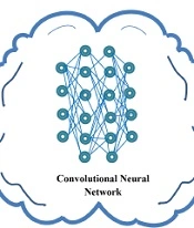
Artificial intelligence algorithms have shown promise in the field of ECG analysis. One notable application is the identification of left ventricular systolic dysfunction (LVSD) in the emergency department (ED), which can evaluate the treatment of heart failure and predict mortality in patients with acute heart failure. Increased mortality in both heart failure and non-heart failure patients is associated with LVEF (33). Adedinsewo et al. conducted a study utilizing a convolutional neural network (CNN) model and a DL approach to recognize patients with LVSD (20). The study included a total of 1 606 patients, and its primary strength lies in accurately identifying LVSD in the ED. Figure 1 depicts a CNN model based on the DL method.
 model based on DL method")
A convolutional neural network (CNN) model based on DL method
2.3. Two-Dimensional (2D) CNN Models
Jun et al. (34) introduced a deep two-dimensional (2D) CNN that utilized grayscale ECG beat images for arrhythmia detection. They compared the performance of their proposed architecture with well-known structures, such as AlexNet and VGGNet. Similarly, Jeong and Lim (29) employed a 2D CNN to rank eight different types of arrhythmias using standard 12-lead ECG records. Papageorgiou et al. (21) proposed a detection method for processing ECG indicators, which involves a dynamic digitization process. The study aimed to develop a systematic approach for assessing the vital degree characterizing the diagnosis of a high-risk coronary heart disorder. In this type of coronary disease, the assessment relies on only 2 of 12 ECG leads to calculate a patient’s risk of arrhythmogenic right ventricular cardiomyopathy (ARVC). These researchers recommend a low-complexity CN that gets highly reliable classification performance with an accuracy of 99.98% and 98.6%, a value percentage of 99.98, and a specificity of 98.25% for the training and retrieval processes, respectively.
Ullah et al. (27) introduced a CNN scheme that was evaluated with freely available arrhythmia data. They achieved a 99.11% accuracy. The model utilized the output layer with eight neurons to obtain high accuracy.
A 12-layer deep one-dimensional (1D) CNN in the MIT-BIH Arrhythmia database is presented for classifying five micro-classes of heartbeat types. They included five heartbeat features and were accurately classified using the proposed model. The wavelet self-adaptive threshold denoising method was employed during the experiments. Comparative analysis with other classification approaches, such as the backpropagation (BP) neural network and random forests, demonstrated that the proposed CNN model achieved significantly higher accuracy. Its precise classification not only contributed to the conservation of medical resources but also had a positive impact on clinical practice (2). Figure 2 illustrates the applications of 2D CNN models.

The applications of 2D CNN models
2.4. CNN-Based Autoencoder Methods
Raza et al. (22) presented an ECG Monitoring Healthcare System that utilizes CNN-based autoencoder (AE) methods. Firstly, they presented a classifier that reconstructed the raw input signals and improved overall performance within a federated learning setting to diagnose raw ECG signals from patients. The results showed that the proposed framework had better performance than other models. Additionally, they achieved a high accuracy in classification.
Obaidi et al. (31) proposed an approach that combines convolutional autoencoders (CAEs) and transfer learning (TL) for ECG signal classification. Their approach demonstrates a promising way of learning. Through experiments conducted on datasets collected from three well-known medical centers in Baghdad, Iraq, their proposed design successfully classified four types of ECG signals. Another study by Ojha et al. (24) also utilized CAEs and focused on abnormality analysis of ECG signals. Specifically, their research aimed at classifying arrhythmia heartbeats. By employing classifiers on the MIT-BIH Arrhythmia database, they identified the arbitrarily connected network (ACN) model and validated it on a test dataset using 10-fold cross-validation strategies for arrhythmia heartbeat classification. These promising results highlight the potential of their model as an effective cardiac diagnostic system for automatic arrhythmia detection.
Kim et al. (25) conducted a cohort study to investigate the ability of an AI model to detect occult AF. The aim of this study was to diagnose AF disease using an AI model trained on the Helter data. The dataset comprised two different settings: Setting 1, which included supraventricular ectopy (SVE) events, and setting 2, which also included SVE events. The results showed that the area under the receiver operating characteristic curve (AUROC) setting 1 was 0.85, which included SVE events, and setting 2 was 0.84, which included SVE events. These results demonstrated the effectiveness of their AI model in utilizing SVE burden from Holter examination to detect paroxysmal AF. Figure 3 shows the general architecture of a CNN-based AE.

The general architecture of a CNN-based Autoencoder.
2.5. Convolutional Neural Network+Long Short-Term Memory Model
Oh et al. (35) employed a hybrid CNN-long short-term memory (LSTM) approach. In this study, they could classify five different classes of arrhythmia in the MIT-BIH dataset. To ensure consistent input information, a normalization method was applied to standardize the data. Alamatsaz et al. (26) used MIT-BIH databases which is a publicly available dataset that provides standard investigation material for the detection of heart arrhythmia. This designed Convolutional Neural Network+Long Short-Term Memory (CNN+LSTM) model was able to classify 8 different types of arrhythmia. The method utilizes CNNs to extract features and data from the heart signals; nevertheless, temporal features are captured using an LSTM layer. By leveraging the morphology of the heartbeats to detect arrhythmia and incorporating other signal properties, the average accuracy of the test is 98.24% with an inference time of 5.127 ms for a single unseen rhythm. Training the model exhibited the potential to enhance performance and yield improved results. A few of these traits are RR intervals and QRS intervals (period). For this model, CNNs were taken after by an LSTM layer to gain better accuracy in arrhythmia detection. In any case, the classification accuracy of a few arrhythmias still needs improvement in their research.
Madan et al. (18) proposed an innovative approach utilizing the 2D-CNN-LSTM model for automated cardiac arrhythmia analysis. This approach stands out due to the following key features in comparison to traditional methods: (1) It employs continuous wavelet transform (CWT) to transform ECG signals into 2D Scalogram colored images, which are well-suited inputs for this network; (2) the data undergoes processing through a 2D-CNN-LSTM architecture, where the CNN component handles feature engineering, although the LSTM component performs classification. The model has been trained on a substantial dataset of labeled images, contributing to its robustness and accuracy.
Nurmaini et al. (13) proposed a computer-aided approach for locating AF using a 13-layer 1D-CNNs model with 10-fold cross-validation. Their study evaluated four datasets: MIT-BIH Atrial Fibrillation, Physionet Atrial Fibrillation, and MIT-BIH Threatening Ventricular Ectopy. By combining discrete wavelet transform (DWT) with 1D-CNNs, their model was able to automatically extract meaningful features from raw ECG signals and improve the overall classification performance. The study’s findings demonstrated outstanding accuracy, sensitivity, specificity, precision, and F1-score values of 99.98%, 99.91%, 99.91%, 99.99%, and 99.95%, respectively, for the two classes (normal sinus rhythm [NSR] and AF). For the three-class classification (NSR, AF, and non-atrial fibrillation [NAF]), the model achieved accuracy, sensitivity, specificity, precision, and F1-score values of 99.17%, 98.90%, 99.17%, 96.74%, and 97.48%, respectively. These results highlight the effectiveness of their proposed approach in AF detection and classification.
Hassan et al. (28) proposed a CNN-Bi-LSTM model, which is a hybrid bidirectional LSTM, to enhance diagnostic accuracy in a medical setting. Their study aimed to classify five categories of ECG signals and develop an independent computer-aided diagnosis system. The model achieved high accuracy rates of 100%, 98.0%, and 98.0% for training, validation, and testing, respectively, using the MIT-BIH dataset. Additionally, when applied to the St-Petersburg dataset, the model achieved impressive accuracies of 98.0%, 95.0%, and 95.0% for training, validation, and testing, respectively, in the detection of arrhythmia. Figure 4 shows the overall flowchart of the CNN-LSTM model.
 model")
The overall flowchart of convolutional neural network+Long short-term memory (CNN+LSTM) model
2.6. Convolutional Neural Network-Support Vector Machine (CNN-SVM) Classifier
In their article, Jamil & Rahman (32) proposed an attention-based model called ArrhythmiaNet, which incorporates a clump of features (CoF) module for classifying 17 classes of heartbeats. The ArrhythmiaNet model achieved a high classification accuracy of 99.84% when combined with a support vector machine (SVM) classifier. It also exhibited a sensitivity of 100% and an F1 score of 99%. When combined with a K-nearest neighbors (KNN) classifier, the ArrhythmiaNet model achieved a classification accuracy of 98.64%.
The authors conducted a comparison of their proposed system with existing strategies, and the test results demonstrated that ArrhythmiaNet outperforms all the existing methods in terms of accuracy. This finding highlights the superior performance of ArrhythmiaNet in the classification of heartbeats and its potential for improving arrhythmia diagnosis.
Murawwat et al. (30) classified arrhythmia into three classes (i.e., normal, bradycardia, and tachycardia). They proposed a technique that outperforms earlier methods, such as wavelet transform (WT), SVM, and empirical mode decomposition (EMD). Previous techniques used WT for denoising and feature extraction of ECG signals; however, this required a change of signal domain. Support vector machine, on the other hand, served as a classifier but was computationally intensive, demanding significant memory resources and exhibiting slower processing speeds. Empirical mode decomposition was employed for denoising signals; nevertheless, it suffered from mode mixing issues and was not suitable for multichannel signals.
In contrast, the proposed technique showcased efficiency by eliminating the need to change the signal domain for processing. It was simple, required no large memory resources, and exhibited faster processing times. The analysis of the signals indicated no evidence of mode mixing, making it suitable for multichannel signal processing. The results of their study demonstrated that the proposed approach achieved higher accuracy in detecting and classifying arrhythmia than previous techniques. This finding highlights the effectiveness of their technique in improving the accuracy of arrhythmia detection and classification.
3. Results
As evident, AI methods are a new way of classifying different types of arrhythmias. For example, DL algorithms, including CNN, LSTM networks, convolutional recurrent neural networks (CRNN), and CNN-based AEs, have been extensively utilized for ECG signal analysis and arrhythmia detection. Deep learning proves to be highly effective in detecting various heart diseases, such as hypertrophic cardiomyopathy, LVEF, and hyperkalemia, and predicting revascularization in patients with suspected coronary artery disease (14). Sarker broadly categorizes DL techniques into three major categories: (1) discriminative learning, (2) generative learning, and (3) other hybrid learning approaches (36). The findings of this study demonstrated the potential of AI models in the detection of different types of arrhythmias, prevention of heart attacks, and diagnosis of latent heart disease.
This study explored different DL techniques for classifying arrhythmias. The 2D CNN model achieved an accuracy of 97.42% in classifying five different arrhythmias. The CNN-based AE and TL models achieved a high accuracy. The method can classify four types of ECG. The CNN-Bi-LSTM model achieved an accuracy of 98.0% in categorizing five categories of ECG signals. The CNN+LSTM model achieved an accuracy of 98.24% in classifying five classes of arrhythmias. The CNN-SVM classifier model demonstrated an accuracy of 98.64% in detecting seventeen classes of heartbeats. The results indicated that the CNN-based AE and TL models perform exceptionally well with high accuracy in detecting ECG signals.
4. Conclusions
This review aimed to showcase the applications of AI in ECG signal analysis and arrhythmia detection, as explored in previous studies. Various DL methods, such as LSTM, CNN, AE, CRNN, and gated recurrent unit (GRU), have been utilized, and CNN has emerged as the most popular and high-performing approach. Researchers have also combined CNN with other methods to enhance classification accuracy. For instance, Oh et al. (35) used a combination of CNN and LSTM to classify five classes of the MIT-BIH dataset, which includes normal heartbeats.
The current study highlights the superiority of DL models over conventional machine learning strategies in modeling ECG data. The present study examines various aspects of biometric ECG-based frameworks, including DL algorithms, dataset sources, preprocessing strategies, and training designs and tasks.
The current study demonstrates the growing interest in utilizing DL for ECG signal detection in medical and healthcare applications over the past decade. Deep learning models have been shown to outperform experienced cardiologists, delivering state-of-the-art and more accurate results. Deep learning presents a novel approach to diagnose and manage heart disease. The combination of wearable technologies and accessible platforms for deploying DL models offers a unique opportunity for early detection and treatment of various cardiovascular diseases.
