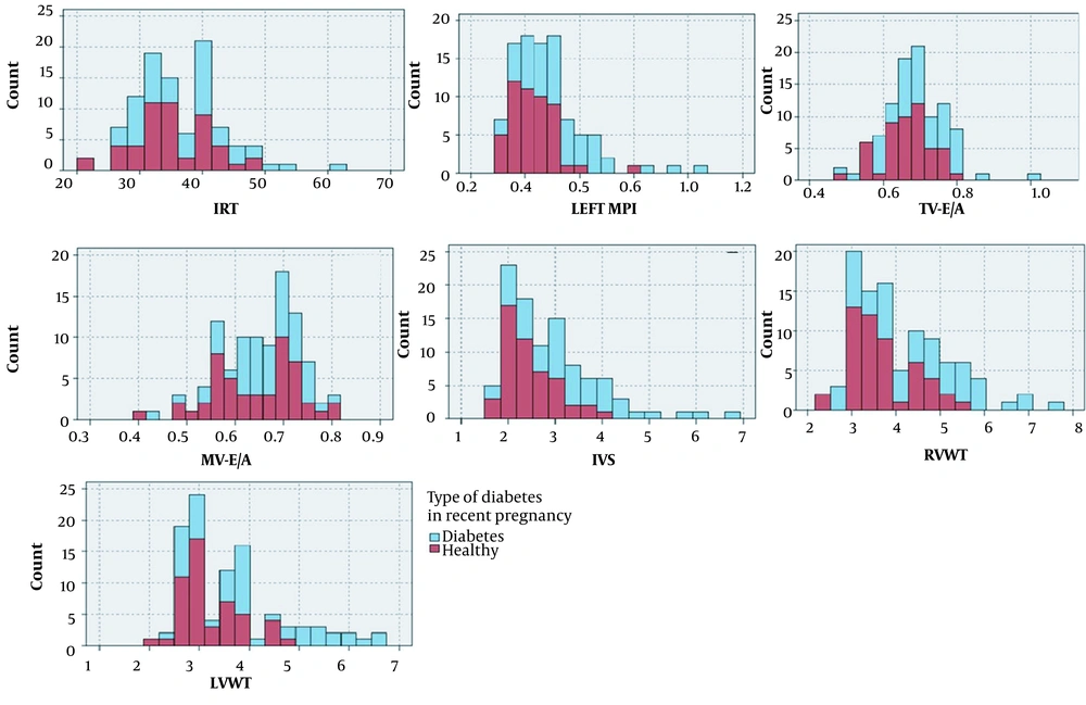
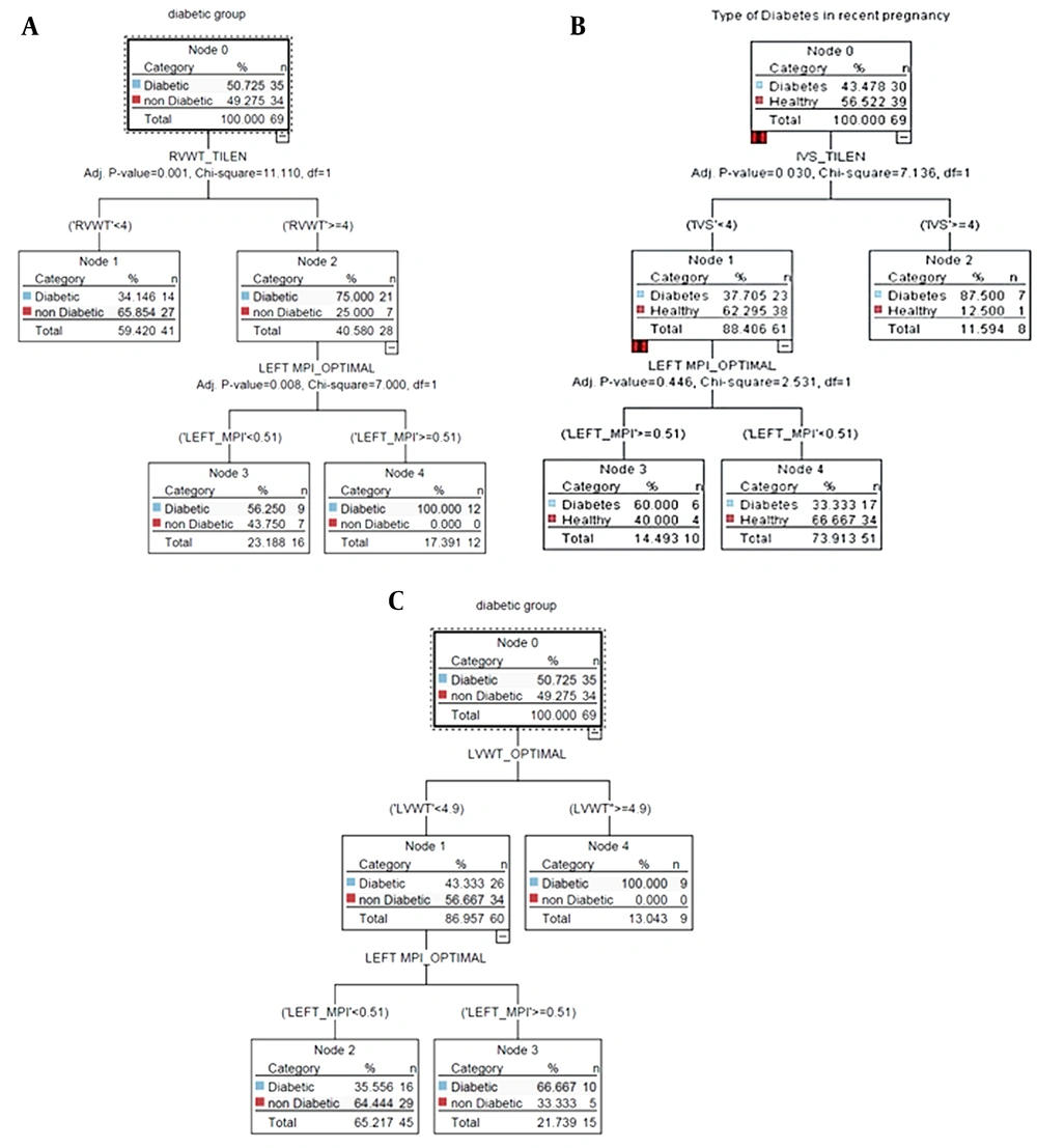
In the current case-control observational study, women with singleton pregnancy in their second or third trimester (18 - 40 or 28 - 40 weeks of gestation) underwent a single additional cardiac scan as they had attended the ultrasound departments for routine prenatal or anomaly ultrasound examination at Mahdiyeh and Shohada hospitals, Tehran, Iran, within March 2020 to February 2021 and were enrolled for participating in this study.
The study protocol was approved by the Ethics Committee of Shahid Beheshti University of Medical Sciences, Tehran, Iran (IR.SBMU.MSP.REC.1399.545). Before enrollment, the researcher explained the design of the study to the eligible mothers and asked them to read and sign the written informed consent. The selection of patients to complete the questionnaire and then undergo an ultrasound in both groups was completely blind and random.
Mothers who gave consent for participation were enrolled in the study using the census method. Mothers with a positive history of overt DM (pregestational) or GDM in the current pregnancy were considered the case group, and mothers without GDM or DM were considered the control group. The inclusion criteria were women with singleton pregnancies in the second trimester (18 - 28 weeks of gestation) or the third trimester (28 - 40 weeks of gestation). The exclusion criteria were multifetal pregnancy, hypertension (pregnancy-induced or essential), intrauterine growth retardation (defined as weight for gestation less than 10th percentile), and polyhydramnios in the study.
According to the above-mentioned criteria, a total of 100 mothers were enrolled, 50 subjects in the case and 50 subjects in the control group. All mothers underwent aneuploidy screening test in the first trimester, three of whom were identified as high-risk combined tests, and one had increased nuchal translucency and underwent amniocentesis. All mothers had normal results of anomaly scans within 18 - 20 weeks of gestation.
The physician collected the mothers’ information by taking a medical history from the mothers, including age, parity, body mass index (BMI), type of antidiabetic medication, use of drugs, and underlying medical diseases. A previous result of their fasting blood sample was evaluated. The mothers were categorized into the case or control groups according to their fasting blood sugar (FBS) or 2-hour oral glucose tolerance test (OGTT). The analysis results were obtained from all cases to differentiate between diabetic and control cases, considering the cut-off levels of < 92 mg/dL for FBS and < 153 mg/dL for OGTT.
Furthermore, mothers in the case group were categorized into poor control DM and normal glycemic level (good control DM) according to the American Diabetes Association and American College of Obstetricians and Gynecologists, considering the target glucose concentrations at FBS < 95 mg/dL, 1-hour postprandial blood glucose concentrations < 140 mg/dL, and 2-hour postprandial glucose concentrations < 120 mg/dL. The researchers initiated insulin therapy (or increased doses of glycemic control medications) when one-third of fasting or postprandial glucose levels exceeded the target in a given week.
The imaging protocol performed for all mothers consisted of ultrasound, performed by one fellowship of perinatology using machine GE Vivid E8 Ultrasound System, which included the following steps:
(1) Fetal echocardiography was performed using the echocardiographic device, as described by Alfred Abuhamad as cited in Carvalho (
20), for the measurement of the end-diastolic interventricular septal thickness (IVST) and myocardial free walls (inferior to atrioventricular valves), including right ventricular wall thickness (RVWT), left ventricular wall thickness (LVWT), recorded in lateral subcostal, apical, or basal four-chamber view (based on the fetal position at the time of scan).
(2) Doppler waveform was performed to measure the left myocardial performance index (MPI), as described by Hernandez-Andrade et al. (
21). After obtaining a five-chamber view of the fetal heart, the ascending aorta was aligned with an angle of insonation at < 20°, and after visibility of the mitral and aortic valves, the Doppler sample gate was opened to about 3 - 4 mm and applied over leaflets of mitral and aortic valves to identify valve clicks along the Doppler waveforms after minimizing noise and artifacts. The procedure was repeated three times to enhance reproducibility.
The pulsed Doppler sample volume was placed on the inner wall of the ventricular septum above the mitral valve and below the aortic valve in the four-chamber view with a basal or an apical projection allowing simultaneous inflow and outflow display from the left ventricle. Then, time periods for the left MPI were measured as follows:
The time period from the mitral valve closure clicks to the opening of the aortic valve click (in milliseconds) was considered isovolumic contraction time (IVCT).
The time period from the aortic valve closure clicks to the opening of the mitral valve click (in milliseconds) was considered isovolumic relaxation time (IVRT).
The time period from the opening to the closure of the aortic valve click (in milliseconds) was considered ejection time.
Fetal left ventricular MPI was calculated by the following equation:
(3) Pulsed Doppler interrogation of atrioventricular valves was performed in the standard apical four-chamber view with sampling volume from distal to tip of the valve. The mean peak values of three early diastolic waves (E) and of three late diastolic or atrial filling waves (A) were recorded, indicating tricuspid and mitral inflow velocities (depicting passive and active ventricular filling); the mean ratio between the peak velocities of the E and A waves in each position (E/A ratio) was determined at the mitral and tricuspid valves, indicating diastolic function.
According to the reported mean duration of IVRT as 34 milliseconds with a range of 26 - 41 milliseconds (
20), IVRT values > 41 milliseconds were considered prolonged. According to the same reference, the normal range of fetal left ventricular MPI was considered within 0.26 - 0.44 (mean: 0.36) and MPI > 0.44 as prolonged (
20). In addition, the cut-off values of IVST and RVWT were considered at 4 mm and LVWT at 4.9 mm (
20).
Finally, the intraobserver reproducibility of the MPI, IVST, E/A ratios, interventricular thickness, and free wall ventricle thickness were analyzed in 100 women within 18 - 40 weeks of gestation.
3.1. Statistical Analysis
The baseline characteristics of the participants are presented as mean ± standard deviation. The
t-test was used to compare two groups of diabetic and nondiabetic mothers. The machine learning method was used to extract important predictors of fetal myocardial function in the case and control groups. Three types of decision tree (DT) algorithms, including classification and regression tree (CART) algorithms, chi-square automatic interaction detection (CHAID), and quick, unbiased, efficient, statistical tree (QUEST), were used for analysis (
22). The accuracy of the models is attached in Appendix 1.
The target variable belonged to case and control groups defined by diabetes (i.e., diabetic and nondiabetic). The input variables (predictors) included the fetal left ventricular MPI, mitral valve (MV) E/A ratio, tricuspid valve (TV) E/A ratio, RVWT, LVWT, and IVST. The total data were divided into 70% (training data) and 30% (test data) for developing and testing DT algorithms, respectively. The performance of DT models was measured on the test data with accuracy, sensitivity, and specificity. IBM SPSS Modeler software (version 18) and Payton software (version 1) were used for statistical and DT analysis. A P-value less than 0.05 was considered statistically significant.