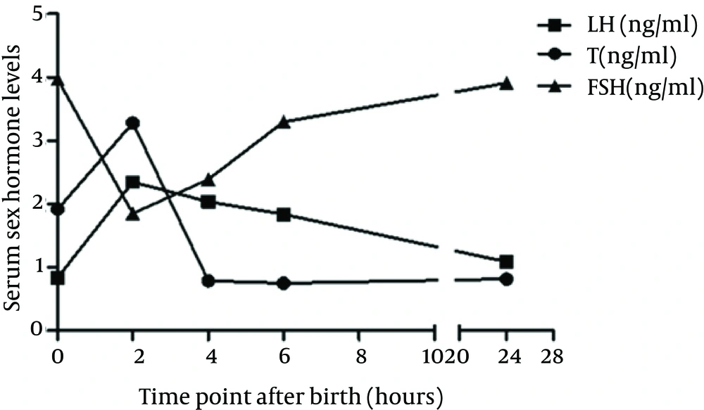
We chose Wistar rats to confirm the existence of mini-puberty and to investigate the impacts of hormone inhibition during this phase on adolescent gonadal axis function. The concentrations of serum sex hormones revealed a transient surge, lasting about six hours, in the hypothalamic–pituitary–gonadal (HPG) axis of male pups; these findings were similar to that reported in Corbier’s study (
5). Meanwhile, the testis weight peaked at four hours after birth following the surge of sex hormones; this may be caused by the increasing effect of sex hormones on neonatal testis tissue. As reported by Svechnikov (
11), postnatal differentiation of testicular cells (including Leydig cells, Sertoli cells and spermatogenic cells) involved a variety of cellular cascades, resulting in mature testicular cells with well-developed steroidogenic machinery required for testosterone biosynthesis and adolescent gonadal axis function. There are two periods of activities in the HPG axis before the onset of puberty: the first during the fetal life and the second during the postnatal months in human (
19). After the postnatal HPG axis activity, the axis is silenced for several years until puberty. Therefore, mini-puberty provides a window of opportunity to study the normal mechanisms that occur from prepuberty to sexual maturity and to get an early assessment of testicular function.
In mammals such as rats, the regulation of testicular function, fundamentally involving complex developmental genetics and endocrinology, can be subdivided into chromosomal, genetic, and hormonal stages, where each stage influences the next, resulting in not only sex differences in the reproductive system but also sexual differentiation of the central nervous system (CNS) (
20). The brain is particularly sensitive to the differentiating effects of androgen only during a critical period of exposure to the supraphysiological androgen levels (
21). Lamminmaki (
22) has found early postnatal testosterone levels showed predicted associations with later behavior after measuring testosterone in 48 full-terminfants by monthly urinary sampling from day seven postnatal to six months old and analyzing their sex-typed behavior. In our study, the serum T levels peaked at two hours, reaching a value of four times of that at birth; this may indicate that the sudden secretion in the neonatal period plays an important role in CNS sexual differentiation as well as the masculinization process at adulthood.
Vega (
23) observed that the postnatal T surge was inhibited in male rat pups anesthetized with ether inhalation immediately after delivery, and that this inhibition resulted in permanent alteration of adulthood sexual behavior, as the male rats showed elevated levels of feminine behaviors and impaired masculine sexual behaviors. However, ether inhalation anesthesia performed at four hours after birth resulted in no significant impact on adulthood sexual behavior in the rats. These findings suggest that the postnatal T surge may be involved in sexual differentiation of the CNS and influence the masculinization processes. Arena (
24) investigated the effects of ether inhalation on male rats immediately after birth and found alterations in sexual behavior along with a decrease in the number of spermatids and spermatozoa at adulthood, which suggested that ether delayed or reduced the testosterone peak of the sex differentiation period, consequently altering the processes of masculinization and defeminization of the CNS.
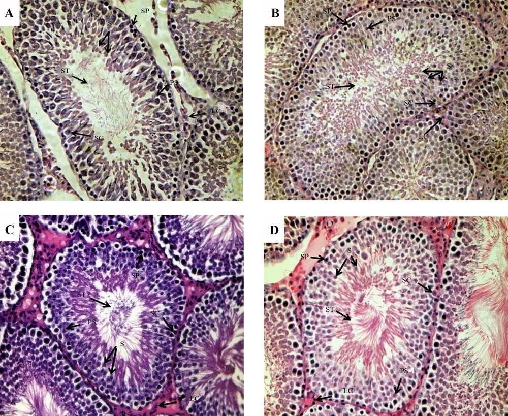
On the basis of the above observations, we investigated the long-term effects of hormone inhibition during the mini-pubertal period on testicular function in male rats at a molecular and morphologic level. In the present study, the levels of serum T, LH, and FSH concentrations in the model group were significantly lower than the corresponding levels in the control group, indicating that ether inhalation was an inhibitory factor for pituitary gonadotropin secretion and reduced T surge, which in turn means that mini-puberty was inhibited in the model mice. The morphological analysis of the testes on PND45 revealed damage of spermatogenic cells in the model mice. The ghrelin mRNA expression level showed a decreasing trend in the model group compared with the control group. Evidence supports that ghrelin is expressed and operates at different levels in the gonadotropic axis and in other reproductive tissues (
10). It is also involved in the secretion of gonadotropin and prolactin and is highly selectively expressed in interstitial mature Leydig cells of the human testis. Therefore, the decreased expression level may indicate a degree of damage of Leydig cells and their function. On PND75, there was a significant decrease in the AMH mRNA expression level as well as a decreasing trend of the AR gene. As AMH is produced by Sertoli cells and is responsible for the regression of the Mullerian ducts, the anlagen of the uterus and Fallopian tubes (
9,
25), it is an excellent marker of Sertoli cells function. Therefore, the significant decrease in the AMH mRNA expression level indicated damage to testicular Sertoli cells function in our adult male rat model. A research (
9) has shown that seminiferous tubules are the major components of the testis, while seminiferous tubule volume depends mainly on Sertoli cells during the whole prepubertal period, especially during mini-puberty; thus, hormone inhibition during mini-puberty can be expected to mainly affect Sertoli cells, which is consistent with our results. Acting through AR, androgens are responsible for the development of the male phenotype and for male sex maturation and the maintenance of male reproductive function and behavior (
18). The decrease in the AR mRNA expression level suggests that the masculinization of the model rats in our study may be partly damaged. INSL3 is a major secretory product of Leydig cells (
26) which reflects their number, differentiation status and ability to produce various factors including steroids. INSL3 is not acutely regulated by the HPG axis (
15), making it an excellent marker for Leydig cells differentiation and functional capacity. INSL3 is important for the process of testicular descent, specifically in transabdominal testis translocation (
27,
28). However, in our research, we did not find any significant differences in the INSL3 mRNA level on PND45 and PND75, suggesting that hormone inhibition in neonatal rats does not get its long-term effects through INSL3. The testicular normal development relies on many regulatory factors and sex hormones, along with their interactions and balance. The decrease of mRNA expression of ghrelin, AMH, AR and INSL3 indicated the damage of the function of Leydig cells and Sertoli cells at different periods and that the balance between them may be broken.
The fact that inhibition in the transient hormone surge of the HPG axis in neonatal male rats had a long-term effect on testicular function at puberty and sexual maturity, confirmed that mini-puberty is a special phase in the male sexual defeminization and masculinization process. The optimized therapy of sexual disorders, especially of congenital hypogonadotropic hypogonadism remains to pose a challenge to pediatric endocrinologists, which may partly be caused by an incomplete medication treatment plan; but, delays in diagnosis should be taken seriously. As mini-puberty has been found to play a vital role in the testicular function, it is meaningful to place a high value on this period to establish an early diagnosis of hypogonadotropic hypogonadism, so that it can be treated at an appropriate time.