1. Background
Sex steroids are the key regulators of skeletal growth and maturation both in females and males (1). Since osteoporosis affects more females than males, most of the research studies on gonadal sex steroids and bone focus on the effect of estrogen on bone (2). It is well established that estrogen is critical in the regulation of bone metabolism, as illustrated by the considerable loss of bone mass in females following natural menopause (3). Loss of androgens in males following the surgical orchidectomy, chemical castration, or age-related hypogonadism evokes the same adverse effect on the skeleton (4).
It is generally believed that expression of androgen and estrogen receptors on the bone compartment and the stimulation of these receptors are the important determinants of the effects of androgens or their metabolites on the skeleton (5). Testosterone, which is the major gonadal androgen in males, is also converted to estrogen by aromatization (6). Thus, the impact of testosterone on skeletal growth and maintenance is contributed via signaling through the androgen and estrogen receptors on bone (7).
Surgical castration by orchidectomy is the most widely used method to produce animal model of male osteoporosis due to testosterone deficiency (8, 9). The use of pharmaceutical agents or chemical castration such as gonadotropin releasing hormone (GnRH) agonists, estrogen receptor antagonists, and aromatase inhibitors, could be an alternative way to induce bone loss in intact experimental rats (10). These pharmaceutical agents are used in humans to treat endometriosis or breast cancer, and are associated with accelerated bone loss (11). Buserelin, which is an agonist to GnRH, induces estrogen-deficiency and causes profound bone loss in female rats (12). To the best of authors` knowledge, the effects of GnRH antagonist on bone and their use in inducing testosterone-deficiency osteopenia in male rats are not studied.
GnRH antagonists block the release of gonadotropins by binding competitively to pituitary GnRH receptors, and therefore, suppressing gonadal steroids secretions in males (13-15). Blocking the GnRH receptor with an antagonist in the pituitary gland was reported the most appropriate way to achieve a fast and reversible decrease of luteinizing hormone (LH), follicle-stimulating hormone (FSH), and testosterone levels (16). Hence, GnRH antagonists may be expected to cause bone loss on the basis of the suppression of the gonadal steroids. Degarelix is a newer series of potent and long-acting GnRH antagonist capable of suppressing pituitary-gonadal axis (17). It produces a pharmacological profile, which closely resembles to those of bilateral orchidectomy (18).
The current study aimed at identifying whether administration of degarelix could induce changes in bone turnover, bone mineral density, and bone mechanical strength in intact male rats. In the current study, the osteopenic effect of degarelix was compared with that of surgical orchidectomy. Since the impact of GnRH antagonist on bone is not studied in intact male rats, this could be the first study that demonstrated the short-term effect of degarelix on bone. It is hoped that the current study can provide a new method to induce testosterone deficiency and osteoporosis without having to remove the testes.
2. Methods
2.1. Animals and Treatments
Eighteen eight-week old intact-male Sprague-Dawley rats were obtained from the laboratory animal resource unit, faculty of Medicine, Universiti Kebangsaan Malaysia (UKM). They were allowed to acclimate to an environmentally controlled room (12:12 hour light/dark cycle, room temperature) in an animal care facility for one week. The rats were housed in plastic cages with free access to standard pellet diet and water. Experimental protocols concerning the use of laboratory animals were reviewed and approved by the UKM animal ethics committee (FP/FAR/2015/NAZRUN/25-MAR/665-MAR-2015-DEC-2017).
In the beginning of the study, rats weighing 300 - 350 g were randomly assigned to three groups. A sham-operated (SHAM) group underwent similar surgical procedure, but the testes were not removed. Surgical castration was performed on the orchidectomized rats in (ORX) group, while the other group was chemically castrated with subcutaneous injection of degarelix (DGX) around the scapular region at the dose of 2 mg/kg of body weight. The rats were monitored daily for six weeks. At the end of the experimental period, blood was collected from the orbital sinus and the rats were euthanized with ether. The serum was extracted by centrifugation (3000 rpm for 10 minutes) and stored at -80°C until biochemical analyses. The right femora were dissected and wrapped in gauze, soaked in phosphate buffered saline (PBS), and stored at -20°C for biomechanical analyses.
2.2. Dual Energy X-ray Absorptiometry Analyses
In vivo whole-body and femoral DXA scans were performed using DXA equipped with regional high-resolution scan software for small animal (Hologic QDR series, Hologic). Bone mineral area (BMA), bone mineral content (BMC), and bone mineral density (BMD) of the whole body and femur were evaluated. Before measurements, a tissue calibration scan was performed with the Hologic phantom for the small animal. Total BMD was calculated as bone mineral content/area. The same operator scanned all animals at baseline and six weeks of experimental period.
2.3. Biochemical Analyses
Serum biomarkers for bone turnover, osteocalcin, and carboxy-terminal of type 1 collagen crosslinks (CTX-I) were assessed using an enzyme-linked immunosorbent assay (ELISA) kit (Immunodiagnostic Systems, Tyne and Wear, UK). The detection limit of the assay was 50.0 ng/mL for osteocalcin and 2.0 ng/mL for CTX-1, and the intra-assay variation of these kits were 4.0% and 6.9%, while the inter-assay coefficients of variation were 6.6% and 12%, respectively. Testosterone level in the serum was also measured by commercially available ELISA kit (Biovendor, Laboratomi Medicina, Czech Republic). The sensitivity of the assay was 0.066 ng/mL, and the intra- and inter-assay coefficients of variation of the kit were 8.54% and 9.97%, respectively.
2.4. Bone Mechanical Testing
Biomechanical properties of femoral bone were evaluated using three-point bending test method (Shimadzu, Autograph AGS-X 500 N, Japan) controlled by proprietary software (Trapezium Version 1.4.2, Shimadzu). The femurs were cleaned from soft tissues and the total length and width of the midshaft were measured with digital caliper. The left femur from each rat was placed on an adjustable two-point block jig spaced 5 mm apart, with a load sensor attached to a bending punch on the crosshead. Force was applied at a rate of 10 mm/minute to the mid-shaft of the femur diaphysis until a break was determined by measuring a reduction in force. The recorded testing parameter included both extrinsic and intrinsic properties. Whole-bone mechanical properties were determined by the extrinsic parameters (force and displacement), while the measurement of the bone material were determined by intrinsic parameters, stress and strain.
2.5. Statistical Analysis
All data were expressed as mean ± standard error of the mean (SEM). Intergroup differences were analyzed by one-way analysis of variance (ANOVA) with SPSS, version 21.0 (Chicago, US). A P value < 0.05 was considered statistically significant.
3. Results
3.1. Testosterone Level and Biochemical Markers of Bone Turnover
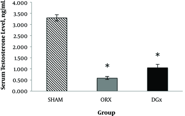
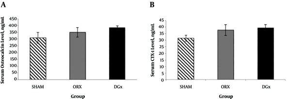
Both orchidectomy and subcutaneous administration of degarelix significantly suppressed serum testosterone to castration level (P < 0.05) following six weeks of initial injection. There was no significant difference between the testosterone levels in DGX and ORX groups (Figure 1). As shown in Figure 2, the DGX and ORX groups showed marginally higher osteocalcin and CTX-1 level when compared with the SHAM group, but the difference was not statistically significant (P > 0.05).
Effects of orchidectomy and degarelix administration on serum testosterone level after 6 weeks of experimental period. Data are presented as mean ± SEM. The number of rats per group was 6. *P <0.05 compared with SHAM group. Abbreviations: SHAM, sham-operated group; ORX, orchidectomized group, DGX; degarelix-induced group.
Effects of orchidectomy and degarelix administration on A, serum osteocalcin; and B, serum CTX-1 level after 6 weeks of experimental period. Data are expressed as mean ± SEM. The number of rats per group was 6. Abbreviations: SHAM, sham-operated group; ORX, orchidectomized group, DGX; degarelix-induced group.
3.2. Whole Body and Femoral DXA Analyses
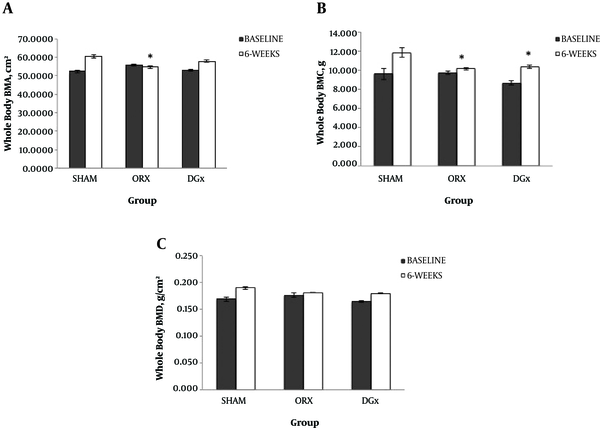
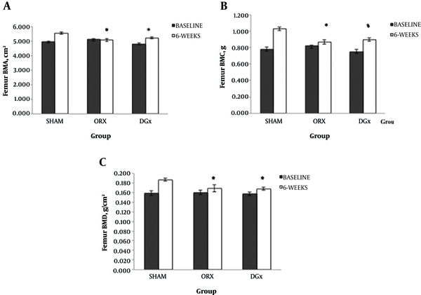
In the beginning of the study, the difference in whole body BMA, BMC, and BMD were not statistically significant between the groups (P > 0.05). After six weeks, chemical castration with degarelix and orchidectomy resulted in significant reductions of the whole body BMC, but the whole body BMA only reduced in ORX group compared with SHAM group (P < 0.05). There were no differences in the whole body BMD among the three groups (Figure 3). The differences in femoral BMA, BMC, and BMD in the beginning of the study between the groups were not statistically significant (P > 0.05). Femoral BMA, BMC, and BMD significantly reduced after six weeks in both DGX and ORX groups (P < 0.05; Figure 4). There were no differences in the femoral parameters between the DGX and ORX groups.
Effects of orchidectomy and degarelix administration on whole body A, bone mineral area; B, bone mineral content; and C, bone mineral density after 6 weeks of experimental period. Data are expressed as mean ± SEM. The number of rats per group was 6. *P < 0.05 compared with SHAM group. Abbreviations: SHAM, sham-operated group; ORX, orchidectomized group, DGX; degarelix-induced group.
Effects of orchidectomy and degarelix administration on femoral A, bone mineral area; B, bone mineral content; and C, bone mineral density after 6 weeks of experimental period. Data are expressed as mean ± SEM. The number of rats per group was 6. *P < 0.05 compared with SHAM group. Abbreviations: SHAM, sham-operated group; ORX, orchidectomized group, DGX; degarelix-induced group.
3.3. Femoral Bone Biomechanical Properties
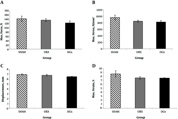
The differences in mechanical properties among the three groups in terms of maximum force, maximum stress, displacement, and maximum strain were not significantly different following six weeks of experimental period (P > 0.05; Figure 5). Although insignificant, the biomechanical parameters for the ORX and DGX groups were lower than the SHAM group.
Effects of orchidectomy and degarelix administration on bone biomechanical strength parameters A, force; B, stress; C, displacement; and D, strain after 6 weeks of experimental period. Data are expressed as mean ± SEM. The number of rats per group was 6. Abbreviations: SHAM, sham-operated group; ORX, orchidectomized group, DGX; degarelix-induced group.
4. Discussion
Osteoporosis in males is an important public health problem worldwide (19). Hence, the need to develop new preventive and therapeutic approaches to treat osteoporosis in males is desirable. In order to promote the research on this subject, new methods to assess bone loss in experimental animals are necessary. Surgically orchidectomized animal is a typical experimental model to study male osteoporosis due to androgen deficiency (20). Densitometry, biochemical markers, histomorphometry, and mechanical testing are the methods of choice to evaluate osteopenic skeleton in animal models (11).
The current study compared chemically and surgically castrated male rats as models of osteoporosis in males by assessing the bone biochemical markers, densitometry, and mechanical strength. This may be the first study on the effect of a single subcutaneous injection of degarelix, a GnRH antagonist, on bone in intact male rats. Administration of degarelix at the dose of 2 mg/kg significantly suppressed serum testosterone to the castration level six weeks after the subcutaneous injection. This result was consistent with an earlier finding, which found that administration of degarelix at 2 mg/kg resulted in the suppression of testosterone up to 42 days (17). The testosterone levels of DGX and ORX groups in the current study were not significantly different, indicating that degarelix was capable of suppressing gonadal steroid hormone in intact male rats to the same degree as that of bilateral orchidectomy.
The increase in bone formation marker, particularly serum osteocalcin observed in hypogonadal males, indicated an increase in bone turnover that led to accelerated bone loss (21). The changes in plasma osteocalcin levels reflected the changes in bone turnover (22). CTX-1 is the degradation product of C-terminal part of telopeptide of collagen I; thus, it is considered as a sensitive marker of bone resorption. Bone loss induced by androgen deficiency in mature rats is associated with increased bone turnover in the first few months after orchidectomy (23). In the current study, although the changes in bone biochemical markers were not significant, marginal increase in such markers both in DGX and ORX groups might indicate increased bone turnover.
DXA is a non-invasive technique to measure time related differences in bone densitometry (24). Orchidectomy induced a significant reduction in femoral BMD detectable with DXA after four weeks in adult rats (25). In the current study, bone loss was detected with DXA, six weeks after orchidectomy or degarelix administration. The reduction in the whole body mineral content and femoral mineral area, content, and density could further elucidate the effects of a GnRH antagonist (degarelix) on bone. Femoral BMA, BMC, and BMD were equally affected by orchidectomy and degarelix administration. Mechanical test was conducted to assess the whole bone strength, but the results did not show any significant reduction of bone strength both in DGX and ORX rats. This result suggested that the six-week follow-up period might be too short to monitor changes in biomechanical properties of bone tissue. Longer than six weeks may be needed to induce significantly lower bone strength in response to androgen deficiency.
In summary, administration of degarelix, a GnRH antagonist, affected bone densitometric parameter to an extent similar to that of orchidectomy. However, the changes in bone biomarkers and mechanical strength were less prominent after six weeks of a single administration of degarelix or bilateral orchidectomy. The marginal elevation in bone markers, which did not reach statistical significance, may be due to small sample size used in the current study. Another reason may be that bone remodeling is a dynamic process; thus the measurement of serum biomarkers is subjected to a constant state of fluctuation (26). The assessment of bone remodeling might be better illustrated by dynamic histomorphometric parameters, since findings are more stable in bones compared with serum findings. Further studies on bone mRNA expression should also be conducted to provide mechanistic overview of how degarelix castration could affect bone remodeling. There were trends towards lower bone mechanical strength and higher bone turnover after six weeks, suggesting that long term and multiple injection of degarelix should exhibit more prominent effects on bone.
The current study had some limitations that should be addressed. Sexually matured young rats were used though aged rats more accurately reflect the target population for osteoporosis study. However, the skeletal effects in androgen-deficient rat model seemed to follow the same pattern as the hypogonadal human in relation to testosterone deficiency. There is extensive literature studying the effect of orchidectomy including evaluation of bone biochemical markers, bone densitometry, histomorphometry, and bone fragility. The current preliminary study on the effect of short-term chemical castration with degarelix on bone turnover, densitometric and biomechanical properties of male rats was compared with the well-established orchidectomized rat models. Therefore, a study on long-term effect of chemical castration with degarelix is warranted in order to provide the most adequate animal model to study male osteoporosis.
Previous studies utilized orchidectomy model as the model of male osteoporosis by surgical removal of testes bilaterally. Administration of degarelix could be an alternative method to induce testosterone deficient-bone loss in intact rats. This method is more convenient and would not expose the rats to surgical stress and wound infections. In comparison with the orchidectomy model, the degarelix model is a better resemblance to aging males with osteoporosis and intact testes. Thus, the degarelix model can be recommended for studies on the prevention and treatment of bone loss due to androgen deficiency. Further studies are warranted to evaluate the long-term effects of androgen deficiency including degarelix on bone metabolism. In conclusion, degarelix is as effective as orchidectomy, but provides a more convenient method to create an androgen-deficient osteoporosis model.

