An Overview of Celiac Disease in Childhood Type 1 Diabetes

authors:

avatar Iraj Shahramian ORCID 1 , avatar Ali Bazi ORCID 2 , * , avatar Alireza Sargazi 3

Pediatric Ward, Amir - Al - Momenin Hospital, Zabol University of Medical Sciences, Zabol, Iran
Clinical Research Development Unit, Amir - Al - Momenin Hospital, Zabol University of Medical Sciences, Zabol, Iran
Student Research Committee, Zabol University of Medical Sciences, Zabol, Iran

How To Cite Shahramian I, Bazi A, Sargazi A. An Overview of Celiac Disease in Childhood Type 1 Diabetes. Int J Endocrinol Metab. 2018;16(3):e66801. https://doi.org/10.5812/ijem.66801.

Abstract

Context:

Celiac disease (CD) is a common phenomenon in children with Type 1 diabetes (T1D). In the present review, we have discussed the pathogenesis, diagnostic biomarkers, risk factors, and prognosis of CD in the context of pediatric T1D.

Evidence Acquisition:

Literature published in Web of Science, PubMed, Scopus, Google Scholar, and Cochrane Library were scrutinized up to the end of 2017. The keywords of celiac disease, Type 1 diabetes, children, and pediatric were used in different combinations.

Results:

Immune cytotoxic reactions along with dampen immune regulatory functions contribute to CD in the context of pediatric T1D. Many children with simultaneous CD and T1D do not represent with the clinical signs of the enteropathy rendering a diagnostic challenge. The most common screening tests in these children are routine serological tests of CD, anti - endomysial, anti - transglutaminase, and anti - deamidated gliadin peptide antibodies. Typing for human leukocyte antigens of DQ - 2 and DQ - 8 may assist in the diagnosis of silent CD in children with T1D. The most significant shared non - HLA genetic loci of CD and T1D comprise CTLA - 4, TAGAP, IL - 18RAP, PTPN2, RGS1, SH2B3, CCR5. Interactions between these loci can be important in susceptibility to CD in T1D. Some new biomarkers have been suggested for diagnosis of CD including ischemia-modified albumin (IMA), soluble syndecan-1 (SSDC-1), regenerating gene Iα (REG-Iα), Neurotensin, and Zonulin, which can be useful for diagnosis and screening of CD in childhood T1D.

Conclusions:

Overall, active seropositive CD seems to be of clinical importance in T1D with significant impacts on the quality of life and predisposition to diabetes associated complications. It is important to detect CD in the context of T1D to prevent potential risks contributing to morbidities and mortalities associated with either CD or T1D.

1. Context

Celiac disease (CD), gluten - induced atrophy of the small intestine, is an autoimmune condition, which can be seen in the context of other autoimmune disorders including Type 1 diabetes (T1D). T1D may be diagnosed in association with CD as high as six times of healthy individuals. On the other hand, T1D patients may be seen with concurrent CD in 8% of cases (1). Children with T1D represent higher propensity to CD. Geographical distributions, consumption of gluten - containing regimes, ethnical origins, and environmental factors are among CD contributing factors in T1D patients. Here we have reviewed the pathogenesis, diagnostic biomarkers, risk factors, and prognosis of CD in the context of pediatric T1D.

2. Evidence Acquisition

Literature published in the Web of Science, PubMed, Scopus, Google Scholar, and Cochrane Library between 1990 up to the October 2017 where studied. The main keyword used were celiac disease, Type 1 diabetes, and pediatrics. The star; “*” truncation was applied as “C*eliac” to recruit the differentially spelled form; coeliac disease.

3. Results

3.1. CD and T1D Juxtaposition, the Role of Immune System

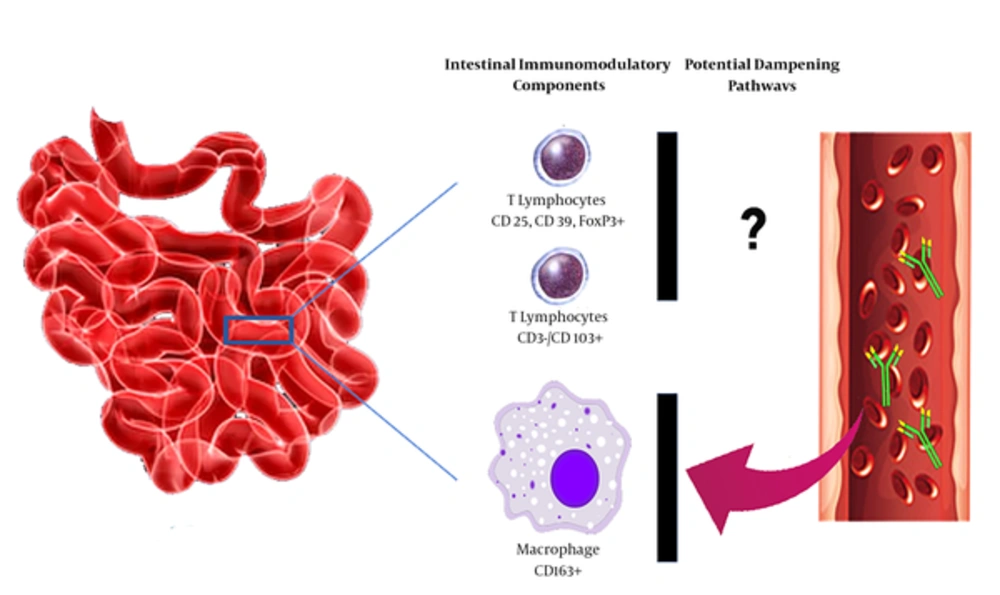
Gluten - induced auto - reactive antibodies and cell mediated cytotoxicity orchestrate the main pathological events in CD (2). Of the all intraepithelial T lymphocytes (IELs) in patients with concurrent CD and T1D, nearly 12.5% have shown CD 25+, CD 39+, and Forkhead box P3 (FoxP3) + T regulatory phenotype (3, 4). Another characterized regulatory lymphocytic population in children with concurrent CD and T1D is CD3-/CD103+ cells, which further highlights the pivotal role of immunoregulation in the development of CD in the context of T1D (5).

This higher regulatory function, however, seems to be functionally incompetent to prevent tissue damage in CD (4). Depressed local immunoregulatory function may be in part due to decreased activity of regulatory intestinal macrophages (CD163+). Some unspecific antibodies have been identified in patients with concurrent CD and T1D to represent binding specificity to these macrophages facilitating tissue damage by depleting these cells (Figure 1) (6). The role of immunomodulatory and inflammatory mediators in progress of CD in the context of T1D needs further evaluations.

Potential intestinal immunomodulatory components executed in patients with T1D and CD. Suppression of CD163+ macrophages by high - affinity auto - reactive antibodies can suppress activity of these cells. Incompetent immunoregulation can result in higher level intestinal mucosa damage in patients with concurrent T1D and CD. Abbreviations; T1D; type 1 diabetes, CD; celiac disease.

3.2. Clinical Features of CD in Children with T1D

Isolated childhood CD presents with malnutrition and malabsorption, vitamin deficiencies, iron deficiency anemia, growth failure, short statue, diarrhea, anorexia, constipation, nausea, and abdominal distention. These clinical features can help in better identification of CD in the context of T1D. However, gastrointestinal symptoms could be very mild in T1D patients with CD, it can hinder the growth in affected children (7). Growth failure and malabsorption have been suggested as well representatives of possible CD in the context of pediatric T1D (8). In those children who present none of the classic signs of CD, the diagnosis is amenable using serological assessments.

3.3. Screening of CD in Children with T1D

3.3.1. Recommended Intervals

Although routine screening for CD has been recommended in T1D, there is no consensus on the appropriate intervals for performing such tests. The American Diabetes Association and the International Society for Pediatric and Adolescent Diabetes recommended CD screening at the time of diabetes diagnosis (9, 10). In the follow up, however, the American Society has proposed performing screening tests whenever suggesting symptoms are evident. On the other hand, the International Society has suggested the screening program to be carried out annually for the first five years and biannually thereafter (10). The European and North American Societies for pediatric gastroenterology, hepatology, and nutrition have noted the ages of 2 and 3 years old, respectively, as the recommended time points in which the first CD screening tests should be performed in children with T1D (11, 12). For following up, however, the recent two societies have stated no specific requirements (11, 12).

3.4. Screening Biomarkers

There is a rough estimation by Australian Gastroenterology Society on the rate of undiagnosed CD ranging from 75% - 83% (13). Currently, the screening tests fall into either serological evaluation, biopsy examination, and human leukocyte antigen (HLA) typing.

3.5. Serological Markers of CD

The most commonly used serological tests designed to detect gluten induced autoantibodies include anti - gliadin (anti - GA), anti - endomysial (anti - EM), anti - transglutaminase (anti - tTG), and anti - deamidated gliadin peptide (anti - DGP) antibodies (1). The most sensitive and specific serological tests for CD include IgA antibodies against either tTG or EMA, and both IgA and IgG antibodies for DGP (14). It seems that detection of IgG isotypes of anti - tTG or anti - DGP does not augment sensitivity for CD diagnosis in the conditions with high titer IgA isotypes (15). Overall, serologic tests are useful to exclude CD in children with T1D with a negative predictive value of 98% (16).

Anti - tTG antibody is the most commonly ordered test for screening CD recommended by American, European, British, and North American societies for both diagnosis and follow up purposes (10-12, 17). Anti - tTG rendered a 90% positive predictive value, which was correlated with the antibody titer with higher titers correlating with higher predictive values (15, 18). In addition, Elitsur et al., noted that anti - tTG antibody titer is three times higher than upper limits is not a good representative of CD in children (19). Anti - tTG antibodies have been reported in a close relationship with HLA-DQB1*02 (20) and HLA - DQ2.5 (21).

IgA anti - EM antibodies are also highly specific for diagnosis of CD associated with childhood T1D (22). IgG1 isotypes of anti - EMA have been strongly associated with biopsy diagnosed CD in T1D (23). These antibodies may also be used as predicting factors for future CD development (24). On the other hand, anti - GA antibodies can be useful for diagnosis of late - onset CD in high risk populations (24). Some new serological markers have been proposed such as antibodies against serum albumin (25) and reticulin (26), which may be useful in screening the progression of CD in at risk populations.

3.6. Intestinal Biopsy Examination

Diabetic children with positive CD serology may demonstrate different histopathological features as compared with their non - diabetic counterparts. Intestinal histological changes in children with concurrent CD and T1D showed erythematous alternations resembling reflux esophagitis, which was different from the pattern seen in isolated CD (27). Intestinal biopsy, as the gold standard, could assist in CD diagnosis in asymptomatic cases.

3.7. HLA Typing

HLA - DQ2 has been mapped in 95% of patients with CD (28, 29), while HLA - DQ8 is seen in the remained 5% (30). HLA - DQ inheritance has been observed in one - third of patients with T1D (28). Accordingly, HLADQ2 comprised the most common allele identified in children with co - existence of CD and T1D (31). In fact, the inheritance of HLA - DQ is one of the main predisposing factor for development of CD in childhood T1D (32). The most significant association between HLA haplotypes and concurrent CD and T1D has been noted for HLA - DR3 - DQ2 and DR4 - DQ8 combinations, as well as presence of DQB1*06:02 allele (33). The role of HLA molecules, also known as Class II major histocompatibility complex (MHCII) - falls into their gliadin - derived antigens presenting activity to CD4+ lymphocytes (34, 35).

HLA typing has been recommended as the screening test for CD in high risk children by the British (17) and European (11) societies for pediatric gastroenterology, hepatology, and nutrition. Screening for HLA - DQ2/DQ8 alleles is recommended as a reliable negative predictor of CD in individuals with suggesting symptoms, as well as at risk populations such as T1D (36, 37). Despite these notions, using HLA - DQ2/DQ8 typing as a routine screening test is hindered owing to their high penetrance in general population, which limits their positive predictive values (38). Furthermore, applicability of this approach for screening CD in populations with low penetrance of these alleles is not recommended as results in high rate of undiagnosed cases (39).

3.8. Non - HLA Genetic Risk Factors of CD in T1D

According to a Genome - wide association study (GWAS), CD susceptibility is modified by at least 70 potential genes mapped within 42 non - HLA loci (40). Seven shared genetic loci between CD and T1D have been addressed by Smyth et al., (41). The list of shared genetic loci between CD and T1D are likely to be increased by more comprehensive studies. Some other genetic polymorphisms that have been associated with an elevated risk of CD in the context of T1D include rs10754558 and rs358294199 SNPs of NLRP3 (42), polymorphisms in IL - 6 (43, 44), CD14 (45, 46), and IL12A/SCHIP, CCR1, CCR2, CCr3, LPP, IL-17 (44, 47-49).

3.9. New Potential Biomarkers for Diagnosis and Screening CD

Some new potential markers for diagnosis of CD include Ischemia - modified albumin (IMA), Soluble Syndecan - 1 (SSDC1), Regenerating gene Iα (REGIα), Neurotensin (NT), Zonulin, and n - 3 polyunsaturated fatty acids (n - 3 PUFA). IMA is a metal - binding modified albumin and a marker of oxidative stress. Clinical significance of elevated levels of IMA has been evaluated in some disorders such as ischemic heart disorders, thalassemia, renal failure, and diabetes (50-52). SDC1 roles as a mediator derived from intestinal mucosa glycocalyx involved in maintaining permeability of intestinal epithelium. The role of SDC1 in regulating intestinal permeability makes it a potential marker with a central role in pathogenesis of CD (53-55). REGIα is a molecule with a substantial role in tissue remodeling processes that its levels have been high in active CD (56, 57). NT is an intestinal hormone synthesized and released by specialized cells located mainly in jejunum and ileum of small intestine (58). NT can promote inflammatory responses through inducing nuclear factor kappa B (NF ‐ κB) signaling pathway and production of proinflammatory cytokines (59, 60). Zonulin is another mediator directly involved in regulation of intestinal permeability by dissociating tight junction complexes (61-63). Belonging to n - 3 long - chain polyunsaturated fatty acids (n - 3 LCPUFA) family, n - 3 PUFA and its abnormal abdominal absorption in CD could pave the route for recruiting the molecule as a diagnostic marker in this condition (64). Table 1 has summarized studies assessing diagnostic applicability of these markers.

Table 1.

Newly Introduced Markers for Detecting Celiac Disease in the Context of Type 1 Diabetes

BiomarkersAuthors, YearPopulationImplicationsRef
IMAYuksel et al., 201765 patients with CD and 65 healthy controlIMA levels were higher in celiac patients respective to controls. Among CD patients, those with positive serological test had significantly higher IMA levels(54)
Sayar et al, 201537 children with CD and 29 healthy controlsIMA levels were significantly higher in patients. IMA level showed moderately positive correlation with t - TGA and Marsh Score. The level of IMA dropped significantly following a GFD.(55)
SD - 1 Yablecovitch et al., 201749 children with CD and no history of treatment, 15 children with abdominal pain and no evidences of CDThe levels of soluble SD - 1 were significantly higher in CD children at the time of diagnosis compared to controls. The levels were significantly correlated with the degree of villous inflammation(53)
REGIa Planas et al., 201140 patients with CD, 35 healthy controlsREGIa showed significantly elevated levels compared to control subjects. The levels decreased following GFD which was correlated with reduction in tTG antibodies. I comparison, those patients with T1D showed normal levels of REG Ia.(57)
ZonulinVorobjova et al., 201740 subjects with CD, and 40 individuals with small intestine functional syplasia as controlSerum zonulin levels were significantly higher in CD patients and were associated with Marsh score III. This correlation was prominent in CD patients with concurrent T1D.(63)
Pro NTMonten et al., 201696 children with CD and 89 healthy controlsThe plasma level of pro - NT was higher in CD compared to non - diseased cases. The levels were correlated with intestinal damage degree, and anti - tTG antibodies(60)
n - 3 PUFATarnok et al., 201520 children with CD, 8 children with coexistence of CD and T1D, and 21 healthy childrenThe plasma levels of docosapentaenoic, docosahexaenoic, n - 3 PUFA, and long chains (n - 3 LCPUFA) were significantly lower in CD+T1D group compared to CD alone or controls.(64)

3.10. Prognostic Impacts of CD on Childhood T1D

Abnormal lipid profile such as decreased HDL - C and elevated LDL - C in patients with concurrent CD and T1D has been noted, which may contribute to the higher risk of cardiovascular incidents in these patients (65). CD can also exaggerate complications such as nephropathy, retinopathy, as well as other auto immune disorders in children with T1D (37). CD may contribute to lower bone density, renal insufficiency, and quality of life of affected children and adults with T1D (66-68). Both Vitamin D deficiency (69) and antibodies against bone regulatory hormone - osteoprotegerin - participate in lower than optimal bone density in patients with CD (70). CD may also increase the risk of gastrointestinal tumors (71).

On the other hand, neither nutritional status, nor life quality of children with concurrent CD and T1D were different compared to individuals with T1D alone (72). In another study, patients with T1D who developed CD revealed no increase in complication rate in comparison to their counterparts without CD (73, 74). Accordingly, diagnosis of CD in the context of T1D showed no significant impact on HbA1c levels in another study (75). However, Leeds et al., argued that children with T1D associated with active CD or high levels of anti - tTG antibodies have a higher risk for diabetes associated complications (76). In general, timely diagnosis and management of CD in the context of T1D could protect patients from reduction in bone density and anemia, as well as deterioration in gastrointestinal functions, and provide them with a better quality of life.

4. Conclusions

Immune cytotoxic reactions with dampen immune regulatory functions can contribute to CD pathogenies in the context of T1D. The most common screening tests for CD include anti - GA, anti - EM, anti - tTG, and anti - DG antibodies. Typing for HLA of DQ - 2 and DQ - 8 can assist in diagnosis of silent CD in children with T1D. Newly proposed biomarkers of CD including IMA, SSDC1, REG Iα, pro - NT, Zonulin, and n - 3 PUFA can be used for diagnosis and screening CD in childhood T1D. Overall, active seropositive CD seems to be of clinical importance in T1D with significant impacts on the quality of life of the patients and occurrence of diabetes associated complications. Adherence to GFD is recommended in T1D children with active or silent CD to ameliorate related complications.

References

  • 1.

    Shahramian I, Dehghani SM, Haghighat M, Noori NM, Teimouri AR, Sharafi E, et al. Serologic evaluation of celiac disease in patients with beta thalassemia major and control. Gastroenterol Hepatol Bed Bench. 2015;8(2):153-9. [PubMed ID: 25926941]. [PubMed Central ID: PMC4403028].

  • 2.

    Kivling A, Nilsson L, Falth-Magnusson K, Sollvander S, Johanson C, Faresjo M. Diverse foxp3 expression in children with type 1 diabetes and celiac disease. Ann N Y Acad Sci. 2008;1150:273-7. [PubMed ID: 19120312]. https://doi.org/10.1196/annals.1447.018.

  • 3.

    Cook L, Munier CML, Seddiki N, van Bockel D, Ontiveros N, Hardy MY, et al. Circulating gluten-specific FOXP3(+)CD39(+) regulatory T cells have impaired suppressive function in patients with celiac disease. J Allergy Clin Immunol. 2017;140(6):1592-1603 e8. [PubMed ID: 28283419]. https://doi.org/10.1016/j.jaci.2017.02.015.

  • 4.

    Uibo R, Panarina M, Teesalu K, Talja I, Sepp E, Utt M, et al. Celiac disease in patients with type 1 diabetes: a condition with distinct changes in intestinal immunity? Cell Mol Immunol. 2011;8(2):150-6. [PubMed ID: 21317917]. [PubMed Central ID: PMC4003136]. https://doi.org/10.1038/cmi.2010.66.

  • 5.

    Valle J, Morgado JMT, Ruiz-Martin J, Guardiola A, Lopes-Nogueras M, Garcia-Vela A, et al. Flow cytometry of duodenal intraepithelial lymphocytes improves diagnosis of celiac disease in difficult cases. United European Gastroenterol J. 2017;5(6):819-26. [PubMed ID: 29026596]. [PubMed Central ID: PMC5625870]. https://doi.org/10.1177/2050640616682181.

  • 6.

    Sonier B, Strom A, Wang GS, Patrick C, Crookshank JA, Mojibian M, et al. Antibodies from a patient with type 1 diabetes and celiac disease bind to macrophages that express the scavenger receptor CD163. Can J Gastroenterol. 2011;25(6):327-9. [PubMed ID: 21766094]. [PubMed Central ID: PMC3142606].

  • 7.

    Cohn A, Sofia AM, Kupfer SS. Type 1 diabetes and celiac disease: clinical overlap and new insights into disease pathogenesis. Curr Diab Rep. 2014;14(8):517. [PubMed ID: 24952108]. [PubMed Central ID: PMC4156157]. https://doi.org/10.1007/s11892-014-0517-x.

  • 8.

    Holmes GK. Screening for coeliac disease in type 1 diabetes. Arch Dis Child. 2002;87(6):495-8. [PubMed ID: 12456547]. [PubMed Central ID: PMC1755832].

  • 9.

    American Diabetes Association. 12. Children and Adolescents: Standards of Medical Care in Diabetes-2018. Diabetes Care. 2018;41(Suppl 1):S126-36. [PubMed ID: 29222383]. https://doi.org/10.2337/dc18-S012.

  • 10.

    Couper JJ, Haller MJ, Ziegler AG, Knip M, Ludvigsson J, Craig ME, et al. ISPAD Clinical Practice Consensus Guidelines 2014. Phases of type 1 diabetes in children and adolescents. Pediatr Diabetes. 2014;15 Suppl 20:18-25. [PubMed ID: 25325095].

  • 11.

    Husby S, Koletzko S, Korponay-Szabo IR, Mearin ML, Phillips A, Shamir R, et al. European Society for Pediatric Gastroenterology, Hepatology, and Nutrition guidelines for the diagnosis of coeliac disease. J Pediatr Gastroenterol Nutr. 2012;54(1):136-60. [PubMed ID: 22197856]. https://doi.org/10.1097/MPG.0b013e31821a23d0.

  • 12.

    Hill ID, Dirks MH, Liptak GS, Colletti RB, Fasano A, Guandalini S, et al. Guideline for the diagnosis and treatment of celiac disease in children: recommendations of the North American Society for Pediatric Gastroenterology, Hepatology and Nutrition. J Pediatr Gastroenterol Nutr. 2005;40(1):1-19. [PubMed ID: 15625418].

  • 13.

    Rubio-Tapia A, Ludvigsson JF, Brantner TL, Murray JA, Everhart JE. The prevalence of celiac disease in the United States. Am J Gastroenterol. 2012;107(10):1538-44. quiz 1537, 1545. [PubMed ID: 22850429]. https://doi.org/10.1038/ajg.2012.219.

  • 14.

    Waisbourd-Zinman O, Hojsak I, Rosenbach Y, Mozer-Glassberg Y, Shalitin S, Phillip M, et al. Spontaneous normalization of anti-tissue transglutaminase antibody levels is common in children with type 1 diabetes mellitus. Dig Dis Sci. 2012;57(5):1314-20. [PubMed ID: 22173747]. https://doi.org/10.1007/s10620-011-2016-0.

  • 15.

    Ermarth A, Bryce M, Woodward S, Stoddard G, Book L, Jensen MK. Identification of Pediatric Patients With Celiac Disease Based on Serology and a Classification and Regression Tree Analysis. Clin Gastroenterol Hepatol. 2017;15(3):396-402 e2. [PubMed ID: 27847281]. [PubMed Central ID: PMC5316297]. https://doi.org/10.1016/j.cgh.2016.10.035.

  • 16.

    Bannister EG, Cameron DJ, Ng J, Chow CW, Oliver MR, Alex G, et al. Can celiac serology alone be used as a marker of duodenal mucosal recovery in children with celiac disease on a gluten-free diet? Am J Gastroenterol. 2014;109(9):1478-83. [PubMed ID: 25070050]. https://doi.org/10.1038/ajg.2014.200.

  • 17.

    Murch S, Jenkins H, Auth M, Bremner R, Butt A, France S, et al. Joint BSPGHAN and Coeliac UK guidelines for the diagnosis and management of coeliac disease in children. Arch Dis Child. 2013;98(10):806-11. [PubMed ID: 23986560]. https://doi.org/10.1136/archdischild-2013-303996.

  • 18.

    Jora R, Raghuvanshi V, Payal V, Sharma P, Vishnoi SK. Correlation of Tissue Transglutaminase with Modified Marsh Grading in Celiac Disease: A Prospective Cohort Study. Indian J Pediatr. 2017;84(7):515-20. [PubMed ID: 28321610]. https://doi.org/10.1007/s12098-017-2323-3.

  • 19.

    Elitsur Y, Sigman T, Watkins R, Porto AF, Leonard Puppa EL, Foglio EJ, et al. Tissue Transglutaminase Levels Are Not Sufficient to Diagnose Celiac Disease in North American Practices Without Intestinal Biopsies. Dig Dis Sci. 2017;62(1):175-9. [PubMed ID: 27778203]. https://doi.org/10.1007/s10620-016-4354-4.

  • 20.

    Agardh D, Nilsson A, Tuomi T, Lindberg B, Carlsson AK, Lernmark A, et al. Prediction of silent celiac disease at diagnosis of childhood type 1 diabetes by tissue transglutaminase autoantibodies and HLA. Pediatr Diabetes. 2001;2(2):58-65. [PubMed ID: 15016199]. https://doi.org/10.1034/j.1399-5448.2001.002002058.x.

  • 21.

    Arregui MV, Urmeneta JM, Brito HL, De Esteban JP, Martinez CP, Llenas LF, et al. The role of flow cytometry in celiac disease screening using human leukocyte antigen in adult patients with type 1 diabetes mellitus. Ann Gastroenterol. 2017;30(2):179-85. [PubMed ID: 28243038]. [PubMed Central ID: PMC5320030]. https://doi.org/10.20524/aog.2016.0113.

  • 22.

    Dretzke J, Cummins C, Sandercock J, Fry-Smith A, Barrett T, Burls A. Autoantibody testing in children with newly diagnosed type 1 diabetes mellitus. Health Technol Assess. 2004;8(22):iii-xi. 1-183. [PubMed ID: 15191683].

  • 23.

    Picarelli A, Sabbatella L, Di Tola M, Vetrano S, Casale C, Anania MC, et al. Anti-endomysial antibody of IgG1 isotype detection strongly increases the prevalence of coeliac disease in patients affected by type I diabetes mellitus. Clin Exp Immunol. 2005;142(1):111-5. [PubMed ID: 16178863]. [PubMed Central ID: PMC1809494]. https://doi.org/10.1111/j.1365-2249.2005.02866.x.

  • 24.

    Grodzinsky E, Falth-Magnusson K, Hogberg L, Jansson G, Laurin P, Stenhammar L. IgA endomysium antibodies--an early predictor for celiac disease in children without villous atrophy. Acta Paediatr. 2008;97(7):972-6. [PubMed ID: 18489624]. https://doi.org/10.1111/j.1651-2227.2008.00881.x.

  • 25.

    Rodriguez-Juan C, Sala-Silveira L, Perez-Blas M, Valeri AP, Aguilera N, Lopez-Santalla M, et al. Increased levels of bovine serum albumin antibodies in patients with type 1 diabetes and celiac disease-related antibodies. J Pediatr Gastroenterol Nutr. 2003;37(2):132-5. [PubMed ID: 12883297].

  • 26.

    Maki M, Huupponen T, Holm K, Hallstrom O. Seroconversion of reticulin autoantibodies predicts coeliac disease in insulin dependent diabetes mellitus. Gut. 1995;36(2):239-42. [PubMed ID: 7883223]. [PubMed Central ID: PMC1382410].

  • 27.

    Tanpowpong P, Broder-Fingert S, Katz AJ, Camargo CJ. Features of children with positive celiac serology and type 1 diabetes mellitus. Pediatr Int. 2015;57(5):1028-30. [PubMed ID: 26508189]. https://doi.org/10.1111/ped.12766.

  • 28.

    Ludvigsson JF, Ludvigsson J, Ekbom A, Montgomery SM. Celiac disease and risk of subsequent type 1 diabetes: a general population cohort study of children and adolescents. Diabetes Care. 2006;29(11):2483-8. [PubMed ID: 17065689]. https://doi.org/10.2337/dc06-0794.

  • 29.

    Tye-Din JA, Cameron DJ, Daveson AJ, Day AS, Dellsperger P, Hogan C, et al. Appropriate clinical use of human leukocyte antigen typing for coeliac disease: an Australasian perspective. Intern Med J. 2015;45(4):441-50. [PubMed ID: 25827511]. [PubMed Central ID: PMC4405087]. https://doi.org/10.1111/imj.12716.

  • 30.

    Kaur A, Shimoni O, Wallach M. Celiac disease: from etiological factors to evolving diagnostic approaches. J Gastroenterol. 2017;52(9):1001-12. [PubMed ID: 28631048]. https://doi.org/10.1007/s00535-017-1357-7.

  • 31.

    Ciacci C, Zingone F. Dietary gluten and the development of celiac disease and type 1 diabetes. Nutr Diet Suppl. 2016;8:51-6. https://doi.org/10.2147/nds.s74713.

  • 32.

    Adlercreutz EH, Svensson J, Hansen D, Buschard K, Lernmark A, Mortensen HB, et al. Prevalence of celiac disease autoimmunity in children with type 1 diabetes: regional variations across the Oresund strait between Denmark and southernmost Sweden. Pediatr Diabetes. 2015;16(7):504-9. [PubMed ID: 25131687]. https://doi.org/10.1111/pedi.12200.

  • 33.

    Lernmark A. Environmental factors in the etiology of type 1 diabetes, celiac disease, and narcolepsy. Pediatr Diabetes. 2016;17 Suppl 22:65-72. [PubMed ID: 27411439]. [PubMed Central ID: PMC5473290]. https://doi.org/10.1111/pedi.12390.

  • 34.

    Liu E, McDaniel K, Case S, Yu L, Gerhartz B, Ostermann N, et al. Exploring T cell reactivity to gliadin in young children with newly diagnosed celiac disease. Autoimmune Dis. 2014;2014:927190. [PubMed ID: 24724018]. [PubMed Central ID: PMC3958769]. https://doi.org/10.1155/2014/927190.

  • 35.

    Lundin KE, Scott H, Hansen T, Paulsen G, Halstensen TS, Fausa O, et al. Gliadin-specific, HLA-DQ(alpha 1*0501,beta 1*0201) restricted T cells isolated from the small intestinal mucosa of celiac disease patients. J Exp Med. 1993;178(1):187-96. [PubMed ID: 8315377]. [PubMed Central ID: PMC2191064].

  • 36.

    Diaz-Redondo A, Miranda-Bautista J, Garcia-Lledo J, Gisbert JP, Menchen L. The potential usefulness of human leukocyte antigen typing for celiac disease screening: A systematic review and meta-analysis. Rev Esp Enferm Dig. 2015;107(7):423-9. [PubMed ID: 26140635].

  • 37.

    Weiss B, Pinhas-Hamiel O. Celiac Disease and Diabetes: When to Test and Treat. J Pediatr Gastroenterol Nutr. 2017;64(2):175-9. [PubMed ID: 27574884]. https://doi.org/10.1097/MPG.0000000000001388.

  • 38.

    Hadithi M, von Blomberg BM, Crusius JB, Bloemena E, Kostense PJ, Meijer JW, et al. Accuracy of serologic tests and HLA-DQ typing for diagnosing celiac disease. Ann Intern Med. 2007;147(5):294-302. [PubMed ID: 17785484].

  • 39.

    Fukunaga M, Ishimura N, Fukuyama C, Izumi D, Ishikawa N, Araki A, et al. Celiac disease in non-clinical populations of Japan. J Gastroenterol. 2018;53(2):208-14. [PubMed ID: 28389733]. https://doi.org/10.1007/s00535-017-1339-9.

  • 40.

    Dubois PC, Trynka G, Franke L, Hunt KA, Romanos J, Curtotti A, et al. Multiple common variants for celiac disease influencing immune gene expression. Nat Genet. 2010;42(4):295-302. [PubMed ID: 20190752]. [PubMed Central ID: PMC2847618]. https://doi.org/10.1038/ng.543.

  • 41.

    Smyth DJ, Plagnol V, Walker NM, Cooper JD, Downes K, Yang JH, et al. Shared and distinct genetic variants in type 1 diabetes and celiac disease. N Engl J Med. 2008;359(26):2767-77. [PubMed ID: 19073967]. [PubMed Central ID: PMC2840835]. https://doi.org/10.1056/NEJMoa0807917.

  • 42.

    Pontillo A, Brandao L, Guimaraes R, Segat L, Araujo J, Crovella S. Two SNPs in NLRP3 gene are involved in the predisposition to type-1 diabetes and celiac disease in a pediatric population from northeast Brazil. Autoimmunity. 2010;43(8):583-9. [PubMed ID: 20370570]. https://doi.org/10.3109/08916930903540432.

  • 43.

    Mysliwiec M, Mysliwska J, Zorena K, Balcerska A, Malinowska E, Wisniewski P. Interleukin 6 -174(G>C) gene polymorphism is related to celiac disease and autoimmune thyroiditis coincidence in diabetes type 1 children. Diabetes Res Clin Pract. 2008;82(1):108-12. [PubMed ID: 18692934]. https://doi.org/10.1016/j.diabres.2008.07.004.

  • 44.

    Akbulut UE, Cebi AH, Sag E, Ikbal M, Cakir M. Interleukin-6 and interleukin-17 gene polymorphism association with celiac disease in children. Turk J Gastroenterol. 2017;28(6):471-5. [PubMed ID: 28928101]. https://doi.org/10.5152/tjg.2017.17092.

  • 45.

    Dezsofi A, Szebeni B, Hermann CS, Kapitany A, Veres G, Sipka S, et al. Frequencies of genetic polymorphisms of TLR4 and CD14 and of HLA-DQ genotypes in children with celiac disease, type 1 diabetes mellitus, or both. J Pediatr Gastroenterol Nutr. 2008;47(3):283-7. [PubMed ID: 18728522]. https://doi.org/10.1097/MPG.0b013e31816de885.

  • 46.

    Catamo E, Segat L, Lenarduzzi S, Petix V, Morgutti M, Crovella S. CD14 polymorphisms correlate with an augmented risk for celiac disease in Italian patients. Genes Immun. 2012;13(6):489-95. [PubMed ID: 22648004]. https://doi.org/10.1038/gene.2012.23.

  • 47.

    Amundsen SS, Rundberg J, Adamovic S, Gudjonsdottir AH, Ascher H, Ek J, et al. Four novel coeliac disease regions replicated in an association study of a Swedish-Norwegian family cohort. Genes Immun. 2010;11(1):79-86. [PubMed ID: 19693089]. https://doi.org/10.1038/gene.2009.67.

  • 48.

    Romanos J, Barisani D, Trynka G, Zhernakova A, Bardella MT, Wijmenga C. Six new coeliac disease loci replicated in an Italian population confirm association with coeliac disease. J Med Genet. 2009;46(1):60-3. [PubMed ID: 18805825]. https://doi.org/10.1136/jmg.2008.061457.

  • 49.

    Guo CC, Wang M, Cao FD, Huang WH, Xiao D, Ye XG, et al. Meta-Analysis on Associations of RGS1 and IL12A Polymorphisms with Celiac Disease Risk. Int J Mol Sci. 2016;17(4):457. [PubMed ID: 27043536]. [PubMed Central ID: PMC4848913]. https://doi.org/10.3390/ijms17040457.

  • 50.

    Sbarouni E, Georgiadou P, Voudris V. Ischemia modified albumin changes - review and clinical implications. Clin Chem Lab Med. 2011;49(2):177-84. [PubMed ID: 21083441]. https://doi.org/10.1515/CCLM.2011.037.

  • 51.

    Cichota LC, Moresco RN, Duarte MM, da Silva JE. Evaluation of ischemia-modified albumin in anemia associated to chronic kidney disease. J Clin Lab Anal. 2008;22(1):1-5. [PubMed ID: 18200583]. https://doi.org/10.1002/jcla.20226.

  • 52.

    Awadallah SM, Atoum MF, Nimer NA, Saleh SA. Ischemia modified albumin: an oxidative stress marker in beta-thalassemia major. Clin Chim Acta. 2012;413(9-10):907-10. [PubMed ID: 22336636]. https://doi.org/10.1016/j.cca.2012.01.037.

  • 53.

    Yablecovitch D, Oren A, Ben-Horin S, Fudim E, Eliakim R, Saker T, et al. Soluble Syndecan-1: A Novel Biomarker of Small Bowel Mucosal Damage in Children with Celiac Disease. Dig Dis Sci. 2017;62(3):755-60. [PubMed ID: 28025744]. https://doi.org/10.1007/s10620-016-4415-8.

  • 54.

    Yuksel M, Kaplan M, Ates I, Ozin YO, Kilic H, Kuzu UB, et al. The relation between ischemia modified albumin level and autoimmunity/chronic inflammation in celiac disease. Turkish J Biochem. 2017;42(3). https://doi.org/10.1515/tjb-2016-0296.

  • 55.

    Sayar E, Ozdem S, Uzun G, Islek A, Yilmaz A, Artan R. Total oxidant status, total antioxidant capacity and ischemia modified albumin levels in children with celiac disease. Turk J Pediatr. 2015;57(5):498-503. [PubMed ID: 27411418].

  • 56.

    Brusca I. Overview of Biomarkers for Diagnosis and Monitoring of Celiac Disease. Adv Clin Chem. 2015;68:1-55. https://doi.org/10.1016/bs.acc.2014.12.006.

  • 57.

    Planas R, Pujol-Autonell I, Ruiz E, Montraveta M, Cabre E, Lucas-Martin A, et al. Regenerating gene Ialpha is a biomarker for diagnosis and monitoring of celiac disease: a preliminary study. Transl Res. 2011;158(3):140-5. [PubMed ID: 21867979]. https://doi.org/10.1016/j.trsl.2011.04.004.

  • 58.

    Egerod KL, Engelstoft MS, Grunddal KV, Nohr MK, Secher A, Sakata I, et al. A major lineage of enteroendocrine cells coexpress CCK, secretin, GIP, GLP-1, PYY, and neurotensin but not somatostatin. Endocrinology. 2012;153(12):5782-95. [PubMed ID: 23064014]. https://doi.org/10.1210/en.2012-1595.

  • 59.

    Zhao D, Pothoulakis C. Effects of NT on gastrointestinal motility and secretion, and role in intestinal inflammation. Peptides. 2006;27(10):2434-44. [PubMed ID: 16872719]. https://doi.org/10.1016/j.peptides.2005.12.016.

  • 60.

    Monten C, Torinsson Naluai A, Agardh D. Role of proneurotensin as marker of paediatric coeliac disease. Clin Exp Immunol. 2016;186(3):387-92. [PubMed ID: 27612962]. [PubMed Central ID: PMC5108069]. https://doi.org/10.1111/cei.12864.

  • 61.

    Fasano A. Zonulin and its regulation of intestinal barrier function: the biological door to inflammation, autoimmunity, and cancer. Physiol Rev. 2011;91(1):151-75. [PubMed ID: 21248165]. https://doi.org/10.1152/physrev.00003.2008.

  • 62.

    Fasano A. Zonulin, regulation of tight junctions, and autoimmune diseases. Ann N Y Acad Sci. 2012;1258:25-33. [PubMed ID: 22731712]. [PubMed Central ID: PMC3384703]. https://doi.org/10.1111/j.1749-6632.2012.06538.x.

  • 63.

    Vorobjova T, Raikkerus H, Kadaja L, Talja I, Uibo O, Heilman K, et al. Circulating Zonulin Correlates with Density of Enteroviruses and Tolerogenic Dendritic Cells in the Small Bowel Mucosa of Celiac Disease Patients. Dig Dis Sci. 2017;62(2):358-71. [PubMed ID: 27995404]. https://doi.org/10.1007/s10620-016-4403-z.

  • 64.

    Tarnok A, Marosvolgyi T, Szabo E, Gyorei E, Decsi T. Low n-3 long-chain polyunsaturated fatty acids in newly diagnosed celiac disease in children with preexisting type 1 diabetes mellitus. J Pediatr Gastroenterol Nutr. 2015;60(2):255-8. [PubMed ID: 25207475]. https://doi.org/10.1097/MPG.0000000000000561.

  • 65.

    Salardi S, Maltoni G, Zucchini S, Iafusco D, Zanfardino A, Confetto S, et al. Whole lipid profile and not only HDL cholesterol is impaired in children with coexisting type 1 diabetes and untreated celiac disease. Acta Diabetol. 2017;54(10):889-94. [PubMed ID: 28639064]. https://doi.org/10.1007/s00592-017-1019-5.

  • 66.

    Simmons KM, McFann K, Taki I, Liu E, Klingensmith GJ, Rewers MJ, et al. Reduced Bone Mineral Density Is Associated with Celiac Disease Autoimmunity in Children with Type 1 Diabetes. J Pediatr. 2016;169:44-8 e1. [PubMed ID: 26561381]. [PubMed Central ID: PMC4849876]. https://doi.org/10.1016/j.jpeds.2015.10.024.

  • 67.

    Bakker SF, Tushuizen ME, von Blomberg BM, Bontkes HJ, Mulder CJ, Simsek S. Screening for coeliac disease in adult patients with type 1 diabetes mellitus: myths, facts and controversy. Diabetol Metab Syndr. 2016;8:51. [PubMed ID: 27478507]. [PubMed Central ID: PMC4966870]. https://doi.org/10.1186/s13098-016-0166-0.

  • 68.

    Mollazadegan K, Fored M, Lundberg S, Ludvigsson J, Ekbom A, Montgomery SM, et al. Risk of renal disease in patients with both type 1 diabetes and coeliac disease. Diabetologia. 2014;57(7):1339-45. [PubMed ID: 24663809]. https://doi.org/10.1007/s00125-014-3223-y.

  • 69.

    Topal E, Catal F, Yildirim Acar N, Ermistekin H, Sinanoglu MS, Karabiber H, et al. Vitamin and mineral deficiency in children newly diagnosed with celiac disease. Turkish J Med Sci. 2015;45:833-6. https://doi.org/10.3906/sag-1408-94.

  • 70.

    Real A, Gilbert N, Hauser B, Kennedy N, Shand A, Gillett H, et al. Characterisation of osteoprotegerin autoantibodies in coeliac disease. Calcif Tissue Int. 2015;97(2):125-33. [PubMed ID: 26092508]. https://doi.org/10.1007/s00223-015-0023-4.

  • 71.

    Naderi M, Shahramian I, Delaramnasab M, Bazi A. Coincidence of celiac disease with nongastrointestinal tumors in children. Pediatr Hematol Oncol. 2017;34(8):478-82. [PubMed ID: 29219666]. https://doi.org/10.1080/08880018.2017.1404171.

  • 72.

    Nunes-Silva JG, Nunes VS, Schwartz RP, Mlss Trecco S, Evazian D, Correa-Giannella ML, et al. Impact of type 1 diabetes mellitus and celiac disease on nutrition and quality of life. Nutr Diabetes. 2017;7(1). e239. [PubMed ID: 28067892]. [PubMed Central ID: PMC5301040]. https://doi.org/10.1038/nutd.2016.43.

  • 73.

    Pham-Short A, Donaghue KC, Ambler G, Chan AK, Hing S, Cusumano J, et al. Early elevation of albumin excretion rate is associated with poor gluten-free diet adherence in young people with coeliac disease and diabetes. Diabet Med. 2014;31(2):208-12. [PubMed ID: 24117620]. https://doi.org/10.1111/dme.12329.

  • 74.

    Reilly NR, Lebwohl B, Mollazadegan K, Michaelsson K, Green PH, Ludvigsson JF. Celiac Disease Does Not Influence Fracture Risk in Young Patients with Type 1 Diabetes. J Pediatr. 2016;169:49-54. [PubMed ID: 26589343]. [PubMed Central ID: PMC4729630]. https://doi.org/10.1016/j.jpeds.2015.10.032.

  • 75.

    Mackinder M, Allison G, Svolos V, Buchanan E, Johnston A, Cardigan T, et al. Nutritional status, growth and disease management in children with single and dual diagnosis of type 1 diabetes mellitus and coeliac disease. BMC Gastroenterol. 2014;14:99. [PubMed ID: 24885742]. [PubMed Central ID: PMC4046848]. https://doi.org/10.1186/1471-230X-14-99.

  • 76.

    Leeds JS, Hopper AD, Hadjivassiliou M, Tesfaye S, Sanders DS. Potential coeliac disease in Type 1 diabetes mellitus: does a positive antibody lead to increased complications? Nutr Metab Cardiovasc Dis. 2014;24(4):378-83. [PubMed ID: 24393392]. https://doi.org/10.1016/j.numecd.2013.09.005.