
Fulltext
Extremely low birth weight (ELBW) is defined as a birth weight less than 1000g. Acute kidney failure (AKF) is registered in 6%-24% of critically ill neonates treated at the intensive care units (ICU). The primary causes of AKF in these neonates are prerenal mechanisms (around 85%), which include hypovolemia, hypotension and hypoxemia. A medication therapy is applied first and in case it does not yield the desired results, a renal replacement therapy is applied[1,2].
Peritoneal dialysis (PD) is generally regarded as the optimal dialysis modality for neonates[3]. PD allows for the slow removal of fluid and solutes while avoiding hemodynamic instability. Peritoneal dialysis is a relatively safe, simple and effective procedure, which can be accomplished with a minimum of equipment and for its efficient use a highly trained team is not essential[4]. There are two main types of PD: continuous ambulatory peritoneal dialysis (CAPD) where the fluid exchanges are made manually. Gravity makes the filling and draining process possible. More sophisticated variant of CAPD is continuous cycling peritoneal dialysis (CCPD) commonly referred to as automated peritoneal dialysis (APD) uses an automated machine called cyler to perform fluid exchanges. APD is not used in ELBW so far, because the minimum loading volume that supports this machine is 100 ml, and because of low body weight, filling volume in ELBW neonates are much smaller.
Due to the limited space in the peritoneal cavity in neonates with extremely low birth weight, it is very difficult to place a rigid peritoneal catheter, which is why alternatives are exploited, such as the suction catheter tip, plastic catheter, angiocath, neonatal chest drain, IV cannula, Wallace catheter, Cook 5F catheter, and Pendelburys catheter[3,4]. We present a case report about a new modality of using IV cannula for peritoneal dialysis in ELBW neonate.
First child from first pregnancy (following a ten-year sterility treatment and myomectomy done ten years ago). Due to preeclampsia, an emergency Cesarean section was performed in 27+1 GA, birth weight 470 g, length 29 cm, head circumference 20 cm, Apgar score 2/6. Endotracheal intubation was performed after the labor, accompanied by mechanical ventilation and dual antibiotic therapy, and transported by our team in its first day of life from the city maternity clinic to our Institute for further treatment.
Mechanical ventilation (SIMV/PS) and antibiotic therapy were continued upon admittance. A hypotension treatment was started (bolus crystalloid infusion, followed by inotropic medicaments). Hypotension was refractory to the treatment. A natural surfactant preparation was administered by means of endotracheal insufflations, but even that did not help decrease the mechanical ventilation parameters. Blood derivatives were used as a substitute due to anemia, thrombocythopenia and hypop¬roteinemia.
Due to extreme immaturity, perinatal asphyxia, hypothermia, severe form of respiratory distress syndrome which requires high parameters of mechanical ventilation, hypotension and the onset of neonatal sepsis, the neonate was anuric from the birth and the amount of nitrogen in the blood was gradually increasing. Creatinine 2.32 mg/dl, urea nitrogen 29.13 mg/dl, acidum uricum 609.0 µmol/l.
Kidney ultrasound: both kidneys of normal size, more echogenic parenchyma found bilaterally, no signs of the pyelocaliceal system dilatation, indistinct corticomedullary borders. A con¬ser vative AKF therapy was administered (Henley-loop diuretic, liquid intake restriction, Dopamin 3ug/kg/min). Since the therapy did not result in diuresis and the neonate gained weight (+130g), on the fourth day of hospitalisation we decided to start acute peritoneal dialysis.
Following abdomen skin disinfection and subcutaneous administration of local anesthetic (1% Xylocaine), an IV cannula (24G, flow 18ml/min. KD-FIX, KD Medical GmbH Hospital Products, Berlin, Germany) was placed in the peritoneal cavity in the left part of the umbilical region, using the blind technique. After the cannula insertion, the tissue adhesive, Epiglu (Mayer-Haake GmbH, Wehrheim, Germany) was applied to the entry point (Fig. 1).
A three-way cannula was fixed to the cannula. One end was attached to the baby dose and a bag containing the dialysis liquid, while the other was attached to a drainage bag. Peritoneal dialysis was started – dialysis solution with 1.5% glucose, fill volume10 ml/kg of dialysis solution, dwell time 10 minutes, drain time 10 min. We used prophylactic antibiotic in the dialysate - ceftazidime 500mg/1 of the dialysis liquid. There was no leakage of liquid, bleeding or other mechanical complica¬tions. An ultrafiltration of 6 ml/kg/h was achieved.
Throughout the treatment, the neonate’s condition was very difficult, a multiorgan dysfunction appeared and exitus lethalis occurred 24 hours after initiation of peritoneal dialysis.
Kohli et al compared three types of peritoneal access in infants with acute renal failure. They used an intravenous cannula, stylet catheter and guide wire inserted femoral vein catheter. They concluded that the complications (intraperi¬toneal bleed, catheter blockade and other mechanical complications, peritonitis) were least (20%) frequent when a guide wire inserted femoral vein catheter was used, in comparison with an intravenous cannula (33%) and stylet catheter (66%). In another study, the same authors compared the frequency of complications during the PD in neonates with AKF. One group of neonates were placed a guide wire-inserted
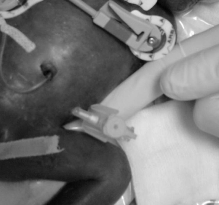
Fig. 1: IV cannula was placed in the peritoneal cavity in the left part of the umbilical
region, using the blind technique. After the cannula insertion, the tissue adhesive, Epiglu was applied to the entry point
femoral vein catheter in the abdominal cavity, while the others were inserted an intravenous cannula. Dialysate leak and catheter blockade were more common with intravenous cannula (23.1% and 61.5%, respectively) then with wire-inserted femoral vein catheter (10% and 40%, respectively). The peritonitis incidence was not significantly different between the two groups of patients[4].
Stylet- or trocar-induced injuries can cause considerable morbidity and occasional mortality in neonates. Permanent catheters, such as Tenckhoff and silicone catheters can be used over a longer period of time and their length is adapted to suit a neonate. However, they are very difficult to obtain, there is little clinical experience regarding their insertion and they require total anaesthesia, which is why they are seldom used in ELBW neonates for early PD[5]
IV cannula placing has its advantages, such as the following: the insertion is less traumatic for the patient, the damages to visceral organs in the abdomen cavity are less likely, and they are cheaper than the catheters for peritoneal dialysis. The skin of the ELBW neonates is very thin, without subcutaneous fat tissue and the greatest problem is that sooner or later the dialysis liquid leaks into the cannula entry point. Mechanical complications are also more frequent with IV cannula, because they have very thin walls that get kinked very easily[4,6]. The incidence of peritonitis is greater when IV cannulas are used, most likely because of the liquid leakage from the cannula entry point.
In our experience so far (unpublished results), in all ELBW neonates with an IV cannula placed in the intraperitoneal region for the purpose of the PD, there has been a very early leakage of the dialysate and the cannula got kinked. The problem might be solved by applying a tissue adhesive, as we did with our patient. This enables the IV cannula to be firmly fixed and immobile when the child is moved or manipulated by the nurses. The dialysis in this case was very efficient and we experienced no technical difficulties.
Yu et al presented a case with a group of very low birth weight neonates who underwent PD and the average net ultrafiltration was 0.6 ml/kg/h[7]. The ultrafiltration in our case was 6 ml/kg/h.
The patient passed away due to a multiorgan dysfunction, so the dialysis did not last long, but during the process no procedural complication was registered. Further clinical experience shall confirm or reject the benefits of this modality of using an IV cannula for the PD in ELBW neonates.