
Fulltext
Necrotizing fasciitis (NF) is rare in infants, and abdominal compartment syndrome (ACS) resulting from NF in an infant has not previously been reported. Proper management is challenging, including the optimal time for treatment. The authors report an infant with Pseudomonas aeruginosa and Staphylococcus epidermidis-mediated NF complicated with ACS and its successful management with combined application of negative pressure wound therapy (NPWT) and split-thickness skin grafts (STSG). A 7-month old boy initially had a rigid red rash on the back of the left lower leg. His parents did nothing until five days later when a similar rash appeared on the right lower extremity, anterior trunk, right upper arm and face. He also developed a fever (38.5áµÂC- 39.0áµÂC), swelling and ecchymosis on the posterior aspect of both legs, oliguria and anorexia. Both legs were severely swollen. Blood and wound swab cultures yielded Pseudomonas aeruginosa. Repeat wound swab two days following admission to the Department of Pediatrics yielded Staphylococcus epidermidis. Immediately, open decompression was performed by pediatric surgeons. However, swelling of both legs did not subside and open decompression was attempted again on the third day following admission. Following day seven of hospitalization, erythema, high fever, dyspnea and oliguria persisted along with soft tissue necrosis occurring in both legs, hypogastrium and both iliac regions, accompanied by massive abdominal distention. Tachypnea (respiratory rate 50/min),
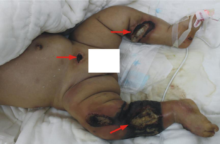
Fig. 1: The necrotic tissue in both legs (circumferential) and hypogastrium (arrows). Both lower limbs including feet were severely swollen.
and cyanosis of the lips and extremities further appeared, and retractions in the suprasternal, supraclavicular and intercostal spaces were noticed. Therefore, the infant was transferred to our department for a definitive treatment. Abdominal ultrasound revealed gas accumulation in the gastrointestinal tract and minimum fluid. Meanwhile following hospitalization, erythema and swelling of the legs was found to be rapidly progressing (Fig. 1). Fasciectomy and extensive surgical debridement of the two legs, hypogastrium and bilateral iliac regions was performed immediately and hand- made NPWT was applied simultaneously (Fig. 2). Pressure was applied at -50 mmHg continuously for the duration of therapy.
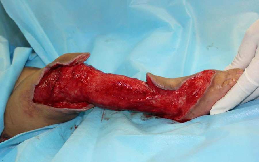
Fig. 2: The defect following surgical debridement. It was not suitable for immediate closure with flap or skin graft
Meropenem (0.1 gr, three times a day) was administered intravenously and changed to ceftazidime (0.5 gr, twice a day) upon receiving the antimicrobial susceptibility test report. Albumin transfusion, nutrition support, and continuous gastrointestinal decompression via nasal tube were performed postoperatively. The wound was assessed and dressings were changed on the third day. Abdominal distention, oliguria and fever subsided, the granulation tissue was activated and edema of the right foot and lower leg resolved rapidly with NPWT therapy. STSG was performed when the granulation tissue was fresh and no secretions were observed (Fig. 3). Hand-made NPWT was used again to fix the skin. The skin graft survived intact with a satisfactory outcome. Despite advances in medical and surgical therapy complications of NF could be inevitable. ACS, whether primary or secondary, is also a fatal condition. Management of these two diseases is challenging. NF caused by Staphylococcus epidermidis[1] and Pseudomonas aeruginosa[2], were described earlier but ACS as a complication of NF in a infant has not previously been reported. ACS is defined as a sustained intra-abdominal pressure (IAP) greater than 20 mmHg that is associated with new organ dysfunction or failure[3,4]. The diagnosis of ACS in our patient was considered mainly by clinical symptoms. Infant had massive abdominal distention, respiratory compromise, cardiovascular compromise (tachycardia), acute renal insufficiency and evidence of systemic infection. We did not attempt to measure and record IAP since the infant was critically ill and required a since the infant was critically ill and required an emergent operation. A
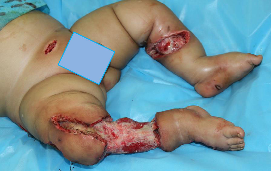
Fig. 3: Healthy granulation tissue of the debrided wound before proceeding with the reconstruction
urinary catheter was inserted only following adequate general anesthesia in the operating room. Reconstruction of wounds of the infant is more challenging because of special characteristics[5]. Although numerous studies of NPWT have been published regarding outcomes and methods of therapy used for managing difficult-to-treat pediatric wounds or as an effective adjunct to expedite granulation tissue in wound preparation[6], no report on combined usage of NPWT with STSG in infants has been found. Wound size of this infant was extensive and across the knee joint. There is also difficulty in immobilization in infants following skin grafting. Due to these reasons, we adopted NPWT that can provide continuous adequate pressure to the recipient site of the skin graft to obtain a higher take rate, and to accelerate the epithelialization of the "blank space" following meshed STSG. Our report highlights the importance of early diagnosis, treatment with modern techniques including negative pressure therapy, prompt surgical debridement, appropriate antibiotics and aggressive supportive care in successful management and improved survival and cosmetic outcome in children with NF, even those complicated with ACS. However, we also know that it is hard to draw any convincing evidence from a single case and therefore, further research is required to verify the effect of NPWT in management of NF before wide acceptance of this method.
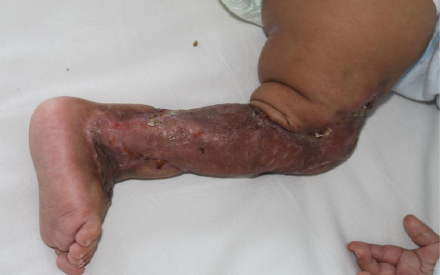
Fig 4: One month following placement of meshed split-thickness skin graft on the right leg