

1. Background
It is a well-known fact that, vitamin B12 has a very important role in neuronal development of children because it is necessary for DNA synthesis, neurotransmitter metabolism, methylation reactions, and homocysteine-methionine cycle. Neurologic dysfunctions such as peripheral neuropathy and orthostatic hypotension associated with vitamin B12 deficiency are widely described (1, 2). In addition, autonomic nervous system dysfunction which can be hidden by compensating mechanisms can also accompany vitamin B 12 deficiency, despite the patient being still asymptomatic (3). However, there is limited data about this effect of vitamin B12 deficiency in pediatric age group (4). Therefore, determining the presence of autonomic dysfunction has a crucial role in these patients to prevent further neurologic disorders. Some special techniques, such as Valsalva maneuver or the tilt table test can be used to evaluate cardiac autonomic dysfunction but these tests are evidently not only hard to perform in children but also difficult to standardize (5). Among these tests, HRV is a selective, reliable and reproducible method to test sympathetic and parasympathetic function of autonomous nervous system (6).
2. Objectives
We aimed in the present study to evaluate autonomic function in children with vitamin B12 deficiency by analysis of HRV with rhythm Holter monitoring.
3. Methods
3.1. Study Population
Thirty-seven consecutive patients who were newly diagnosed and untreated with vitamin B12 deficiency comprised the study group. Randomly chosen, age and sex matched 25 healthy children with normal vitamin B12 levels comprised the control group. Children with structural heart disease, dysrhythmia, hematologic disease (thalassemia, sickle cell anemia, iron deficiency anemia, etc.), chronic illness or children who were taking any regular medications were excluded from the study. All patients and their parents were asked about suggestive autonomic neuropathy symptoms such as urinary urgency and limb weakness, and none of them reported any similar symptom. Also, none of the cases in both the study and control groups had abnormal neurological or cardiac findings on physical examination.
The present study was approved by a scientific committee comprised of the hospital administration and the lecturers (protocol number: 43278876-929). All participants’ parents gave their informed written consent.
3.2. Diagnosis of Vitamin B12 Deficiency
Serum vitamin B12 was measured by chemiluminescent immunoassay method (Architect, Abbott, IL, USA). The diagnosis of vitamin B12 deficiency was made according to the serum level of vitamin B12, homocysteine, and methyl malonic acid. Vitamin B12 deficiency was defined as serum level of vitamin B12 that was lower than 200 pg/mL, accompanied with the increase of serum level of homocysteine and/or methyl malonic acid (higher than 12 µmol/L and 0.45 µmol/L, respectively) (7-9).
3.3. Analysis of HRV
All participants were evaluated by two-dimensional and colour-Doppler echocardiography in order to assess cardiac functions, cardiac structures and additional cardiac abnormalities. HRV analysis was made by measuring the alteration in consecutive RR intervals on 24-hour electrocardiography recordings in all cases. Twenty-four hour ambulatory electrocardiogram recordings were taken with DMS 400-3A solid-state recorder. Recordings were evaluated via computer by using the Cardioscan II Series (DM Software, USA) software. Electrocardiographic recordings were also analysed for rhythm and QT and corrected QT intervals.
Time domain and frequency domain method were used in order to evaluate HRV. In time domain analysis; measurements were calculated from the standard deviation of heart rate variation based on the RR distances between consecutive sinus rhythm beats. Firstly, based on the RR distances, average RR and the standard deviation of RR variation were measured then the standard deviation of all the 5-minute normal to normal (NN) interval means and the mean of all the 5-minute standard deviations of NN were calculated and the parameters of the standard deviation of all the RR intervals (SDNN), the standard deviation of 5 minute RR interval means (SDANN), the mean of the 5 minute RR interval standard deviations (SDNNi) were found. Secondly, variables were evaluated on the basis of the differences between two consecutive RR distances: the square root of the mean of the squared differences of two consecutive RR intervals (RMSSD), the percentage of the beats with consecutive RR interval difference of more than 50 ms (pNN50) were calculated (10, 11).
In frequency domain analysis; periodic signals, an average of 500 sequential R-R intervals to be divided into various bands of frequency response: the area under the spectral curve from 0.01 to 1.0 Hz (total power, TP), the area under the spectral curve from 0.0033 to 0.04 Hz (very low-frequency power, VLF) the area under the spectral curve from 0.04 to 0.15 Hz (low-frequency power, LF), the area under the spectral curve from 0.15 to 0.40 Hz (high-frequency power, HF) and the ratio of LF and HF were calculated. LF, HF and their ratio LF/HF are most frequently used in frequency dependent analysis.
The increase in high frequency (HF) is parasympathetic and the increase in LF reflects more sympathetic activity. Total power reflects the entire autonomic nervous system activation. LF/HF ratio was used as a measure of the sympathovagal balance (10-13). There is a strong relationship between time-dependent and frequency-dependent HRV parameters. Time domain parameters’ approximate frequency-domain correlates are SDNN and SDNNi-TP, RMSSD and pNN50-HF.
3.4. Statistical Analysis
The data was recorded with the Statistical Package for the Social Sciences program version 21 (SPSS Inc., Chicago, IL, USA). The distributions of continuous variables were analyzed with the Shapiro-Wilk test. The descriptive statistics were defined as mean ± standard deviation (SD) for normally distributing data and as median (minimum-maximum) for non-normally distributing data. Normally distributed continuous variables were compared by Student’s t-test and the significance of the differences in median values between two independent groups was analyzed with the Mann-Whitney-U test. A multiple linear regression model was used to identify independent factors such as age, gender, body surface area, and serum vitamin B12 level that could affect HRV parameters. Pearson or Spearman correlation analysis was used to compare two continuous variables based on normality of distribution. A P value of less than 0.05 was considered as statistically significant.
4. Results
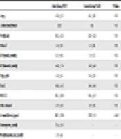
A total of 62 children (37 with vitamin B12 deficient and 25 healthy controls) were enrolled in this study. Table 1 summarizes gender, age, weight, body surface area (BSA), systolic and diastolic blood pressure and complete blood count parameters of the participants. No statistically significant difference was found between groups for these parameters, only serum B12 level was significantly lower in the study group.
| Study Group (N: 37) | Control Group (N: 25) | P Value | |
|---|---|---|---|
| Age, y | 14.3 ± 3.2 | 14.1 ± 2.9 | NS |
| Gender, male/female | 20/17 | 9/16 | NS |
| Weight, kg | 58.2 ± 15.3 | 52.9 ± 13.5 | NS |
| BSA, m2 | 1.6 ± 0.3 | 1.5 ± 0.2 | NS |
| BP (systolic), mmHg | 112 ± 10.5 | 111 ± 7.5 | NS |
| BP (diastolic), mmHg | 64.2 ± 7.6 | 64.1 ± 6.8 | NS |
| Hgb, gr/dL | 14.1 ± 1.4 | 13.6 ± 1.0 | NS |
| Htc, % | 42.6 ± 4.3 | 41.4 ± 4.8 | NS |
| MCV, fL | 83.5 ± 10.0 | 84.3 ± 4.7 | NS |
| RBC, 106/mm3 | 5.0 ± 0.4 | 4.9 ± 0.5 | NS |
| Serum B12 level, pg/mL | 147.5 ± 23.9 | 337.2 ± 93 | < 0.01 |
| Homocystein, µmol/L | 17.6 ± 11.0 | - | - |
| Methylmalonic acid, µmol/L | 1.9 ± 6.9 | - | - |
Abbreviations: BP, blood pressure; BSA, body surface area; Hgb, hemoglobin; Htc, hematocrit; MCV, mean corpusculer volume; RBC, red blood cell.
Echocardiographic evaluation of participants was normal and mean left ventricular ejection fraction (EF) was 73.4% ± 5.7% (range, 64% - 84%), shortening fraction (SF) was 42.6 ± 5.3 (range, 34% - 52%) in the vitamin B12 deficient group, while mean EF was 75.6% ± 5.5% (range, 67% - 87%), and SF was 44.6 ± 5.3 (range, 36% - 56%) in the control group. There was no statistically significant difference between these parameters. Rhythm Holter monitoring recordings were evaluated in all participants. In the study group only one subject had rare ventricular extrasystoles, but in the control group two of the subjects had insignificant rhythm disturbances (one with rare ventricular extrasystoles and the other with rare supraventricular extrasystoles).
All heart rate variability parameters (time-domain and frequency-domain) seemed to be reduced in the vitamin B12 deficient group compared with controls (Table 2). Minimum heart rate, SDNNi, RMMSD, pNN50, total power, and LF were statistically significantly different variables between the two groups (P < 0.05).
| Study Group (N: 37) | Control Group (N: 25) | P Value | |
|---|---|---|---|
| Minimum heart rate, bpm | 49.6 ± 5.9 | 45.8 ± 6.6 | 0.03 |
| Maximum heart rate, bpm | 163.5 ± 14.2 | 155.4 ± 23.4 | NS |
| Average heart rate, bpm | 85.2 ± 9.5 | 83.3 ± 17.5 | NS |
| SDNN, ms | 147.4 ± 40.1 | 167.1 ± 49 | NS |
| SDNNi, ms | 66.7 ± 17.8 | 77.6 ± 18.5 | 0.04 |
| SDANN, ms | 130.2 ± 36.1 | 146.4 ± 50.5 | NS |
| RMSSD, ms | 42 ± 14.1 | 50 ± 13.6 | 0.02 |
| pNN50, % | 17.7 ± 9.1 | 23.2 ± 10.5 | 0.03 |
| Total power, ms | 4611.3 ± 2295.3 | 5995.4 ± 2812.5 | 0.05 |
| VLF, ms | 2892 ± 1581.7 | 3883.3 ± 2169.9 | NS |
| HF, ms2 | 624.6 ± 355.8 | 784.4 ± 345.9 | NS |
| LF, ms2 | 1065.5 ± 517.5 | 1294.3 ± 461 | 0.03 |
| LF/HF | 2.08 ± 1.05 | 1.83 ± 0.53 | NS |
aSDNN, standard deviation of all the RR intervals; SDANN, standard deviation of 5 minute RR interval means; SDNNi, the mean of the 5 minute RR interval standard deviations; RMSSD, the square root of the mean of the squared differences of two consecutive RR intervals; pNN50, the percentage of the beats with consecutive RR interval difference of more than 50 ms; VLF: very low-frequency power; LF, low-frequency power; HF, high-frequency power.
A multiple linear regression model was used to identify independent factors such as age, gender, BSA, and serum vitamin B12 level that could affect HRV parameters. Serum vitamin B12 level was statistically significantly effective on both time-domain and frequency-domain heart rate variability parameters (P < 0.05). A positive correlation was also found between serum vitamin B12 levels and SDNNi, RMSSD, LF parameters (P < 0.05, r: 0.285, 0.262, 0.246, respectively). Whereas no correlation was found between serum homocysteine, methyl malonic acid levels and HRV parameters. In addition, age was found to be of statistically significant effect on almost all HRV parameters except LF, HF, pNN50 and RMSSD.
There was also a strong negative correlation between minimum and average heart rate with age (r: -0.45, P < 0.01; r: -0.36, P = 0.03, respectively).
5. Discussion
B12 deficiency associated with neuropathy and autonomic nervous system dysfunction could occur much earlier than known (1, 2, 6). Therefore, several different diagnostic tests such as Valsalva ratio, tilt table test and blood pressure response have been used to detect this condition earlier. However, the use of these tests in pediatric clinical practice is not only difficult to perform but also difficult to evaluate because of their subjective nature (11). HRV data obtained from Holter monitoring provides to determine an abnormality of autonomic nervous system function, in the form of sympathetic and parasympathetic activity. In addition, this method is easy, feasible and reliable in children (11, 14, 15).
Normal heart rate variability is achieved by autonomic neural regulation of the heart and circulatory system with parasympathetic and sympathetic nervous system balance. Neural pathologies due to vitamin B12 deficiency were attributed to the disruption of methylation in the myelin sheat cells. Cardiac autonomic neuropathy is due to the changes in heart rate control and vascular dynamics as a result of this methylation defect in nerve endings innervating the heart and vessels and also the changes in neurotransmitter metabolism (1, 2, 6, 16, 17).
Several publications showed autonomic nervous system dysfunction by analyzing HRV in adult patients with vitamin B12 deficiency. In these studies, long term (24 hour) and short term (5 minute) measurements of parameters of HRV were found to be significantly lower as compared to healthy control subjects (2, 16, 17). However, there is still limited data about the influence of vitamin B12 deficiency on autonomic nervous system in children. The present study shows that vitamin B12 deficiency may cause autonomic dysfunction in children. We found that all time-domain and frequency-domain heart rate variability parameters reduced in the vitamin B12 deficient group compared with those of healthy controls. However, SDNNi, RMMSD, pNN50, total power, and LF were only the variables with statistically significant difference between the two groups (P < 0.05). With regard to time domain parameters, it has been indicated that parameters calculated based on the RR interval variation average, such as pNN50, RMSSD, SDNNi are less influenced by the cardiac circadian rhythm and these variables are thought to be sensitive to the parasympathetic condition (10, 11). Our findings suggest an impaired parasympathetic activity in children with vitamin B12 deficiency. On the other hand, frequency domain parameters such as the low-frequency spectral analysis informs us about the both sympathetic and parasympathetic activity and total power reflects the entire autonomic nervous system activation (10, 11). Therefore, in the light of our findings, we speculated that vitamin B12 deficiency gives rise to both impaired parasympathetic and sympathetic activity in children.
Limited number of published studies based on the HRV analysis in children has also showed similar findings. Celik et al revealed decreased rMSSD, LF, and HF in children with B12 deficiency all of which is in line with decreased parasympathetic modulation of the autonomic nervous system (14). Another study performed by Sucharita et al. disclosed that young children born to mothers with lower vitamin B12 status have reduced cardiac sympathetic activity (4).
Heart rate variability parameters are affected from different variables such as, age, body surface area and gender, as well as variable diseases (diabetes, cardiovascular diseases, inflammation, obesity and psychiatric disorders). One of these variables is age and HRV values are known to be decreased with age (10). Similarly, in our study, a negative correlation was found between age and average as well as minimum heart rate. However, we found no effect of gender and body surface area on heart rate variability parameters, because gender related differences start in later ages and we did not include obese children into the study. A recent study has disclosed that diet has also an important effect on HRV (18). Vitamin B12 is a major component of diet, and cannot be synthesized in the human body. Our data support this recent findings.
The limitations of this study are related to its small sample size and not evaluating the effect of vitamin B12 treatment on HRV parameters. Therefore further prospective studies with large sample size are required to evaluate autonomic activity before and after B12 treatment. In conclusion; our data suggest that in children with vitamin B12 deficiency autonomic dysfunction can occur via decreased sympathetic and parasympathetic activity similar to adults.
