1. Background
Cerebral predominance alludes to the supremacy of one cerebral hemisphere over the other one. More specifically it is the capability of each cerebral hemisphere to predominantly control specific tasks and functions consequently it is related to handedness (i.e. right-handed versus left-handed) (1). The neurologic functions of Wernicke’s area, other speech centres, and motor control areas, are usually more highly developed in one cerebral hemisphere. Therefore, this hemisphere is called the dominant hemisphere. The left hemisphere is the dominant one, in 95 percent of the population. Even at birth, the area of the cortex that will eventually become Wernicke’s area is approximately 50 percent larger in the left hemisphere than in the right in more than one-half of neonates (2). The non-dominant hemisphere plays an integral role in spatial perception, visual perception, musical talents, identification of body language and emotions, verbal collateral skills, and other esthetical patterns in the environment (3).
A minor percentage of the population are labelled as ambidextrous, in which no preference for performing manual tasks with either hand, right or left. A famous example of ambidexterity is in the unique case of Leonardo da Vinci (an Italian polymath). Ambidexterity occurs when a person has an approximately equal set of skills in both hands and/or both sides of the body. However, true ambidexterity is extremely rare, because the person will still show preference to one side of the body more than the other (4, 5).
From a developmental neurobiology perspective, this lateralization of the brain, initializes in reference to the median longitudinal fissure, this fissure divides the human brain into two well-defined hemispheres, and both are interconnected by a bridge-like structure, the corpus callosum. Each human’s brain grows in a gradually and differently from others, leading to unique cerebral lateralization in the individual. There are some evident macroscopic, microscopic, and biochemical differences in between the right and left cerebral hemisphere, all these are supporting evidence for the lateralization of brain functions. The best case of a set-up lateralization, is that of speech centres known as Broca’s and Wernicke’s, are almost exclusively located on the on the left hemisphere in right-handed individuals. These ranges much of the time compare to handedness (6-9)
This observational study and cross-sectional analysis was conducted in accordance with the Ethical approval no. 620-73, on the 15th of May 2016. The approval is under the authority of the dean office at the faculty of medicine at the University of Baghdad.
2. Objectives
This cross-sectional study will aim to investigate the existence of a potential correlation between three-dimensional visual analytic skills and the lateralization of brain functions. This study is purely observational; it is based on the use of an interactive survey tool. Although purely observational, proper randomization and blinding were implemented to prevent coverage and selection biases among participants.
3. Materials and Methods
The data presented in this paper was derived from a survey directed towards 1st-year undergraduate medical students at the largest medical university in Iraq. A survey, cross-sectional, was created online using Google Forms (10, 11). All the participant students have successfully completed the lower limb module of the Human Anatomy course and were competent enough in identifying sectional images of magnetic resonance imaging (MRI), including cross sections.
This study is considered to be of a randomised single-blinded design, in which participants were randomly selected to participate in the survey, restrictive randomization by stratifying the participants according to age, which was followed by block randomization. Participants were also blinded towards the survey’s nature and its aim. Those who delivered and distributed the survey were knowledgeable of the nature, structure, and objectives of the survey. The resultant data from the survey summed up to 523 pages, upon which this study was based. Each participant in the survey was allowed to take the survey only once. The blinding and randomization were done to prevent coverage error and selection biases among participants in this study.
The survey itself included four cross-sectional MRI images of the lower limb, which were taken from the atlas of human anatomy on MRI (12). Sections were selected by professional Anatomists at three main levels: leg (two sections), knee (one section), and thigh (one section). All these sections can be normally visualised (imagined), by a non-professional Anatomist (students in this case) either from above (a superior view) or below (an inferior view). All images were fully labelled with anatomical landmarks, and the students were only asked about how they would first visually imagine the sections, either from above or from below.
The survey included three main sections: MRI images, parameters of cerebral dominance, and demographic parameters. The survey was electronically delivered via a secured university intranet system (13). The targeted audience was of 297 undergraduate medical students (year-1). The survey participants were oriented about the survey a week in advance, and the survey was later posted online for three successive days from the 30th of April, 2016 to 3rd of May 2016, the majority of participants took the survey in day-1.
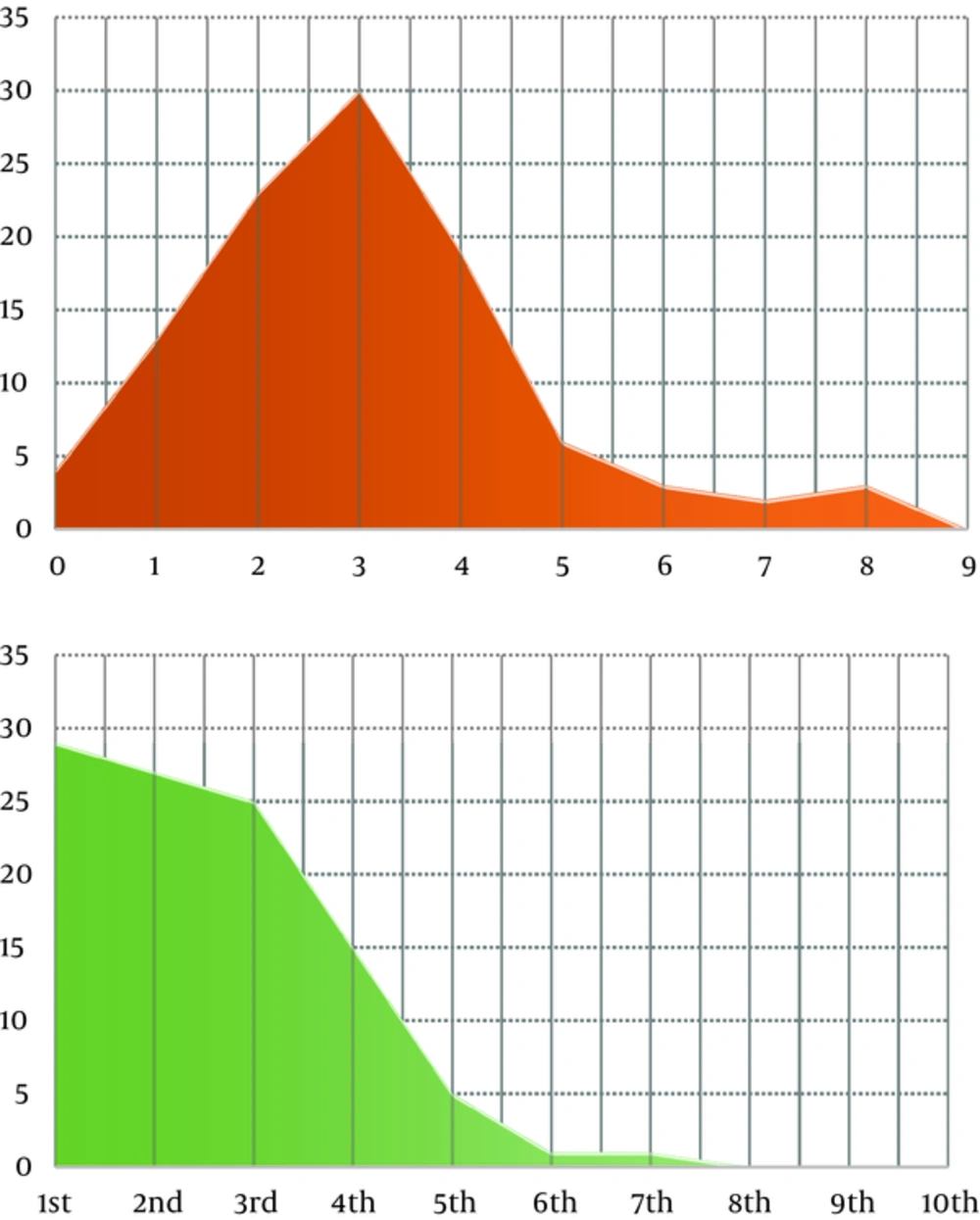
The parameters of Cerebral dominance, which were being correlated against how each participant visualised the MRI images, include: handedness (i.e. dominant hand), the number of spoken and written languages, and subjective (self-assessment) of the 3D-visual analytic skills. The 3D-imaginative abilities were further objectively confirmed by using a specialised 3D-visual IQ testing delivered online via a professional IQ testing society, the international high-IQ society (14). Demographic data included: age, gender, the number of siblings, birth order (rank), in addition to parents’ and siblings’ handedness. Number and position of siblings are visualised in the form of an area-under-curve graph (Figure 1).
Additionally, a systematic literature review was conducted from the 1st of April 2016 to the 10th of June 2016, concerning the concept of cerebral dominance and laterality, and previous studies and surveys on the topic. Specific and pre-determined keywords (including MeSH keywords), were applied across medical and paramedical databases: PubMed, The Cochrane Library, Scopus, OpenGrey, and Google Scholar. This search methodology was aiming to extract the most relevant and up-to-date literature material, which were published in professional and high-impact journals in the past 5 years (2012 - 2016). However, other literature materials and research papers were also considered when finding relevant and of high level-of-evidence.The overall level of evidence of this paper is Level-5, according to the Oxford Centre for Evidence-based Medicine (15).
A total number of papers that were initially mapped were 32. The papers and textbooks were later filtered by reading: the paper title, abstract, and full text. This was followed by critical analysis using critical appraisal tools suitable for each type of retrieved literature papers. Duplicate papers were excluded. Accordingly, only twenty-three reference materials were finally used in the citation of this paper. Statistical analyses of this paper were conducted using the Statistical Package for Social Sciences (SPSS version 20.0), Shodor-Interactivate software (16). Other forms of data analysis and presentations, were done in Microsoft Excel 2016. Further, data were numerically encoded, to perform specialised statistical analysis with SPSS, these were: Chi Square and Pearson’s Correlation test. The significance is considered at an alpha (α) value of 0.05
4. Results
The survey was successfully and fully completed by 103 medical students (34.68% of targeted audience). The majority of participants completed the survey in day-1, with a decline in day-2 and a steeper decline in day-3. Demographics and related parameters (Table 1), showed that 64.1% and 27.2% of the participants were aged 18 and 19 years respectively, while age range of participants was 17 to 21. Female participants were much more compliant to take the survey, as 66.99 % of participants were female.
| Demographics | Results |
|---|---|
| Age, y | |
| 17 | 3 |
| 18 | 66 |
| 19 | 28 |
| 20 | 5 |
| 21 | 1 |
| Gender | |
| Male | 34 |
| Female | 69 |
| Handedness | |
| Right | 91 |
| Left | 12 |
| Ambidextrousa | 0 |
| Number of Siblings | |
| None | 4 |
| 1 | 13 |
| 2 | 23 |
| 3 | 30 |
| 4 | 19 |
| 5 | 6 |
| 6 | 3 |
| 7 | 2 |
| 8 | 3 |
| 9 | 0 |
| Position Among Siblings | |
| 1st | 29 |
| 2nd | 27 |
| 3rd | 25 |
| 4th | 15 |
| 5th | 5 |
| 6th | 1 |
| 7th | 1 |
| 8th | 0 |
| 9th | 0 |
| 10th | 0 |
| Siblings’ Handedness | |
| All are similar | 71 |
| All are different | 14 |
| At least one is different | 18 |
| Parents’ Handedness | |
| Both are similar | 81 |
| Both are different | 8 |
| At least one is different | 14 |
| Average Performance in Human Anatomy | |
| High (< 70%) | 34 |
| Medium (50% - 70%) | 52 |
| Low (> 50%) | 17 |
| Number of Languages | 1 |
| 1 | 3 |
| 2 | 37 |
| 3 | 48 |
| 4 | 14 |
| 5 | 1 |
| 3D Visual Skills | |
| Imagine and Rotate 3D Objects | |
| Yes | 64 |
| No | 39 |
| Frequently play 3D Videogames | |
| Yes | 76 |
| No | 26 |
| Visual and Neuro-psychiatric Disturbances | |
| Myopia | 14 |
| Myopia and Astigmatism | 2 |
| Astigmatism | 1 |
| Hyper-tension | 1 |
| Anxiety | 2 |
| Skin Problems | 1 |
| Sensori-neural Hearing loss | 1 |
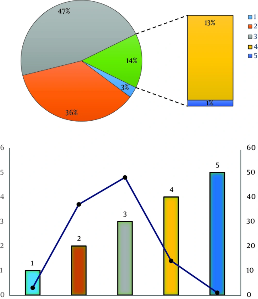
Participants’ handedness was predominantly right-handed (88.35%), and left-handed (11.65%). The participants’ handedness was similar to parents’ handedness in 78.6%, while the participants’ handedness was similar to siblings’ handedness in 68.9%. The number of spoken languages (Figure 2) was in the range of 1 to 5, while the majority spoke either two language (35.9%) or three languages (46.6%). Only one participant (0.97%) spoke five languages. Position and Number of Siblings of participants (Figure 1), visualised that the major part of the area under the curve (AUC) lies between zero and five (number of siblings), and 1st to 5th (Position among siblings).
The three-dimensional visual imagination (visual analytic skills), were estimated in the survey by asking about: self-assessment of the ability to imagine and rotate 3D-figures in their mind, and their frequent involvement in playing 3D video games. This was further consolidated by estimating the visual intellectual quotient (IQ) of those who acclaimed good/high 3D-visual analysis skills, via a professional IQ test (14). Those who failed to pass this IQ test were considered of inferior 3D visual skills, and were labelled as “No” and subsequently dropped out from relevant statistical calculations. Participants with strong visual skills, based on the subjective and objective evaluation, accounted for 64.1% of participants. In relation to this, those who had distinction-level (more than 70%) of cumulative performance in Human Anatomy, accounted for 33%, while 50.4% of participants had a percentile score of performance in the range of 50% - 70%.
To make sure, the participants were free of illnesses that may interfere or lead to biases in this research, each participant was asked about medical problems including visual, neurologic, psychiatric problems, and others. It has been found that 16.5% had visual problems (including myopia and/or astigmatism), while 4.9% had other disorders (anxiety, hypertension, hyperhidrosis, sensorineural hearing loss, and skin problems). However, all these were evaluated and subsequently considered as non-limiting factors in taking this survey.
Concerning data on sectional MRI images, for sections-1, 66% visualized the section from above (a superior view), for section-2, 60.2% visualized the section from above (superior), for section-3, 47.6% visualized the section from above (superior), and for section-4, 51.5% visualized the section from above (superior). Obviously, the rest of the participants visualised these four sections from below (an inferior view). The data obtained from all the participants (103), where mathematically converted into a binary code of 0s and 1s.
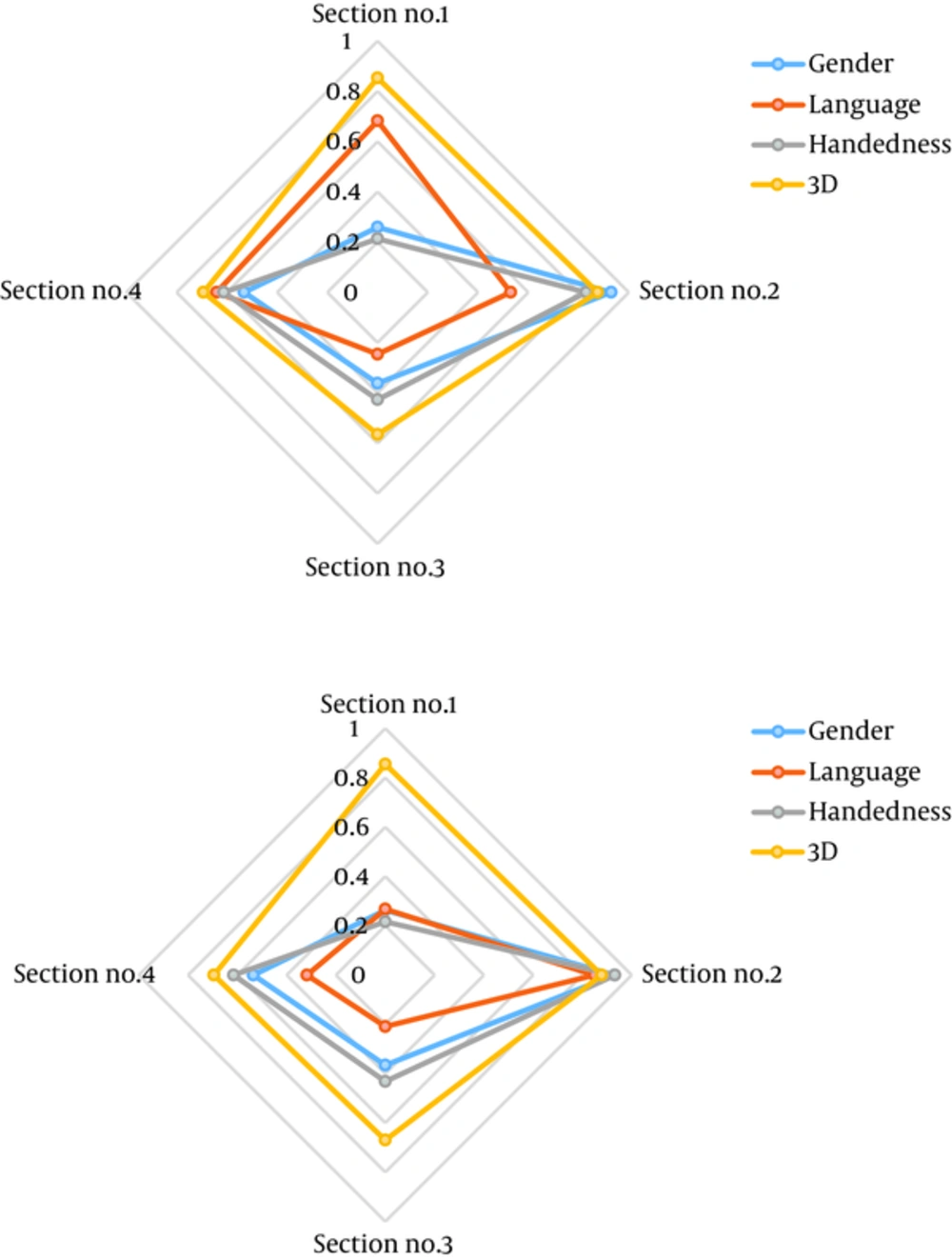
The application of Chi Square and Pearson’s Correlation test (Table 2, Figure 3), revealed that there was no any statistical correlation between the visual analytic skills and any of the tested representative parameters of cerebral dominance. These parameters included: handedness, gender, and the number of spoken-written languages (Table 2). Significance was considered at a P value of 0.05 or less. For example, in relation to subject’s handedness the P values were: 0.213, 0.830, 0.427, 0.612 for sections 1 - 4 respectively while using Chi Square test, and 0.216, 0.932, 0.432, 0.616 for sections 1 - 4 respectively while using Pearson’s Correlation test.
| Chi Square | Section No.1 | Section No.2 | Section No.3 | Section No.4 |
|---|---|---|---|---|
| Gender | 0.259 | 0.930 | 0.362 | 0.531 |
| Language | 0.682 | 0.530 | 0.246 | 0.641 |
| Handedness | 0.213 | 0.830 | 0.427 | 0.612 |
| 3D | 0.853 | 0.876 | 0.565 | 0.693 |
| Pearson’s Correlation | ||||
| Gender | 0.263 | 0.931 | 0.366 | 0.535 |
| Language | 0.267 | 0.849 | 0.209 | 0.318 |
| Handedness | 0.216 | 0.932 | 0.432 | 0.616 |
| 3D | 0.855 | 0.878 | 0.670 | 0.696 |
This study can be further developed in subsequent designs in the future, to correlate the visual analytic skills with cognitive enhancers, a subset of psychoactive and novel psychoactive substances (NPS) (19, 20).
5. Discussion
To the best of the author’s knowledge, this observational cross-sectional study is the 1st of its kind with regards to analysing and correlating the cerebral lateralization and visual analytic skills, while using sectional Anatomical MRI images, which was designed in a randomised sample of Iraqi undergraduate medical students. However, there has been numerous several other studies and attempts, for correlating the effect of cerebral lateralization and visual analytic skills, using other investigative tools, including: tachistoscopic recognition; facial recognition; visuospatial attention; written verbal commands; Japanese kanji; and reaction-time studies; and functional MRI (fMRI) (21-27). Other parallel studies also examined subject with some neurological pathologies, including dyslexia; developmental disorders; and autism (28-30). On the other hand, this observational analytic study was conducted among normal Iraqi subjects from both gender, they were undergraduate medical students with high visual IQ.
In this sample of Iraqi undergraduate medical students, who are evaluated as non-professional Anatomists. It was hypothesised prior to carrying out this survey that there was a positive correlation between visual analytic skills and other parameters of the surveyed subjects. These parameters are neurologic manifestations of cerebral dominance, particularly handedness, gender, and the number of spoken languages. In other words, and in relation to subject’s handedness, it was hypothesised that left-handed participants could potentially visualise objects, specifically sectional Anatomical MRI images, differently from right-handed participants.
5.1. Conclusion
The statistical analyses of this cross-sectional analysis, which was of a randomised and a single-blinded design, establish a confirmatory evidence of the contrary, in which there was an absence of any form of correlation between the studied parametric manifestations of cerebral lateralization and complex visual analytics skills, as tested using the MRI images


