Full Text
Full text is available in PDF
An Innovative Journal in the Field of Radiology
Authors
Noninvasive measurement of bone mineral density (BMD) is a technology that benefits both the patient and society through its potential to decrease the morbidity, mortality, and cost of fractures associated with osteoporosis through early detection and treatment. In general, two sites are assessed, the spine and the hip. If one of these sites cannot be assessed, the protocol should outline the next site to be used. Quantitative computed tomography (QCT) can identify patients with low BMD compared to the QCT reference database and who are at risk for fracture. QCT cannot be used to diagnose osteoporosis based on the quantitative BMD value obtained, since it has never been validated for WHO criteria. It is, however, the only other technology besides DXA that is approved for treatment follow up. Quantitative computed tomography (CT) is a three-dimensional non-projectional technique to quantify bone mineral density (BMD) in the spine, proximal femur, forearm, and tibia with a number of advantages to other densitometric techniques: cortical and trabecular bone can be separated, trabecular volumes of interest (VOI) are largely independent of degenerative changes in the spine; so, QCT can be used to assess/monitor patients with extensive degenerative disc disease, more sensitively than DXA for density changes in the vertebral body and 3D geometric parameters can be determined. Bone mineral density (BMD), as measured by QCT, is a true density measured in g/cm3, in contrast to dual-energy X-ray absorptiometry (DXA), which determines an areal density (BMDa) measured in g/cm2. It has also some disadvantages such as higher radiation dose and lack of applicability with WHO diagnostic criteria, Furthermore, diagnosis of low bone mass is made by using the QCT criteria which is technically more difficult than DXA unless spiral technique is used. In modern spiral CT scanners, various techniques are implemented to significantly reduce radiation exposure by optimally adapting the X-ray tube current to the individual subject being scanned. The level of dose reduction depends on anatomical location. However, due to limited medical evidence, definitive advice on its use in all clinical practices cannot be provided until more data emerge.
© 2014, Author(s). This open-access article is available under the Creative Commons Attribution 4.0 (CC BY 4.0) International License (https://creativecommons.org/licenses/by/4.0/), which allows for unrestricted use, distribution, and reproduction in any medium, provided that the original work is properly cited.
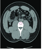
Ganji Jameshouran MA, Abedi AR, Montazeri S, Tasharrofi MA, Mansouri Tehrani MM. CT-Scan as an Opportunistic Osteoporosis Screening Tool in High-Risk Populations; A Diagnostic Study Conducted on a Sample of Urinary Stone Patients. I J Radiol. 2020;17(4):e102177. doi: https://doi.org/10.5812/iranjradiol.102177
Ghorbaniafzal S, Rostami Z, Raei M, Javanbakht M, Jafari R, et al. Comparison of CT Scan and Bone Mineral Densitometry for Diagnosing Osteoporosis and Osteopenia in Patients with Chronic Kidney Disease. Nephro-Urol Mon. 2025;17(4):e161015. doi: https://doi.org/10.5812/numonthly-161015
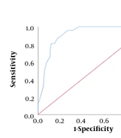
Rajaei A, Mohammadi K, Oroei M, Farsad F. Diagnostic Value of Trabecular Bone Score in Osteoporosis. I J Radiol. 2023;20(2):e131845. doi: https://doi.org/10.5812/iranjradiol-131845
Rajaei A, Dehghan P, Ariannia S, Ahmadzadeh A, Shakiba M, et al. Correlating Whole-Body Bone Mineral Densitometry Measurements to Those From Local Anatomical Sites. I J Radiol. 2016;13(1):e25609. doi: https://doi.org/10.5812/iranjradiol.25609
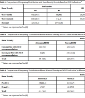
Malekee H, Tabatabaei M, Tasorian B. Assessment of Bone Density of Patients Referred to Arak Densitometry Center from 2016 to 2017: A Cross-sectional Study. Jundishapur J Chronic Dis Care. 2021;10(4):e112744. doi: https://doi.org/10.5812/jjcdc.112744
Last Update: 2 weeks ago
Last Update: 2 weeks ago
Last Update: 2 weeks ago
Last Update: 1 day ago