


1. Background
Retinopathy of prematurity (ROP) is an abnormal retinal neovascularization in infants with a low birth weight (1). ROP is recognized as a cause of blindness in children (2). This disease generally has two phases. Phase 1 is defined as the onset of ROP with delay in retinal vascular development after premature birth, and phase 2 is characterized by hypoxia, inducing the release of some factors (hypoxia factors) that stimulate new blood vessel growth (3). The cause of ROP varies in different countries and is influenced by many factors (2, 4). Plus disease is a major cause of ROP (5). Generally, progression of plus disease and retinal vascular changes are prominent features of ROP (6).
Color Doppler imaging (CDI) is a non-aggressive and safe technique for the clinical management of retinal diseases (7-9). It is applied to characterize several ophthalmic disorders, including diabetic retinopathy, optic atrophy, carotid occlusive disease, and intraorbital arteriovenous malformations. This modality evaluates the circulatory parameters of retrobulbar blood vessels, central retinal vein, central retinal artery (CRA), and ophthalmic artery (OA) (7-14).
The prevalence of ROP seems to vary in different areas of Iran (15), and the number of infants with ROP is increasing due to the incidence of premature births. Also, few studies have been conducted on this subject.
2. Objectives
This study aimed to compare the CDI criteria for OA and CRA vessels in ROP patients with and without plus disease.
3. Patients and Methods
This case-control study was conducted on premature infants with a gestational age < 37 weeks or a birth weight < 1500 g, who were referred to the ROP Clinic of Shahid Sadoughi Hospital, Yazd, Iran, during 2018-2019. After taking informed consent from the infants’ parents, this study was approved by the Ethics Committee of Shahid Sadoughi University (no.: 5175).
The first ophthalmologic examination was performed four to six weeks after birth. The examinations were performed once every week or every two weeks until the retinal vessels were completely examined. Hospitalized and ROP patients with a history of eye treatment, such as laser therapy or intraocular injection, were excluded from the study. After the definitive diagnosis of ROP, information, including age, weight at birth, plus disease, and initial stage of ROP, was extracted from the patients’ medical records.
Next, infants with ROP (21 infants with plus disease and 21 infants without plus disease) underwent CDI using the linear transducers of a Vivid-6 unit (7.5 MHz; Munich, Germany) at a Doppler frequency of 5 MHz. The linear probe was placed in a vertical position perpendicular to the eye globe on the right side of the closed eyelid, using a sterile coupling gel. Arterial Doppler parameters, such as end-diastolic velocity (EDV), pulsatility index (PI), resistance index (RI), and peak systolic velocity (PSV), were measured for the patients. However, the cutoff point could not be measured due to the small sample size.
3.1. Statistical Analysis
Data were entered into SPSS version 22. Independent t-test was used to compare the groups with and without plus disease in terms of parameters, such as PSV, EDV, RI, PI, gestational age, birth weight, and infant’s age. Moreover, Chi-square test was used for evaluating the gender distribution of the patients. P-value < 0.05 was considered statistically significant.
4. Results
The current study was conducted on 42 patients with ROP. In terms of gender, 54.7% of the patients were female, and 45.3% were male (P = 0.63). Parameters, such as gestational age, birth weight, and infant’s age, are presented in Table 1.
| Parameters | Number | Mean ± SD | P-Valuea |
|---|---|---|---|
| Gestational age, wk | 0.29 | ||
| With plus disease | 21 | 28.52 ± 2.27 | |
| Without plus disease | 21 | 29.19 ± 1.72 | |
| Birth weight, g | 0.42 | ||
| With plus disease | 21 | 1153 ± 490 | |
| Without plus disease | 21 | 1260 ± 362 | |
| Infant’s age, wk | 0.46 | ||
| With plus disease | 21 | 35 ± 1.3 | |
| Without plus disease | 21 | 34 ± 1.5 |
Parameters in the Groups with and Without Plus Disease, Including Gestational Age, Birth Weight, and Infant’s Age
As shown in Table 1, no significant difference was observed between the two groups considering the gestational age, birth weight, or infant’s age (P > 0.05). Also, comparison of the two groups regarding the arterial CDI parameters, including PSV, EDV, RI, and PI, is presented in Table 2.
| Parameters | Type of Artery | Number | Groups | Mean ± SD | P-Valuea |
|---|---|---|---|---|---|
| PSV, cm/sec | OA | 21 | With plus disease | 22.04 ± 4.95 | 0.11 |
| 21 | Without plus disease | 25 ± 6.78 | |||
| CRA | 21 | With plus disease | 15.65 ± 3.35 | 0.029 | |
| 21 | Without plus disease | 18.39 ± 4.39 | |||
| EDV, cm/sec | OA | 21 | With plus disease | 4.91 ± 1.5 | 0.1 |
| 21 | Without plus disease | 5.68 ± 1.47 | |||
| CRA | 21 | With plus disease | 4.35 ± 1.00 | 0.005 | |
| 21 | Without plus disease | 5.27 ± 1.02 | |||
| RI | OA | 21 | With plus disease | 0.77 ± 0.05 | 0.97 |
| 21 | Without plus disease | 0.77 ± 0.05 | |||
| CRA | 21 | With plus disease | 0.72 ± 0.05 | 0.65 | |
| 21 | Without plus disease | 0.72 ± 0.05 | |||
| PI | OA | 21 | With plus disease | 1.37 ± 0.22 | 0.82 |
| 21 | Without plus disease | 1.18 ± 0.23 | |||
| CRA | 21 | With plus disease | 1.18 ± 0.23 | 0.37 | |
| 21 | Without plus disease | 1.24 ± 0.2 |
Comparison of Groups with and Without Plus Disease Regarding Arterial Doppler Imaging Parameters
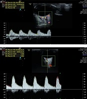
As shown in Table 2, there was a significant difference between the two groups regarding PSV and EDV of CRA (P < 0.05). However, no significant difference was found between the two groups in terms of PSV or EDV of OA (P > 0.05). Also, no significant difference was observed between the groups considering the RI or PI of CRA and OA (P > 0.05). Figure 1A and B show the color Doppler images of CRA in patients with plus disease.
![A and B, Color Doppler images of central retinal artery (CRA) in patients with plus disease. The sample was placed 2 mm behind the sclera at the center of the triangle formed by the optic nerve shadow (peak systolic velocity [PSV] was estimated at 29 cm/s, and end-diastolic velocity [EDV] was estimated at 9 cm/s).](https://services.brieflands.com/cdn/serve/3170b/cf4975275d3dfebc260f71fc1b77b613c2ae5b83/iranjradiol-103451-g001-F1-preview.webp "A and B, Color Doppler images of central retinal artery (CRA) in patients with plus disease. The sample was placed 2 mm behind the sclera at the center of the triangle formed by the optic nerve shadow (peak systolic velocity [PSV] was estimated at 29 cm/s, and end-diastolic velocity [EDV] was estimated at 9 cm/s).")
A and B, Color Doppler images of central retinal artery (CRA) in patients with plus disease. The sample was placed 2 mm behind the sclera at the center of the triangle formed by the optic nerve shadow (peak systolic velocity [PSV] was estimated at 29 cm/s, and end-diastolic velocity [EDV] was estimated at 9 cm/s).
5. Discussion
ROP is a common disease in preterm newborns with a very low birth weight (16). This vascular disorder is related to an altered blood flow (16). CDI has been proposed as a promising tool for the diagnosis of different eye diseases (17). However, some studies have reported that CDI is not clinically beneficial for the management of ROP and plus disease in premature infants (6). Moreover, there is little information regarding the hemodynamic characteristics of retinal perfusion in preterm infants (14). In this regard, some studies have revealed that the value of CDI is lower in preterm infants than in healthy adults (14). Besides, evaluation of CDI in premature infants is more difficult than adults (14).
In the present study, the CDI criteria were not evaluated among patients with and without ROP, because diagnosis of retinopathy and its stages can be well made by an ophthalmologist. Accordingly, we assessed the CDI criteria in ROP patients with and without plus disease and observed that the mean Doppler findings were lower in CRA than in OA. In this regard, Papacci et al. (18) assessed the Doppler sonography findings of blood flow velocity in CRA and OA during the neonatal period and found the decreased Doppler value for CRA as compared to OA. On the other hand, some studies reported that the value of CDI was higher in OA than in CRA (18-22); the findings of these studies are consistent with our study.
Moreover, the present results showed that the mean PSV of CRA was significantly higher in patients without plus disease as compared to those with plus disease. Besides, the mean PSV of OA was higher in the absence of plus disease. Holland et al. reported similar results and found that PSV was higher in the absence of plus disease. Hauch et al. (23) assessed the ocular blood flow changes in ROP patients with plus disease and observed significant differences in the PSV of CRA at baseline and at the time of plus disease. Also, Kaiser et al. (22) reported that the mean PSV was significantly lower in CRA than in OA. Additionally, Ozcan et al. (24) reported that the mean PSV of OA in patients with ROP was significantly higher than those without ROP. The difference between studies by Ozcan et al. (24) and Keyser et al. and our study is that we did not include a control group.
In the current study, no significant differences were found in the RI of CRA and OA in premature infants with and without plus disease. In this regard, Niwald and Gralek (25) evaluated the blood flow of CRA and OA in preterm infants and observed that the RI of OA was significantly higher than CRA; however, the difference was not statistically significant. Holland et al. also used CDI for premature infants with ROP and did not observe any significant differences between premature infants with and without ROP regarding RI.
We observed a significant difference between the two groups with plus and without plus disease regarding the EDV of CRA. However, no significant difference was seen in the EDV of OA. Hauch et al. (23) also reported no significant difference regarding this parameter. Olufemi Adeyinka et al. (26) also found that the EDV of OA and CRA in glaucoma patients was significantly lower than the control group. Moreover, Ozcan et al. (24) reported no significant difference between patients with and without ROP regarding EDV. The difference between studies by Ozcan et al. (24) and Olufemi et al. and our study is that we did not include a control group. Also, we did not observe any significant differences between the two groups regarding PI. The mean PI of OA was higher than that of CRA in patients with plus disease. Papacci et al. (18) also reported that PI of CRA was remarkably lower than that of OA in the early neonatal period, which is consistent with our study.
According to the findings of the present study, CDI criteria, such as EDV and PSV of CRA, were significantly lower in infants with plus disease as compared to those without plus disease. Since early detection of plus disease is a challenge for ophthalmologists, assessment of these criteria can be beneficial. However, further studies with a larger sample size are needed to determine the cutoff point.
