1. Background
Appropriate timing is one of the main concerns in orthodontic treatment planning, especially in dentofacial orthopedics for young adolescents. The estimation of maturation stage is influential in a successful growth modification and correction of skeletal discrepancies (1, 2). However, due to variations in the individuals’ chronological age at puberty, it cannot be regarded as a reliable indicator for determining skeletal age (3). Generally, the measurement of bone age is an interesting topic in various fields, especially in orthodontics, forensic science, and surgery (4-6). It is of utmost importance for orthodontists to be informed of the growth rate of bones during adolescence for choosing an appropriate and timely treatment plan. However, the optimal time for maxillary expansion and mandibular growth modification is not at the same chronological age for all patients (7).
Different methods, such as hand-wrist maturation, tooth mineralization, medial clavicular epiphyseal fusion, cervical vertebral maturation (CVM), and spheno-occipital synchondrosis (SOS), have been applied to determine skeletal age in adolescents (8-12). Hand-wrist radiography has been long applied as the method of choice by orthodontics to assess skeletal maturation. Nevertheless, one of the shortcomings of this method is the difficulty of differentiating landmarks, leading to a false prediction of growth.
Previous studies have revealed a significant positive relationship between the hand-wrist skeletal maturity indices and the stage of spheno-occipital fusion in both males and females. The use of SOS fusion for skeletal maturity evaluation can eliminate the need for extra hand-wrist radiation and can be also helpful for the growth assessment of patients (12-14). The assessment of CVM by lateral cephalometric radiography, which is routinely applied in orthodontics, was introduced as an appropriate alternative for the hand-wrist method. However, this method is not always suitable to determine the exact maturity stage. Considering a strong correlation between the fusion of SOS and CVM, measurement of the SOS closure degree can facilitate skeletal age estimation (2, 5, 15, 16).
The SOS plays a significant role in estimating skeletal age in adolescents and affects the craniofacial structure (6, 16). It is a cartilaginous growth center between the basisphenoid and basioccipital bones (17). Shirley and Jantz, by direct inspection of synchondrosis, indicated the occurrence of complete fusion at the basilar synchondrosis before the age of 25 years and revealed that the onset of fusion is highly associated with the onset of puberty (6).
Recently, Sinanoglu et al. evaluated cone-beam computed tomography (CBCT) in a Turkish population and reported that SOS is an appropriate indicator of age in individuals aged around 18 years (18). CBCT is known to exhibit reduced radiation exposure compared to computed tomography (CT) scan. Also, three-dimensional image acquisition in CBCT as compared to two-dimensional conventional radiography has made this modality important in orthodontic clinics (14, 19-21). Previous studies have reported variations in the time of fusion between different populations globally, as genetic, geographical, and socioeconomic status can affect age estimation (16-19). Regarding the late ossification of SOS, clinicians should have adequate knowledge in this area (4).
2. Objectives
Considering a lack of research evaluating the efficacy of SOS fusion in age estimation using CBCT images in the Iranian population, the present study aimed to assess the degree of SOS fusion for age estimation in individuals aged 6 - 25 years, based on CBCT images. This CBCT analysis is the first investigation of SOS closure in the Iranian adolescent population. Also, based on a linear regression analysis, age prediction formulae were presented for this population.
3. Patients and Methods
The medical ethics committee of Shiraz University of Medical Sciences approved this study (IR.SUMS.DENTAl.REC.1399.060). A total of 240 CBCT images of patients (126 females and 114 males), aged 6 - 25 years, were examined in this study. The samples were randomly selected from the patient database of the Maxillofacial Radiology Department of Shiraz Dental School. Written informed consent was also obtained from the participants or their legal guardians. The participants’ sex, date of birth, date of examination, and SOS ossification status were determined in this study. The inclusion criterion was age under 25 years. Individuals were excluded if they had a history of trauma or surgery in the craniofacial region, syndromes affecting craniofacial growth, or orthodontic treatment.
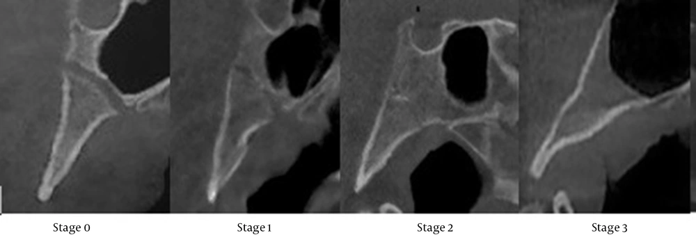
The participants were positioned with the Frankfurt plane parallel to the ground. The CBCT images were acquired using a fixed dental prostheses (FDP)-based CBCT system (NewTom VGi, QR s.r.l., Italy). The measurements were performed at a total exposure time of 1.8 seconds with a field of view of at least 10 × 10 cm2 at 110 kVp. A slice thickness of 1 mm and an interslice distance of 1 mm were considered for the measurements. Two observers independently evaluated all the images. Next, they cooperatively reviewed them and reached consensus regarding the fusion status of SOS. The sagittal view at the mid-sagittal level was used to assess the fusion status of SOS. The four ossification stages of SOS were classified as follows (Figure 1) (5): (1) stage 0, unfused; (2) stage 1, endocranial fusion; (3) stage 2, ectocranial fusion; and (4) stage 3, complete fusion. Finally, the SOS status was determined based on sex and age.
3.1. Data Analysis
Data analysis was performed in SPSS for Windows version 26 (IBM Corp. Released 2019. IBM SPSS Statistics for Windows, Version 26.0. Armonk, NY: IBM Corp.) at a P-value less than 0.05 as the level of statistical significance. Descriptive statistics, including mean, standard deviation (SD), range, and confidence interval, were measured for both groups. Kolmogorov-Smirnov and Shapiro-Wilk tests were also applied to assess the normal distribution of data. Based on the results, the distribution of variables was not normal, and therefore, Mann-Whitney test was performed to indicate differences in age and SOS fusion stage according to sex. Besides, Spearman’s correlation coefficient test and regression analysis were carried out to assess the correlation between age and fusion stage. Moreover, Kruskal-Wallis test was employed to compare the median age and SOS fusion stage based on sex.
4. Results
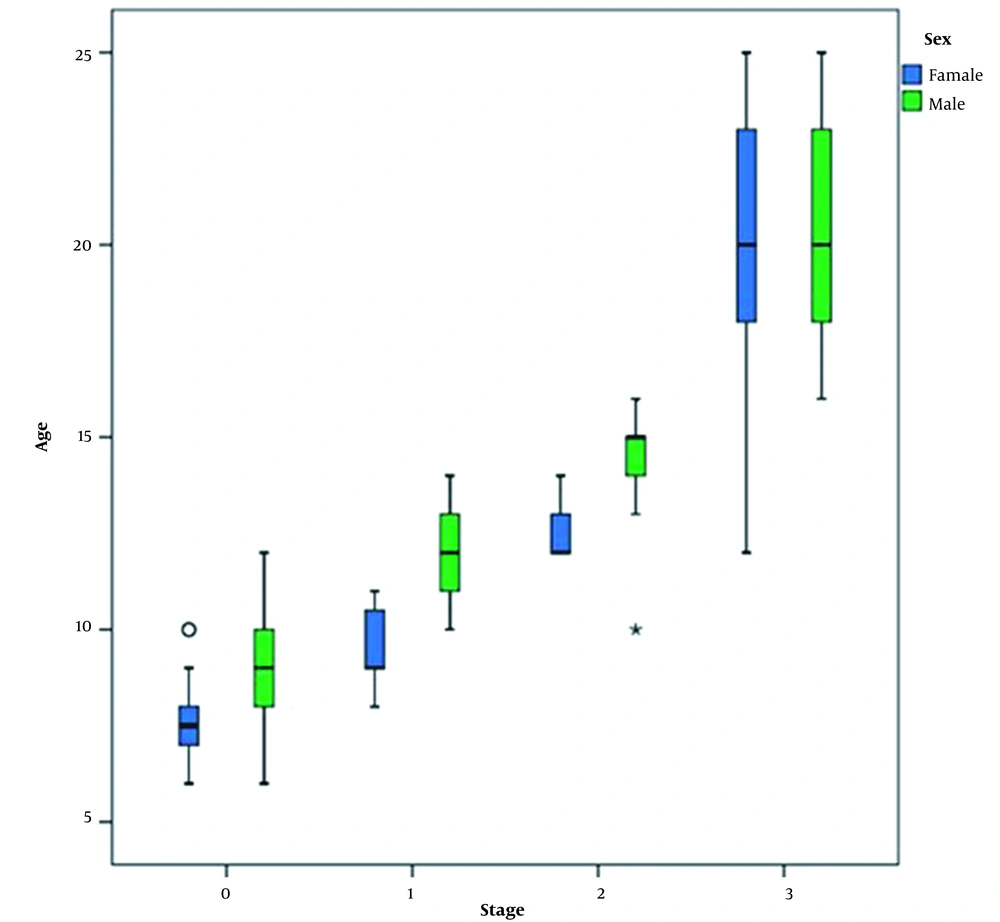
The study population of this study included 126 females and 114 males, aged 6 - 25 years (average age, 17.31 and 15.89 years in females and males, respectively). The participants’ characteristics, such as sex, age, and SOS fusion stage, are presented in Table 1 and Figure 2. There was one 10-year-old male at stage 2 and two 10-year-old females at stage 0. Based on the descriptive statistics in Table 2, the SOS was open at the mean (SD) age of 7.63 (1.25) years in females and at 8.85 (1.5) years in males. Females preceded males regarding the mean age of stage 1, as it occurred about two years earlier in females than males. The oldest age at which fusion persisted as incomplete (stage 2) was 14 years for females (mean, 12.5 years) and 16 years for males (mean, 14.31 years).
| Stages | Age groups | |||||||||||||||||||
|---|---|---|---|---|---|---|---|---|---|---|---|---|---|---|---|---|---|---|---|---|
| 6 | 7 | 8 | 9 | 10 | 11 | 12 | 13 | 14 | 15 | 16 | 17 | 18 | 19 | 20 | 21 | 22 | 23 | 24 | 25 | |
| 0 | ||||||||||||||||||||
| Female | 3 | 5 | 5 | 1 | 2 | - | - | - | - | - | - | - | - | - | - | - | - | - | - | - |
| Male | 1 | 5 | 5 | 5 | 8 | - | 2 | - | - | - | - | - | - | - | - | - | - | - | - | - |
| Total | 4 | 10 | 10 | 6 | 10 | - | 2 | - | - | - | - | - | - | - | - | - | - | - | - | - |
| 1 | ||||||||||||||||||||
| Female | - | - | 1 | 3 | 1 | 2 | - | - | - | - | - | - | - | - | - | - | - | - | - | |
| Male | - | - | - | - | 1 | 4 | 4 | 4 | 3 | - | - | - | - | - | - | - | - | - | - | - |
| Total | - | - | 1 | 3 | 2 | 6 | 4 | 4 | 3 | - | - | - | - | - | - | - | - | - | - | - |
| 2 | ||||||||||||||||||||
| Female | - | - | - | - | - | - | 7 | 4 | 1 | - | - | - | - | - | - | - | - | - | - | - |
| Male | - | - | - | - | 1 | - | - | 2 | 3 | 4 | 3 | - | - | - | - | - | - | - | - | - |
| Total | - | - | - | - | 1 | - | 7 | 6 | 4 | 4 | 3 | - | - | - | - | - | - | - | - | - |
| 3 | ||||||||||||||||||||
| Female | - | - | - | - | - | - | 1 | 1 | 4 | 1 | 2 | 10 | 14 | 5 | 9 | 6 | 7 | 14 | 9 | 8 |
| Male | - | - | - | - | - | - | - | - | - | - | 2 | 10 | 7 | 7 | 7 | 7 | 2 | 6 | 5 | 6 |
| Total | - | - | - | - | - | - | 1 | 1 | 4 | 1 | 4 | 20 | 21 | 12 | 16 | 13 | 9 | 20 | 14 | 14 |
| Stages | N | Age (y) | P value |
|---|---|---|---|
| 0 | < 0.001 | ||
| Male | 26 | 8.85 ± 1.54 | |
| Female | 16 | 7.63 ± 1.25 | |
| 1 | |||
| Male | 16 | 12.25 ± 1.23 | |
| Female | 7 | 9.57 ± 1.13 | |
| 2 | |||
| Male | 13 | 14.31 ± 1.65 | |
| Female | 12 | 12.5 ± 0.67 | |
| 3 | |||
| Male | 59 | 20.34 ± 2.78 | |
| Female | 91 | 20.25 ± 3.27 |
a Values are expressed as mean ± SD unless otherwise indicated.
The results of spearman’s correlation test showed a significant positive correlation between age and fusion stage in both males and females (rs = 0.783 in females and rs = 0.911 in males; P < 0.001). Besides, the results of Mann-Whitney U test for evaluating sex differences in the SOS fusion stage indicated significant differences between males and females at the age of 12 (P = 0.01), 13 (P = 0.024), and 15 (P = 0.04) years; however, no significant difference was found in other age groups.
Additionally, Kruskal-Wallis test was used to compare the median age between SOS fusion stages. The results showed a significant difference between different age groups of females (P < 0.001), except between the mean ages of stage 0 and stage 1 (P = 0.440) and between the mean ages of stage 1 and stage 2 (P = 0.143). On the other hand, there was a significant difference between all age groups of men. With an increase in the mean age, the fusion stage increased. In other words, the mean age at stage 3 was higher than the mean age at stage 2, and the mean age at stage 2 was higher than the mean age at stage 1.
Moreover, a regression analysis was performed separately for males and females by considering age and degree of fusion as dependent (Y) and independent (X) variables, respectively. The R-squared (r2) value generally indicates the fitness of the model and normally ranges from zero to one. When it approximates one, the model can significantly explain the total variance in the dependent variable (y). In this study, the r2 value was estimated at 0.710 in females and 0.809 in males. These findings confirmed the application of SOS fusion degree for age estimation. Based on the results presented in Table 3, the linear regression model indicated the following formulae for age prediction (Y: age, X: SOS stage):
| Model | Unstandardized coefficients | Standardized beta coefficient | R2 | |
|---|---|---|---|---|
| B | Std. error | |||
| Female | 0.710 | |||
| Constant | 6.476 | 0.676 | ||
| Stage | 4.493 | 0.257 | 0.844 | |
| Male | 0.809 | |||
| Constant | 8.492 | 0.404 | ||
| Stage | 3.854 | 0.176 | 0.900 | |
Table 3 presents the linear regression model parameters for age estimation in females and males, based on the SOS fusion degree.
5. Discussion
The present study evaluated the efficacy of SOS fusion degree in age estimation for the Iranian population. Previous studies (17, 22, 23) have reported differences in the time of fusion between different populations due to genetic, geographical, and socioeconomic factors. These factors are known to play a considerable role in age estimation, which can be applied in different related areas, such as forensic science, orthodontics, and surgery, as children undergo different developmental stages until adulthood (24, 25). The present findings demonstrated that the SOS fusion degree is positively correlated with age in both males and females (P < 0.001).
The degree of SOS fusion has been widely investigated in macroscopic, histological, radiographic, and CT scan studies for age estimation (9, 10, 24, 25). Because of detailed examinations in CT scans, they have been used more frequently than macroscopic and two-dimensional radiographic methods (4, 6, 26). Generally, both CT and CBCT scans have advantages, such as high resolution and three-dimensional image acquisition; therefore, the SOS fusion degree can be detected straightforwardly. Besides, CBCT can produce images with good quality and reduced radiation (27).
Different scoring methods have been suggested for evaluating the status of SOS. In this regard, Bassed et al. introduced a five-stage scoring system with scar formation (28). Also, based on macroscopic studies, Shirley and Jantz suggested that a fusion scar might persist for decades following fusion (6); therefore, these scars cannot be regarded as a recent sign of fusion, and they are not applicable for age estimation. Accordingly, in the present study, a four-stage scoring system, recommended by Franklin and Flavel, was employed (23).
In the current study, the oldest female with an open SOS (stage 0) was 10 years old, whereas the oldest male was 12 years old. The fusion of SOS started about two years earlier in females than males. This finding is consistent with the results of previous studies (4, 23, 25, 26), which reported that fusion occurred about two years earlier in females. Shirley and Jantz observed that fusion in females occurred four years earlier than males (6). In another study, the mean age at the onset of puberty was 9.74 years, depending on breast development in the Iranian population (29), which is very close to the age of stage 1 in the current study (9.57 years). Besides, menarche occurred at a mean age of 12.68 years; similarly, the mean age at SOS fusion stage 2 was 12.25 years in the current study; this stage can be considered as a critical point for mandibular growth modification, as the growth spurt is terminated up to SOS fusion stage 2. On the other hand, since the SOS directly affects maxillary growth (7), growth modification of maxilla with appliances, such as facemasks, is mostly predictable in SOS fusion stage 0 and stage 1.
Compared to some previous studies, in the present study, the mean age of complete SOS fusion varied in different populations and age groups. In the modern Australian population, Bassed et al. reported complete fusion at the age of 17 for males and females (28). In another study, the mean age of fusion in the Turkish population was 18.21 years in females and 20.02 years in males (18). Moreover, in the modern American population, the corresponding age was 15.25 years in females and 16.41 years in males (22). In the current study on the Iranian population, the mean age of complete fusion was 20.25 years in females and 20.34 years in males. In line with the results of studies by Can et al. and Sinanoglu et al., the current study reported a similar age range, with a significant difference between females and males, which might be due to sex differences in skeletal growth cessation (4, 18).
The measured r2 value in the present study indicated the fitness of the model. The r2 value normally ranges from zero to one. As discussed earlier, when this value is close to one, it indicates that the model can significantly explain the total variance in the dependent variable (y). In the current study, the r2 values were 0.71 and 0.8 for females and males, respectively, which indicated the better fitness of the model for the data as compared to previous studies (4, 30). Moreover, the results of one-way ANOVA showed a significant difference in males in different age groups; in other words, with an increase in the fusion stage, the mean age increased, which is consistent with the results of a study by Akhlaghi et al. (24). On the other hand, the mean age of individuals with SOS fusion stage 3 was higher than that of individuals with SOS fusion stage 2; also, the mean age of cases with SOS fusion stage 2 was higher than that of cases with SOS fusion stage 1.
According to a cadaveric study on the Iranian population, Akhlaghi et al. found mean ages of 19.44 and 21.17 years for complete fusion in females and males, respectively. Also, the minimum and maximum age for complete fusion was 12 - 26 years in females and 15 - 26 years in males (24). In the same population, based on our CBCT evaluation, a mean age of 20.25 years (age range, 12 - 25 years) for females and a mean age of 20.34 years (age range, 16 - 25 years) for males were reported for complete fusion. Considering the similar ethnicity of the groups, these results may indicate the potential of CBCT to estimate age. According to the current results, it is suggested to use SOS for age estimation when CBCT is available for diagnosis. However, further studies are recommended on the relationship between SOS fusion and skeletal age to approve these findings.
In conclusion, a four-stage system could be used for the Iranian population, aged 6 - 25 years, to estimate the age of SOS fusion. The fusion of SOS was positively correlated with age in the Iranian population, and its fusion started earlier in females than males (about two years). Considering the SOS stage, the linear regression model presented age estimation formulae which are applicable for the Iranian population. This estimation may be also valid for orthodontists and clinicians to plan a reliable treatment for patients.


