1. Introduction
Bronchobiliary fistula (BBF) is an abnormal communication between the bronchial tree and the biliary tract. It occurs as a rare complication following trauma, various hepatobiliary diseases, hepatobiliary surgery, and other procedures. It is also associated with congenital anomalies (1, 2). In the general pathogenic mechanism of BBF, biloma or hepatic abscess, due to biliary obstruction, triggers an inflammatory response and erodes the diaphragm, with subsequent rupture into the bronchial trees (3).
There are many clinical symptoms associated with BBF, including cough, fever, and jaundice; the most pathognomonic symptom is biliptysis (1, 2). BBF can cause severe pulmonary complications that can be fatal; therefore, rapid diagnosis and treatment are important (1, 2, 4). With the development of modern medicine, minimally invasive techniques, such as direct embolization of the BBF, have been employed (1-8). Herein, we report a case of BBF secondary to biloma, which was successfully treated with percutaneous transhepatic embolization using n-butyl cyanoacrylate (NBCA) and microcoils.
2. Case Presentation
A 56-year-old man visited the department of urology as an outpatient due to black-colored urine to undergo an abdominopelvic computed tomography (CT) work-up for suspected hilar cholangiocarcinoma (Bismuth type IIIa). His initial total bilirubin level was 3.5 mg/dL; however, one month later, he showed jaundice, and a follow-up laboratory test revealed that his total bilirubin level had increased to 13 mg/dL. For diagnostic and therapeutic purposes, endoscopic biopsy of the common hepatic duct and endoscopic nasobiliary drainage (ENBD) tube insertion via endoscopic retrograde cholangiopancreatography (ERCP) were performed.
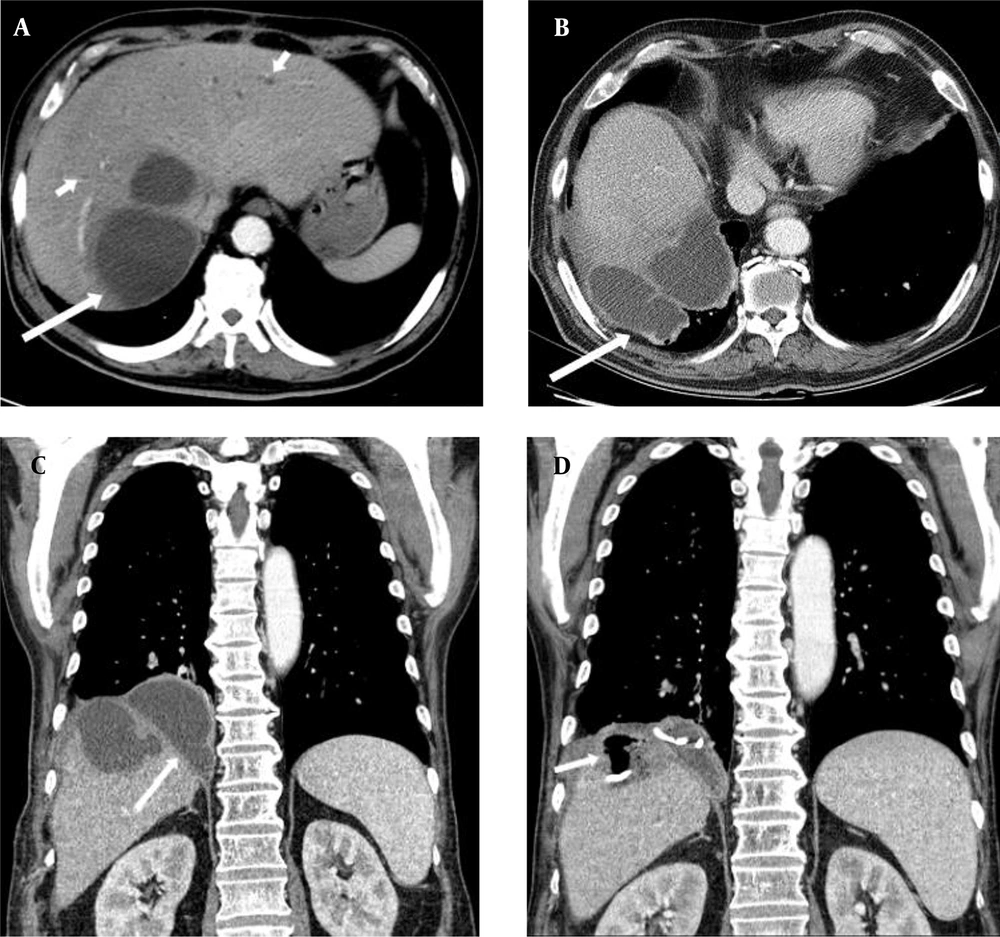
A histopathological examination of the biopsy sample revealed hilar cholangiocarcinoma. Using exploratory laparotomy, invasion of the right hepatic artery and multiple enlarged regional lymph nodes were observed (T4N2M0); accordingly, only palliative cholecystectomy was performed. Two months after the operation, the patient presented with general weakness and fever. An abdominopelvic CT scan demonstrated biloma in hepatic segments 7 and 8 (Figure 1A). Percutaneous transhepatic biliary drainage (PTBD) tube and metallic biliary stent insertion were performed for biliary tract decompression, and percutaneous catheter drainage (PCD) tube insertion was performed for biloma drainage (not shown). The PTBD and PCD tubes were removed as the patient's symptoms improved.
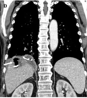
A 56-year-old man with a history of palliative cholecystectomy for cholangiocarcinoma, complaining of severe biliptysis. A, An axial abdominopelvic computed tomography (CT) scan revealing a large biloma (long arrow) in hepatic segments 7 and 8, with intrahepatic bile duct dilatation (short arrows) in both hepatic segments; B and C, Axial and coronal contrast-enhanced chest CT scans, revealing the increasing biloma extension into the right subphrenic space (arrow); D, The follow-up coronal contrast-enhanced chest CT scan revealing a further decrease in the size of biloma, air (arrow), and percutaneous catheter drainage tube in situ.
Three months after the procedure, the patient complained of severe biliptysis (bile-stained sputum), which made it difficult to lie down and sleep. A contrast-enhanced chest CT scan demonstrated biloma extension into the right subphrenic space (Figure 1B and C). Bronchial wall thickening and peribronchial ground-glass opacities were also observed in the right lower lobe (not shown). Bronchoscopy revealed a large amount of biliptysis in the right bronchial tree, but not a definite, visible fistulous tract (not shown). BBF was highly suspected. Therefore, we initially decided to proceed with conservative treatment through biliary tract decompression, expecting the fistulous tract to close naturally.
The reinsertion of a PCD tube for biloma drainage and regular PTBD tube change were performed. Some in-stent restenosis was observed in the follow-up cholangiogram; therefore, balloon dilatation was performed using a 6-mm balloon catheter. However, no significant improvement was observed in the patient’s symptoms during the three-month follow-up. The follow-up contrast-enhanced chest CT scan revealed the appearance of air in the biloma (Figure 1D), suggesting the presence of BBF. Further management was required for the patient; however, he was not in a good condition to undergo surgery; therefore, the decision to perform an interventional procedure was made.
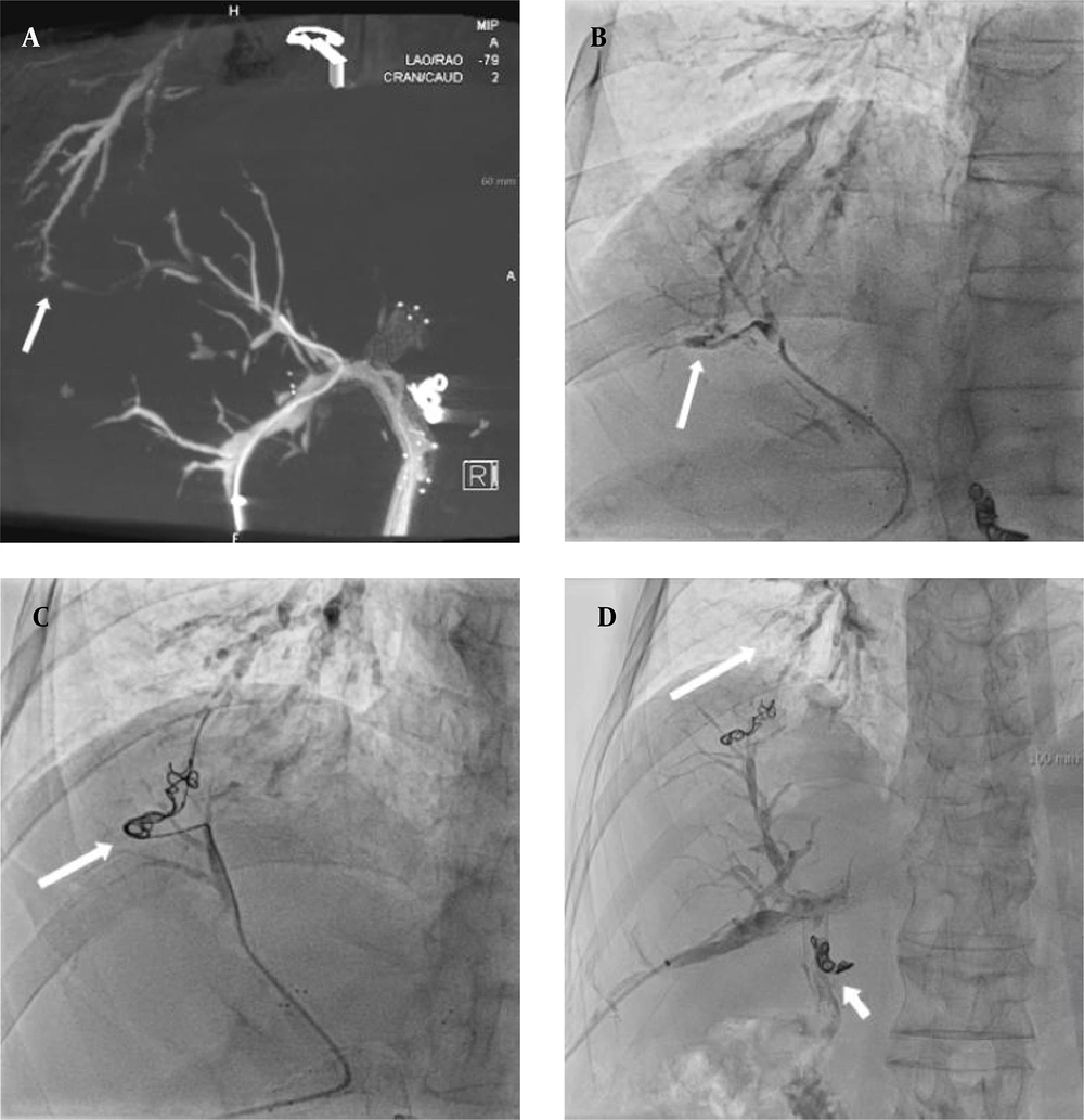
The three-dimensional reconstruction of percutaneous transhepatic cholangiography showed an abnormal communication between the bile duct in hepatic segment 8 and segmental bronchus of the right lower lobe (Figure 2A), which confirmed the presence of BBF. Embolization of the fistulous tract was performed using a 1:3 mixture of n-butyl cyanoacrylate (NBCA; Histoacryl blue, B. Braun; Melsungen, Germany) and ethiodized oil (Lipiodol Ultra-Fluide; Guerbet, Roissy-Charles-de-Gaulle; France and Switzerland) (not shown). Immediately after embolization, his biliptysis and cough improved significantly. Nevertheless, the symptoms recurred occasionally whenever the PTBD tube became clogged; accordingly, additional embolization using NBCA and PTBD tube change was performed several times.
Percutaneous transhepatic cholangiography and the three-dimensional reconstruction image. A & B, A recanalized bronchobiliary fistula between the bile duct in hepatic segment 8 and the posterobasal segmental bronchus in the right lower lobe (arrows); C, Fluoroscopy obtained during embolization indicating microcoils in the fistulous tract (arrow), which were applied through a microcatheter. An additional embolization was performed using a mixture of n-butyl cyanoacrylate and lipiodol (not shown); D, The final cholangiography reveals no contrast medium passage into the bronchobiliary fistula or residual contrast medium in the right lower lobe segmental bronchus (long arrow). Another microcoil may be also observed around the proximal common bile duct due to prior embolization of a ruptured pseudoaneurysm of the gastroduodenal artery stump (short arrow).
After each embolization, biliptysis did not appear for some time, but the patient's symptoms recurred as the BBF was recanalized. In the final procedure, superselective embolization of recanalized BBF (Figure 2B) was performed using microcoils (NESTER: 4 mm × 7 cm, 6 mm × 7 cm; Tornado, 3 mm × 2 cm; Cook Medical; Bloomington, IN, USA) and a 1:1 mixture of NBCA and lipiodol (Figure 2C and D). After the final procedure, the patient’s symptoms did not recur throughout a two-year follow-up, and the right PTBD tube was changed after it was clogged intermittently.
3. Discussion
BBF is a rare disease that can lead to serious complications if not diagnosed and treated promptly. However, there is no consensus regarding the optimal treatment of BBF. Traditionally, treatment of this disease aims to decompress the biliary tract via percutaneous or endoscopic drainage tube insertion. However, natural closure of the fistulous tract requires time, and some patients do not respond well to treatment. Fistulectomy is a definite treatment for BBF, although it is associated with significant morbidity and mortality (1-6). Recently, novel minimally invasive techniques, such as direct embolization of BBF via transhepatic or bronchial approaches using various embolic materials, have been employed (1-8).
In the literature, BBF management using NBCA under bronchoscopic guidance has been reported (5, 7). In the present case, this approach was not employed because the fistulous tract was not visible on bronchoscopy. Moreover, Kim et al. (4) reported the use of percutaneous transhepatic embolization with microcoils and NBCA to treat a BBF secondary to biloma after transarterial chemoembolization for hepatocellular carcinoma. So far, several studies have been carried out using vascular plugs for the embolization of BBF. In this regard, Lee et al. (8) reported the exclusive use of vascular plugs for patients who underwent hemi-hepatectomy due to intrahepatic cholangiocarcinoma. Also, Rice et al. (3) reported a case of BBF secondary to chemotherapy-induced biliary stricture. The patient was treated using vascular plugs and NBCA. However, unlike previous case reports which indicated a relatively short-term follow-up (< 6 months) (4, 8), we showed the effectiveness of percutaneous transhepatic embolization for the treatment of BBF in a relatively long-term follow-up (two years).
Generally, NBCA is an effective endovascular embolic agent that can completely and permanently block the vessels (9). However, the effects vary in different cases of embolization using NBCA for the nonvascular fistulous tract (10-12). Rokni Yazdi et al. (11) reported a successful embolization of BBF, caused by a liver laceration using a mixture of NBCA and lipiodol. However, the use of only NBCA might be ineffective and require additional procedures (12). In our case, biliptysis was not sufficiently resolved when embolization was performed with only NBCA; however, when using microcoils together, the patient's symptoms did not recur.
In conclusion, percutaneous transhepatic embolization is technically more accessible than surgery and may be a promising alternative strategy for BBF. As demonstrated in our case, the use of a combination of microcoils with NBCA could be more effective in long-term follow-ups compared to the use of NBCA alone.


