1. Introduction
Intractable otorrhagia, as a solitary symptom, is not a common manifestation of petrous internal carotid artery (ICA) aneurysms and pseudoaneurysms. Besides idiopathic cases, various etiologies, such as trauma, infection, and radiation, have been suggested for petrous ICA aneurysms. Secured by the petrous bone, these aneurysms are challenging targets for open surgery, making endovascular approaches an advantageous treatment for these cases (1, 2). Considering the variety of endovascular options, such as stenting and coil embolization, besides the rarity of this condition, herein, we report a case of petrous ICA pseudoaneurysm presenting with only otorrhagia, who was treated by coil embolization. Moreover, a review of petrous ICA aneurysms and pseudoaneurysms presenting with otorrhagia was conducted.
2. Case Presentation
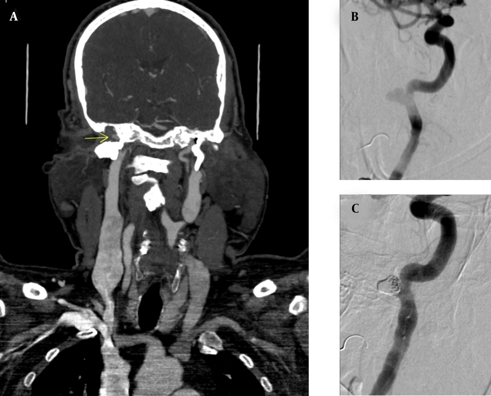
A 58-year-old man, who was a known case of chronic otitis media, was referred from the otorhinolaryngology ward with intractable bleeding from the right ear. He had a history of tympanoplasty and mastoidectomy due to chronic otitis media 23 years ago. Although he never mentioned otorrhagia in his medical history, he had experienced severe hearing loss and episodes of otorrhea since the operation. After hemodynamic stabilization, a computed tomography (CT) angiogram of the head and neck was requested for the patient, which was highly suspicious of a pseudoaneurysmal lesion of the right ICA. Considering the possible location of the ICA lesion, the patient underwent conventional angiography, which indicated a 7 × 5 mm pseudoaneurysm in the right petrous ICA and confirmed our previous findings.
Femoral artery access was replaced with a 7F vascular access sheath (Accura Systems Inc., USA). Using a 7F ConcierGE guiding catheter (Merit Medical Systems, Inc., USA), an Echelon-14 microcatheter (Ev3 Inc., USA) was inserted into the pseudoaneurysm by a SilverSpeed-10 micro-guidewire (Ev3 Inc., USA). The pseudoaneurysm was packed with an Axium Prime 3D coil (6 mm × 20 cm; eV3 Inc., USA) and Axium Prime helical coil (6 mm × 20 cm; eV3, Inc.), with no complications. At the time of this procedure, no flow diverter stent was available in our center. After the procedure, a final angiography of the right ICA was performed, revealing normal blood flow through the ICA, with obliteration of pseudoaneurysm. The patient visited the center twice in the past year, and no signs of aneurysm recurrence, otorrhagia, or other symptoms were found (Figure 1).
3. Discussion
Massive otorrhagia has limited differential diagnoses, such as glomus tumors and vascular lesions, including uncovered jugular bulbs, ICA aneurysmal lesions, hemangioma, and arteriovenous malformations (3, 4). Although few of these differential diagnoses, such as glomus tumors, may appear the same as petrous ICA on physical examination, a CT angiography of the head and neck can provide a definite diagnosis (4). Nearly 40% of aneurysms in the human body originate from the internal carotid artery; however, different parts of this artery are not similarly involved in aneurysm formation (5). Petrous ICA is less affected by aneurysms compared to other intracranial arteries (6). Infection and trauma are among the main etiologies for aneurysm formation (7, 8).
In a systematic review investigating the manifestations of petrous ICA aneurysms, the most common manifestations were cranial nerve palsy for primary aneurysms and bleeding for secondary aneurysms (2). The present case was an aneurysm of petrous ICA, which only presented with intractable otorrhagia, possibly originating from chronic otitis media. The angiography of ICA confirmed the diagnosis of an aneurysmal lesion in the petrous segment of ICA. Since access to this area of the head and neck is challenging, and serious complications may occur during surgery, open surgery is not the primary intervention (9). Today, minimally invasive endovascular techniques, such as coil embolization and stenting, have replaced open surgery (1).
In this study, we also conducted an extensive literature review in the NCBI database for petrous ICA aneurysms and pseudoaneurysms, presenting with massive bleeding from the ears. A total of 10 similar cases were found in 10 articles, as summarized in Table 1. The etiology of aneurysm was infection in four cases (10-12), congenital in two cases (13, 14), idiopathic in two cases (4, 15), radiation-induced in one case (16), and iatrogenic in one case (1). Only three out of 10 cases reported coil embolization. Moreover, Yu et al. used coil embolization for the treatment of petrous ICA aneurysms; nevertheless, nine years after this procedure, the patient presented to the hospital with hematoma and recurrence of otorrhagia. The coil wire had ruptured the aneurysmal lesion, and an iatrogenic pseudoaneurysm was formed. The patient was sent to the operating room, and after removing the coil, extracranial-intracranial bypass was performed, and ICA was occluded (1).
| Authors | Year | Age | Sex | Symptoms | Etiology | Treatment |
|---|---|---|---|---|---|---|
| Anderson et al. (15) | 1972 | 19 | Female | Otorrhagia | Idiopathic | Occlusion with a metallic clip |
| Holtzman and Parisier (13) | 1979 | 35 | Male | Otorrhagia; Hearing loss | Congenital/ infection without a known origin | Common carotid artery ligation and; transection |
| Chiappetta et al. (17) | 1983 | 60 | Man | Otorrhagia | Infection | Common carotid artery ligation |
| Forshaw et al. (4) | 2000 | 20 | n/a | Otorrhagia; Hearing loss; Tinnitus | Idiopathic | Balloon occlusion |
| Cheng et al. (16) | 2001 | 35 | Male | Otorrhagia; | Radiation-induced | Stenting |
| Oyama et al. (12) | 2010 | 60 | Male | Otorrhagia; | Chronic otitis media | Coil embolization, proximal ICA ligation, and EC bypass |
| Mascitelli et al. (11) | 2015 | Middle aged | n/a | Otorrhagia; Hearing loss | Chronic otitis media | Stenting |
| Nemeth et al. (10) | 2017 | 68 | Female | Otorrhagia; Otorrhea; Otalgia | Malignant otitis externa | Stenting |
| Akhtar et al. (14) | 2017 | 13 | Male | Otorrhagia; | Congenital | Superficial temporal artery/middle cerebral artery bypass |
| Yu et al. (1) | 2018 | 58 | Male | Otorrhagia; | Iatrogenic | Extracranial-intracranial (EC-IC) bypass with ICA occlusion |
A Review of Cases Similar to the Present Case
Mascitelli et al. reported a case of mycotic aneurysm of petrous ICA, for whom coil embolization was carried out. However, 12 days after coil embolization, the episodes of otorrhagia were repeated, and stent insertion was performed as the final treatment (11). Additionally, Oyama et al. performed coil embolization, proximal ICA ligation, and external carotid bypass for a patient with a petrous ICA pseudoaneurysm and a history of chronic otitis media, without any complications (12). To the best of our knowledge, this was the first case of petrous ICA pseudoaneurysm, presenting with only otorrhagia, who was treated with coil embolization without any complications. We managed to obliterate the petrosal pseudoaneurysmal sac by coil embolization, with no damage to the main blood flow in ICA and no further need for a bypass.
Based on the present findings, physicians should consider petrous ICA aneurysms and pseudoaneurysms as lethal differential diagnoses after finding no apparent cause for intractable ear bleeding. Besides, CT angiography of the head and neck and confirmatory angiography of ICA can rule out other differential diagnoses. Although some studies have reported complications arising from coil embolization, by using a proper technique, coil embolization can be a less invasive and lifesaving treatment option for the management of petrous ICA pseudoaneurysms. Therefore, coil embolization of a narrowneck petrous ICA pseudoaneurysm can be a safe and suitable treatment option for this type of pseudoaneurysm.

