


1. Background
Ischemic coronary artery disease (CAD) is known as the most common cardiovascular disease due to the plaque buildup in the walls of arteries supplying blood to the heart. It is the leading cause of mortality and morbidity in most industrialized countries (1). Coronary artery calcification (calcified plague score < 100, 100 - 300, and > 300 on computed tomography scan without contrast), higher proportions of non-calcified materials, and higher remodeling indices are associated with a higher risk of CAD in these patients (2). A larger plaque volume is another major risk factor for CAD. In other words, the larger the plaque is, the greater the probability of stenosis will be.
According to previous studies, the coronary plaque volume is larger in patients with stable angina (2, 3). Coronary angiography is the gold standard for diagnosing CAD, providing a good visualization of luminal changes in coronary arteries. However, coronary angiography is an invasive method, associated with morbidity and mortality. Besides, it can only exhibit the coronary lumen in a two-dimensional outline which cannot represent the complex nature of atherosclerotic plaques responsible for the link between the clinical outcomes and angiographic findings (4). Moreover, acute coronary events mainly occur in noncritical lesions (< 50% luminal stenosis), highlighting the need for complementary imaging techniques to characterize the plaque composition and detect vulnerable plaques (2).
Coronary computed tomography angiography (CCTA) and coronary artery calcium score (CACS) are two main techniques used for cardiac CT imaging. CCTA is a non-invasive diagnostic procedure providing imaging of heart chambers, pulmonary vessels, plaque characteristics, and coronary arteries in three dimensions (5). It allows a precise quantification of plaque burden, patent luminal area of plaque, and luminal area (3). The use of contrast agents in CCTA facilitates visualization of calcified and non-calcified plaques, as well as detection of coronary stenosis (6-8). The main characteristics of plaques examined by CCTA include plaque composition, spotty calcifications, and positive remodeling. Therefore, the plaque characteristics may effectively predict the clinical outcomes of CAD (3, 8).
2. Objectives
So far, few studies have been conducted on the relationship between plaque characteristics, such as plaque dimensions, and stenosis in patients undergoing CCTA, and this association is still remained unclear (9). Therefore, the current study is aimed to assess the relationship between the characteristics and dimensions of calcified plaques and coronary artery stenosis in patients undergoing CCTA.
3. Patients and Methods
3.1. Study Design and Sample
This cross-sectional study was conducted on 211 plaques from patients above the age of 18 who were candidates for CCTA in our center. Sampling was performed using a non-random method. The exclusion criteria were allergy to iodine contrast, kidney disorders (creatinine ≥ 1.7 mg/dL), arrhythmia, resting heart rate > 100 bpm, resting systolic blood pressure < 100 mmHg, contraindications to beta-blockers (e.g., concurrent application of calcium channel blockers), pregnancy, and the history of CAD. The Ethics Committee of Isfahan University of Medical Sciences approved this study (IR.MUI.MED.REC.1398.190). The participants were enrolled in this study after signing informed consent forms.
3.2. Image Acquisition and Analysis
The 128-slice prospective and retrospective ECG-gated CT scanning (General Electric Healthcare, USA) was used to perform all CT angiography studies using a standardized protocol via spiral scanning at 120 kV. Beta-blocker (metoprolol, 5 - 20 mg) and nitroglycerin (sublingual, 0.4 mg) tablets were administered before scanning under the supervision of a cardiologist to reduce the heart rate to < 60 bpm and induce the dilation of coronary vasculature. A non-ionic contrast medium was injected into the antecubital vein at a rate of 4.5 - 6 mL/s and then flushed with 20 - 40 mL of saline for contrast-enhanced scanning. The contrast dose was adjusted to the body weight and duration of the study.
Before contrast-enhanced scanning, the test bolus technique was utilized to determine the exact delay time, with the ascending aorta as the point of reference, which was defined as four seconds after the peak time in the ascending aorta. A prospective ECG-gated scan was performed in patients with a heart rate below 65 bpm, with the central point of the triggering window set at 70% of the RR interval, while other patients underwent a retrospective ECG-gated scan. The table pitch was synchronized with the heart rate, scanning through the craniocaudal direction; the starting point was set at the coronary ostia while the ending point was set at the diaphragm to include all cardiac structures. The table pitch in the retrospective ECG-gated scan was 0.18 to 0.24, adjusted for the patient's heart rate and size; however, it was not defined in prospective ECG-gated scanning. The section thickness was 0.625 mm with a gantry rotation time of 300ms. Image reconstruction was defined with a matrix size of 512×512 with 0.4 mm increment using a soft-tissue convolution kernel.
An experienced radiologist evaluated all the images. Only calcified plaques were evaluated in this study. The plaque location was reported according to the coronary artery branches, including the left anterior descending artery (LAD), left circumflex artery (LCX), left main artery (LMA), and right coronary artery (RCA). The length, width, and thickness of the plaque and the luminal diameter were reported in millimeter (mm) while the plaque and luminal areas were reported in mm2. Moreover, the largest end-to-end plaque area was measured to evaluate the plaque luminal area.
The luminal area was measured at proximal and distal to the stenosis caused by the plaque. In other words, if the plaques were located in the middle of the desired vessel, the luminal area was measured considering the proximal part of the plaque as the normal reference, if the plaques were located in a place such as the origin or bifurcation point to the desired coronary vessel, the diameter of the lumen was considering the distal part of the plaque as the normal reference. Luminal stenosis was reported as percentage and calculated based on the following formula:
Luminal stenosis (%) = (Plaque patent luminal area in the lumen)/ (luminal area) × 100
The area proximal to the plaque was typically considered to be a normal lumen. Otherwise, the region distal to the plaque was suggested to be a normal lumen. Stenosis was categorized into four groups in terms of the extent of stenosis. Stenosis with 1 - 24%, 25 - 49%, 50 - 69%, and 70 - 99% obstruction was classified as minimal, mild, moderate, and severe, respectively.
3.3. Statistical Analysis
Data analyses were conducted using SPSS Version 20 (released in 2011, IBM SPSS Statistics for Windows, IBM Corp., Armonk, NY, USA). Qualitative variables are expressed as number and percentage, and quantitative variables are expressed as mean and standard deviation (SD). To compare the dimensions of plaques with the vascular stenosis criterion, a logistic regression analysis was performed, where stenosis (yes/no) was considered as dependent variable, and the characteristics and dimensions of calcified plaques were regarded as independent variables. A logistic regression analysis was carried out to investigate the effects of different characteristics and dimensions of calcified plaques on coronary artery stenosis. In this model, a binary variable indicating stenosis > 50% or < 50% was considered as the dependent variable, while the characteristics and dimensions of calcified plaques were regarded as independent quantitative variables.
4. Results
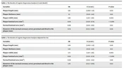
In this study, none of the patients underwent angiographic evaluations, and the possibility of examination could not be evaluated due to the non-random conditions of the plaques. Out of 211 plaques in 54 patients, 26 were in the proximal LAD, four in the LAD origin, 19 in the middle of LAD, eight in the distal LAD, eight in the proximal LCX, five in the distal LCX, three in the middle of LCX, nine in the proximal RCA, four in the middle part of RCA, six in the distal RCA, four in the origin of the left main coronary artery, and five in the distal left main coronary artery. The rest of the plaques were found in both proximal and distal LAD, and a few plaques were detected in the ramus intermedius, diagonal branches (D1 and D2), and obtuse marginal artery (OM). Comparison of atherosclerotic plaque dimensions and luminal stenosis is presented in Table 1.
| Variables | Total (n = 211) | Stenosis < 50% (n = 112) | Stenosis > 50% (n = 99) | P-value |
|---|---|---|---|---|
| Sex male, No. (%) | 122 (57.5) | 61 (50) | 61 (50) | |
| Length (mean ± SD) | 5.3 ± 2.03 | 4.95 ± 2.66 | 5.81 ± 3.35 | 0.092 |
| Thickness (mean ± SD) | 1.79 ± 0.66 | 1.72 ± 0.59 | 1.87 ± 0.72 | 0.017 |
| Width (mean ± SD) | 2.98 ± 0.84 | 2.81 ± 0.85 | 3.1 ± 0.79 | 0.003 |
| Plaque luminal area (mean ± SD) | 6.5 ± 4.43 | 8.52 ± 4.78 | 4.26 ± 2.55 | < 0.001 |
| Normal luminal area (mean ± SD) | 11.56 ± 5.53 | 11.60 ± 5.56 | 11.52 ±5.53 | 0.941 |
| Diameter of the normal coronary artery proximal and distal to the plaque (mean ± SD) | 3.03 ± 1.08 | 3.17 ± 1.25 | 2.86 ± 0.87 | 0.031 |
Comparison of Atherosclerotic Plaque Dimensions and Luminal Stenosis
The results of statistical analysis using Mann-Whitney U test indicated a significant difference in the thickness, width, and luminal area of plaques, as well as the diameter of normal coronary artery proximal and distal to the plaques among individuals with coronary artery stenosis. The mean rate of stenosis was estimated to be 56.1 ± 24.4%. The frequency of stenosis < 50% was 100 (47.3%). A crude model was plotted in this study without confounding factors, followed by an adjusted model including gender. In both models, the evaluated variables were not significant. Therefore, in the adjusted model, the plaque width (OR = 1.73), luminal area of the plaque (OR = 0.64), and diameter of the normal coronary artery proximal or distal to the plaque (OR = 1.70) were significant with the severity of stenosis (P < 0.05) (Tables 2 and 3). In the crude model, the length (OR = 1.10) was also significant. After performing statistical analyses using logistic regression models, a significant relationship was found between stenosis and the luminal area of the normal coronary artery and atherosclerotic plaque and also between stenosis and the plaque width. In other words, with every one-unit increase in any of the significant variables including plaque width (OR = 1.73), plaque luminal area (OR = 0.64), and diameter of the normal coronary artery proximal and distal to the plaque (OR = 1.70), the probability of reduction in plaque stenosis increased by 1.73, 0.64, and 1.7, respectively.
| Variables | OR | CI (0.95%) | P-value |
|---|---|---|---|
| Plaque length (mm) | 1.10 | (1.002 - 1.21) | 0.04 |
| Plaque thickness (mm) | 1.5 | (0.92 - 2.41) | 0.10 |
| Plaque width (mm) | 1.68 | (1.20 - 2.36) | 0.003 |
| Plaque luminal area (mm2) | 0.64 | (0.56 - 0.74) | < 0.001 |
| Normal luminal area (mm2) | 0.99 | (0.95 - 1.04) | 0.92 |
| Diameter of the normal coronary artery proximal and distal to the plaque (mm) | 0.74 | (0.55 - 0.99) | 0.04 |
The Results of Logistic Regression Analysis (Crude Model)
| Variables | OR | CI (0.95%) | P-value |
|---|---|---|---|
| Plaque length (mm) | 1.09 | (1.002 - 1.21) | 0.06 |
| Plaque thickness (mm) | 1.43 | (0.92 - 2.41) | 0.13 |
| Plaque width (mm) | 1.73 | (1.20 - 2.36) | 0.002 |
| Plaque luminal area (mm2) | 0.64 | (0.56 - 0.74) | < 0.001 |
| Normal luminal area (mm2) | 0.99 | (0.95 - 1.04) | 0.88 |
| Diameter of the normal coronary artery proximal and distal to the plaque (mm) | 1.70 | (0.55 - 0.99) | 0.02 |
The Results of Logistic Regression Analysis Adjusted for Sex
5. Discussion
A chest CT scan is a common procedure, which can be used to evaluate calcified plaques in coronary arteries, although it is not the standard protocol for assessing these arteries. According to our study, there was a significant association between the plaque width and thickness and the luminal area of the normal vessel and atherosclerotic plaque with the severity of stenosis. Therefore, plaque dimensions, such as the plaque thickness and width, which were measured in both coronal and longitudinal coronary artery scans (Table 1) as well as the patent luminal area, which could be measured in non-contrast chest CT, could predict the severity of stenosis (more than 50%) with a high likelihood. Additionally, since contrast injection in CT angiography cannot be decisive in some cases, the size of plaques may play a significant role in determining coronary artery stenosis in patients undergoing CCTA.
Invasive coronary angiography is the gold standard for the diagnosis of CAD due to the visualization of the entire coronary tree and coronary luminal changes. However, it is an invasive method associated with mortality and morbidity (2). Studies have reported that high-risk plaques have a larger lipid core, occupying approximately 40% of the plaque volume and demonstrating positive remodeling. These plaques develop into intra-plaque hemorrhage compared to the stable plaques. Therefore, non-invasive detection and analysis in early stages, particularly in low-risk and asymptomatic patients, can improve risk stratification without requiring more invasive methods (10). CCTA affect the coronary artery lumen; moreover, a smaller coronary luminal diameter was related to a higher coronary concentration of IL-1β, which was associated with an increased risk of clinical complications (11). Besides, Dodge et al. demonstrated that knowledge about the normal luminal diameter at the anatomic coronary location could be a more useful quantitative measure of CAD severity compared to stenosis percentage (12).
CCTA is a highly accurate modality for assessing the luminal area, plaque patent luminal area, and percentage of stenosis compared to intravascular ultrasound (13). Gupta et al. assessed the efficacy of plaque thickness by CCTA in carotid artery stenosis (CAS) and demonstrated that an increase in the maximal soft plaque thickness was strongly associated with symptomatic disease status in CAS (9). However, assessment of plaque volume by CTA may be associated with alterations in heart rate, iodine bolus, and/or myocardial motion (14). Moreover, Pasterkamp et al. reported a weak relationship between the intimal thickness and luminal stenosis, which might be attributed to the events of de novo arterial remodeling (15).
Additionally, Ricotta et al. evaluated the plaque characteristics and observed that stenosis diameter and width were predictive of complex atheroma. According to their findings, the plaque width is a valuable parameter for classifying carotid atheroma. Therefore, plaque width should be assessed in future studies on carotid atheroma (16). Although in the present study, we did not evaluate carotid arteries, the plaque width was significantly associated with the stenosis severity of coronary arteries. Finally, based on our findings, the patent luminal area, width, and thickness of plaques could facilitate evaluation of the stenosis severity in coronary arteries. However, available data on this topic is limited, and further studies are essential to obtain more robust conclusions.
In conclusion, our findings demonstrated a significant relationship between stenosis and the width, thickness, and patent luminal area of plaques. Therefore, the plaque dimensions can be considered as a predictive factor for stenosis.
