This retrospective study showed that, in an asymptomatic young population, the frequency of radiological parameters associated with FAI is high. We found that 64.5% of the 400 joints had at least one, and 34.25% of the joints had two or more abnormal morphological parameters associated with FAIs. A review of the literature revealed that there are two types of studies describing the prevalence of the radiological parameters associated with FAIs in symptomatic and in asymptomatic patients. It has been shown in many studies that the prevalence of the radiological parameters is high in symptomatic patients. For example, Nogier et al. (
10) found dysplasia, acetabular pincer effects, or cam effects in 95% of 584 hips in symptomatic patients; and Ochoa et al. (
11) found at least one abnormal parameter associated with FAIs in 135 of 155 patients (87%) with symptoms. In addition, Bowler et al. showed that 65% of 142 patients who had undergone hip arthroplasty had abnormal AAs, indicating cam impingement (
12).
In asymptomatic populations, most studies have focused on cam type morphological features, especially on the measurement of the AA. In only a few studies have both the cam and pincer type radiological parameters been analyzed in asymptomatic patients. For instance, Chakraverty et al. (
2) found at least one abnormal parameter in 66% of 100 joints, and two or more abnormal parameters were present in 29% of the joints without symptoms. In another study, Kang et al. (
13) found that 39% of 100 joints had at least one parameter, and that 11% had more than one parameter associated with FAIs in asymptomatic patients. Both studies used CT images in the assessment of the radiological parameters of the hips.
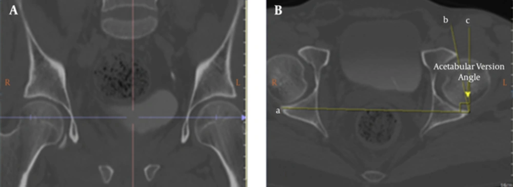
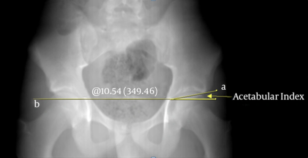
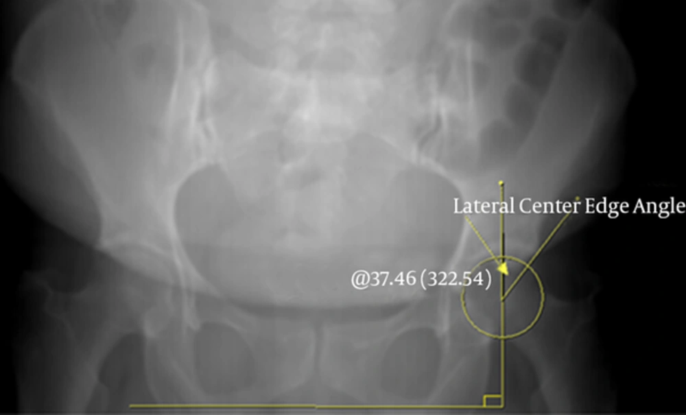
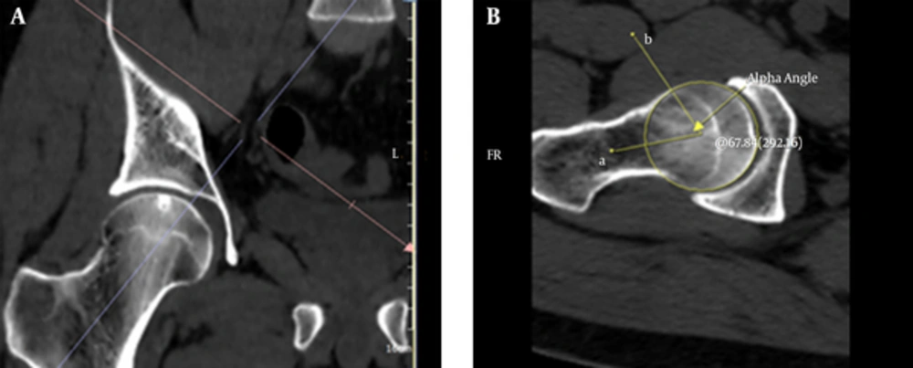
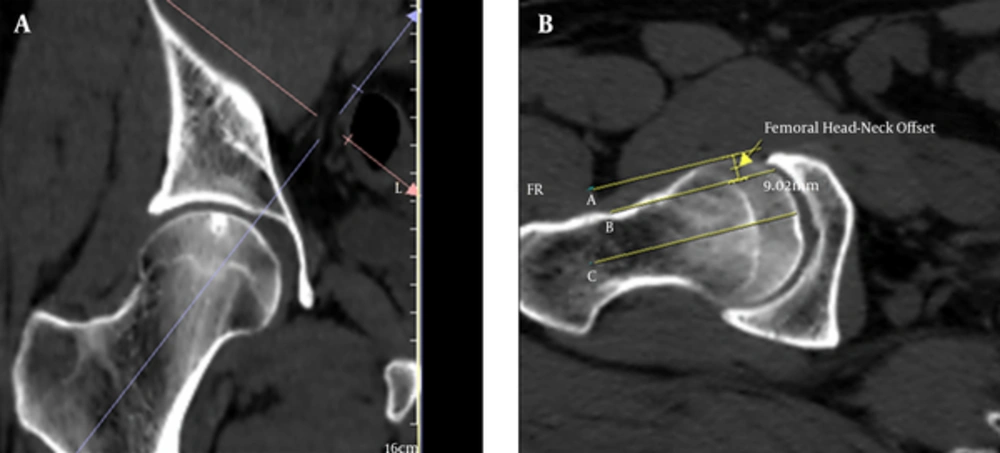
In this study, we evaluated six quantitative parameters (three cam and three pincer) associated with FAI morphology, and used reformatted CT images to calculate the AAs of the femoral head-neck junction in the anterosuperior quadrant, according to the studies of Chakraverty et al. (
2) and Kang et al. (
13). We performed quantitative analyses in only 200 patients (400 joints) and, to our knowledge, this study is the most comprehensive evaluation of FAIs in an asymptomatic population. Seventy-seven of 218 hips (35.3%) in the men displayed at least one abnormality associated with cam FAI, while two or more abnormalities were seen in 48 of the 218 hips (22%). Sixty-six of 182 women (36.3 %) had at least one cam abnormality, and 39 of 182 (21.4%) had two or more. No statistically significant gender difference was detected for the cam FAI morphology, but the overall prevalence was higher than previously reported. Reichenbach et al. (
14) found that 67 of 244 (27.5%) male patients without symptoms had cam type deformities on the hip MRIs. Furthermore, Hack et al. showed that 14% of 200 asymptomatic volunteers had at least one cam type abnormality in an MRI study (
15).
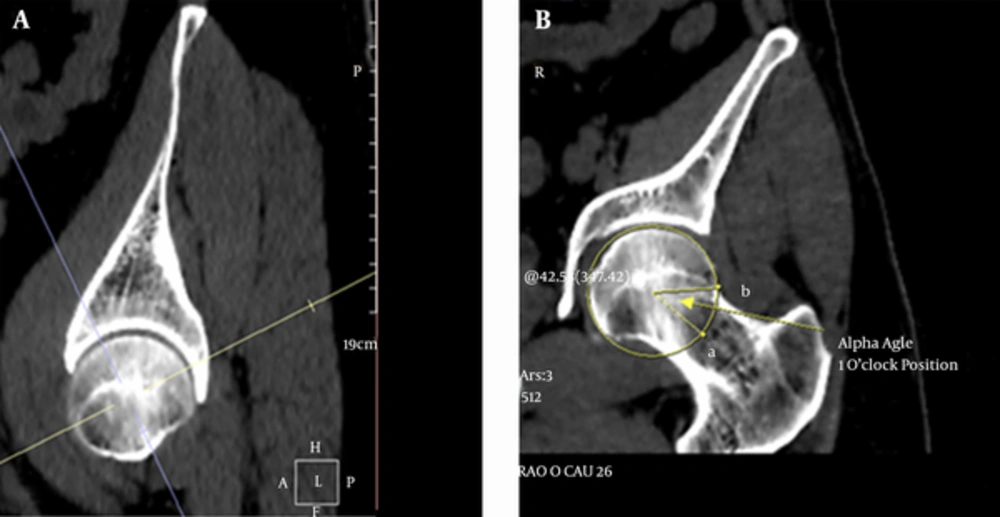
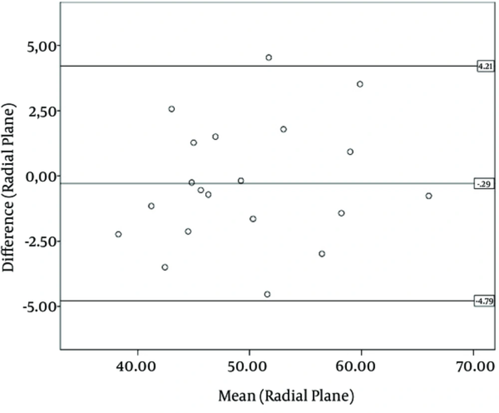
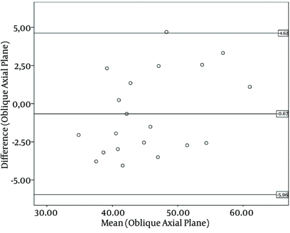
Previous studies (
9,
16,
17) have shown that the measurement of the AA in reformatted radial images is more accurate than the measurement of the angle in the conventional axial oblique plane alone. Therefore, we used reformatted CT images to measure the AA along the anterosuperior quadrant of the femoral head-neck junction in two planes (oblique axial and radial one o’clock position). In addition, we defined an AA greater than 55° to be abnormal in any plane, in accordance with previous studies (
9,
16,
17). We found that measuring the AA in the one o’clock position revealed more abnormalities than in the oblique axial plane, which was a statistically significant finding (P < 0.001). In our study, 79 of 218 hips (36.2%) in the men and 96 of 182 hips (52.7%) in the women had at least one pincer type characteristic. Moreover, twenty-seven hips in the women and 18 hips in the men had two or more characteristics associated with pincer FAIs. Overall, there was a statistically significant gender difference (P < 0.001), and a female predominance in the prevalence of pincer type morphological features, in accordance with the literature (
2,
13).
We found that only 60 of 400 joints (15%) had mixed FAIs in our study. In the literature, an FAI has often been thought to represent mixed type features, but our findings showed little overlap, unlike those of previous studies (
4,
18), and support the results of Cobb et al. (
19) and Laborie et al. (
20). In these studies, the authors concluded that the hips with cam and pincer deformities were distinct pathoanatomical entities; however, the low percentage of mixed FAI features could be due to the number of patients with pincer FAI features in this study. The radiological definitions of the thresholds used for pincer FAIs most often used standard pelvic radiographs, while CTs were less commonly used for this purpose. Therefore, these thresholds may have been larger if standard pelvic radiographs had been used. This study had several limitations. First, we worked on a population without histories or symptoms of hip-related problems with regard to our radiology and hospital databases. Second, we did not perform radial AA measurements through the entire circumference of the femoral head and neck. However, the large number of patients and the use of only quantitative measurements strengthened our study.
In this study, we found that the frequency of the cam and pincer FAI morphologies was high, even in an asymptomatic population, which was similar to the results of other studies. We speculate that the high frequency of abnormal morphologies associated with the FAIs of this healthy young population suggest that the threshold values may have been set too low in the current literature. In addition, the high prevalence of these findings may reflect anatomical variations, rather than true pathological abnormalities. Consensus is required on the threshold values with regard to what is normal and abnormal in all of the imaging modalities. For example, the threshold value of the AA has been suggested to be 50° based on the MRI (
21), and 55° based on the CT (
22). However, the reliability of its measurement and reproducibility have been questioned (
23). We believe that the use of the 55° threshold at the femoral head-neck junction on CT imaging may lead to overestimates of the contour abnormality. Additionally, we do not know how many or which parameters need to be present for the future development of symptomatic FAIs. Therefore, we could not determine what proportion of this population will have symptomatic FAIs in the future, when considering the radiological parameters alone. It is important to understand that the FAI is a morphological and dynamic syndrome. Other variables, such as the BMI (
24), daily activity level, heavy work, and previous trauma, may be important in the development of symptoms. A study (
7) have shown that the rate of hip implantation in ballet dancers at the age of 50 is 25%. Moreover, in a sibling study, Pollard et al. showed that a sibling of a patient with a cam deformity has a relative risk of 2.8, while the siblings of those patients with pincer deformities have a relative risk of 2.0 of having the same deformity, when compared with a control group (
25). In this study, we found that the frequency of radiological parameters associated with cam and pincer FAIs is high, even in asymptomatic populations, as in other studies. This high frequency of abnormal radiological parameters associated with FAIs could be due to the low cutoff values in the current literature. Consensus is needed on the threshold values and the standardized imaging protocols of the hip for all imaging modalities. Those patients presenting with hip pain and indices of femoroacetabular impingement may be suitable candidates for early operative intervention to decompress the impingement, which may potentially delay or even prevent the progression of arthrosis.