1. Introduction
Insufficiency fractures of the sacrum cause pain and limitation of mobility ranging up to complete immobility. Conservative treatment with confinement to bed and analgesic therapy frequently does not achieve a satisfactory reduction in pain and can also lead to the development of decubitus ulcers, venous thrombosis and pulmonary artery embolism as well as pneumonia (1). In contrast to trauma-related fractures, insufficiency fractures mostly affect the area between the iliosacral joint and the neural foramina (type I fracture zone) (2), but they may also radiate into the neural foramina and lead to more severe neurological symptoms, e.g. sphincter dysfunction. Up to 40% of the insufficiency fractures occur bilaterally and may be additionally complicated by horizontal fracture lines running through the sacral vertebrae (3). Percutaneous cement augmentation of the sacrum, analogous to vertebroplasty, has increasingly been used in osteoporosis-related insufficiency fractures over the past few years, successfully treating pain. The more recent use of balloon catheters has made it possible to close fracture gaps and minimize the risk of cement leakage, which has contributed to further improvement of the method (4). In the following case, we report the simultaneous treatment of a bilateral insufficiency fracture of the sacrum with CT-assisted balloon sacroplasty.
2. Case Report
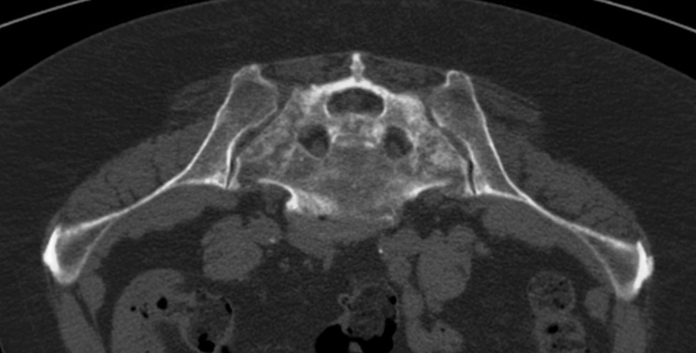
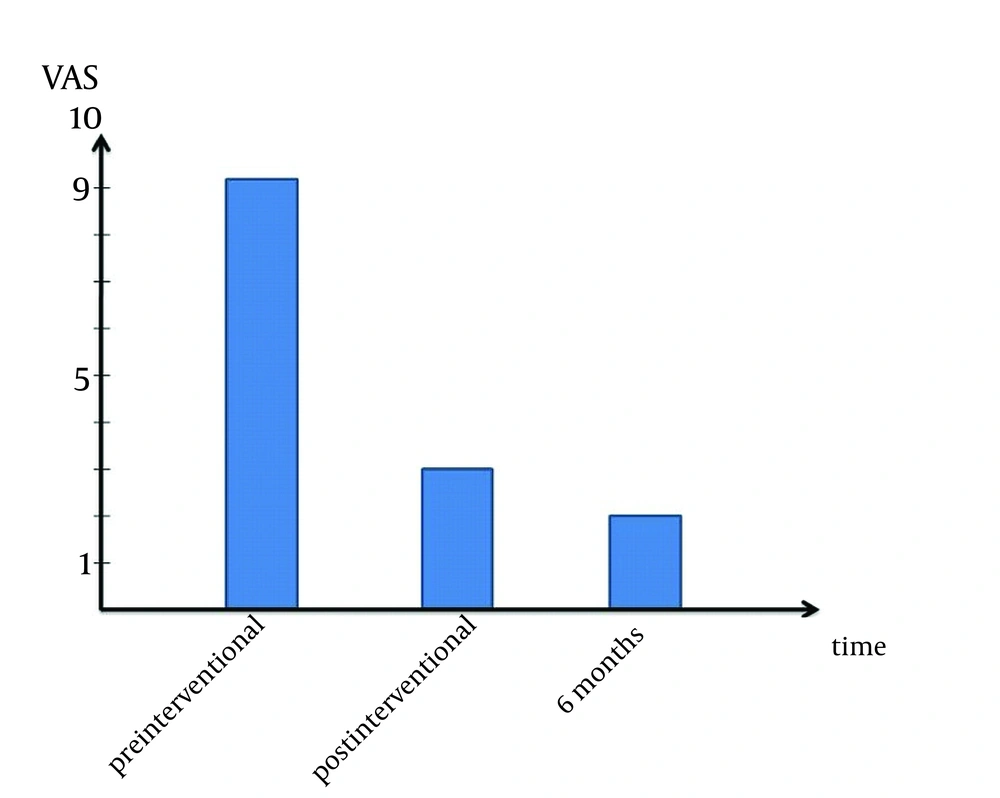
We report a 73-year-old woman, who presented with a sacral fracture that had already been diagnosed in an outpatient setting. In addition, there was strong evidence of a renal cell carcinoma of the right kidney. Despite sufficient pain treatment and accompanying physiotherapy, the patient's mobility was severely restricted by pain at the time of presentation. The patient rated her pain as 9 on a visual analogue scale (VAS) with 10 as the maximum value. Osteoporosis was also suspected and confirmed by bone density measurement, so therapy with bisphosphonates was started in accordance with the guidelines (5). In the preinterventional CT of the pelvis and abdomen, a bilateral fracture line running sagittally through the lateral mass of the sacrum could be seen (Figure 1).
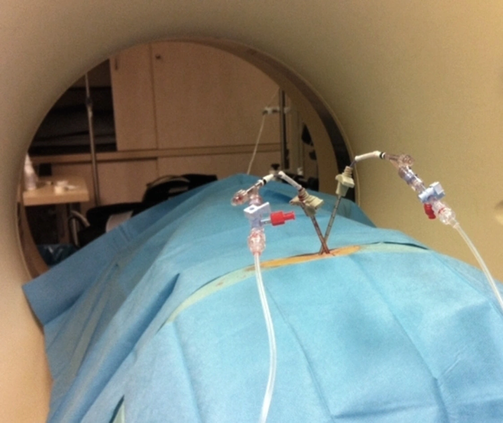
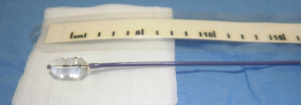
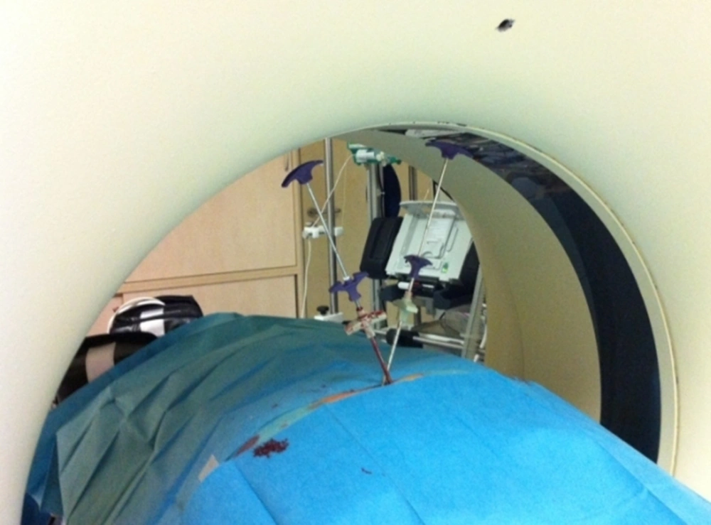
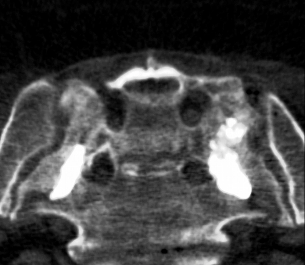
On the left, the fracture line traverses the transforaminal zone at the level of the second sacral vertebra. Both in this examination and in an additionally performed MRI, no metastases of the suspected renal carcinoma were observed in the areas of the body examined. The morphology of the sacral fracture was also most consistent with an insufficiency fracture. In order to enable the further therapy of the renal tumor and to achieve sufficient pain reduction, in an interdisciplinary tumor conference, it was initially decided to treat the sacral fracture by CT-assisted balloon sacroplasty. In order to avoid further fracturing and prolonged hospitalization of the patient, we decided on simultaneous treatment of the bilateral fractures. In addition, a biopsy was to be taken for histopathological analysis for definitive exclusion of metastasis. The intervention was performed under intubation anesthesia and anesthesiological monitoring. The patient was placed in the prone position in the CT scan machine. As is routine practice, a single-shot antibiotic was administered (cefazoline 2 g IV). The short axis of the sacrum was selected as the entry plane. Access to the fracture zones was established from dorsal to ventral by two Kirschner wires, which can be directed and angulated easily without causing further damage in the osteoporotic bone. Along the Kirschner wires, two hollow needles were then placed in the fracture zone. The desired position was observed by single slice CT-images. To begin with, the bone biopsy was performed via the hollow needles. The balloon catheters (Kyphon®, Medtronic, 20 mm length, 6 cc volume) were then inserted (Figure 2 and 3).
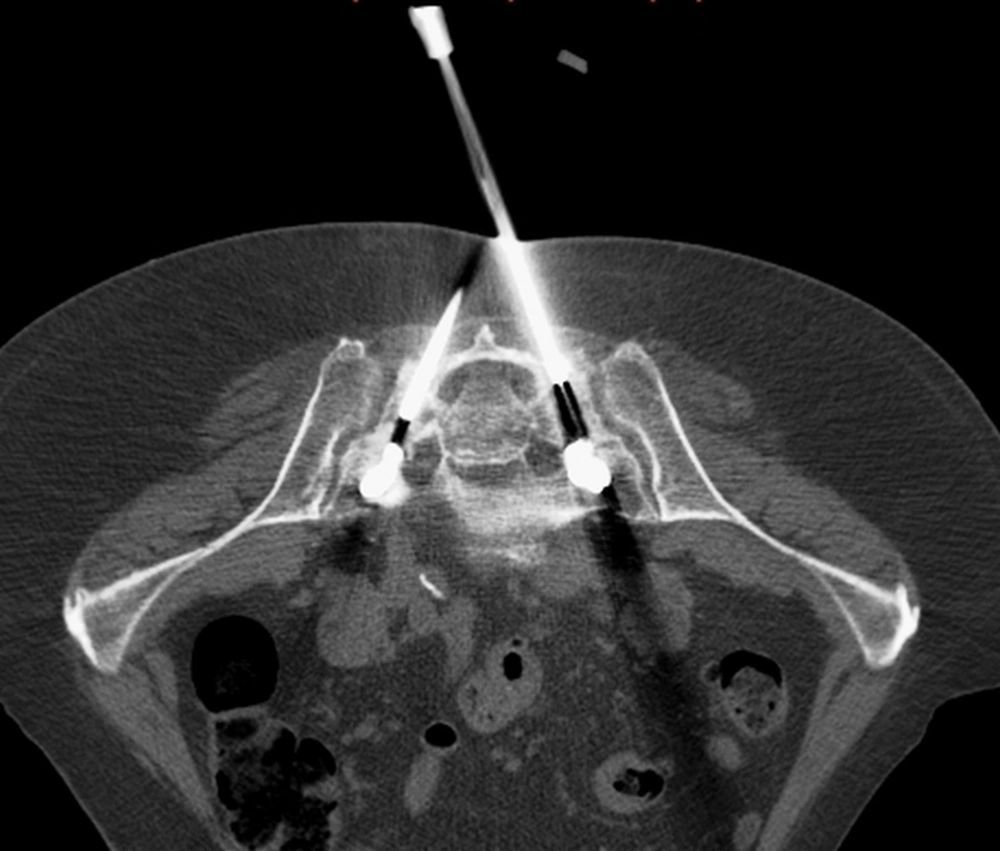
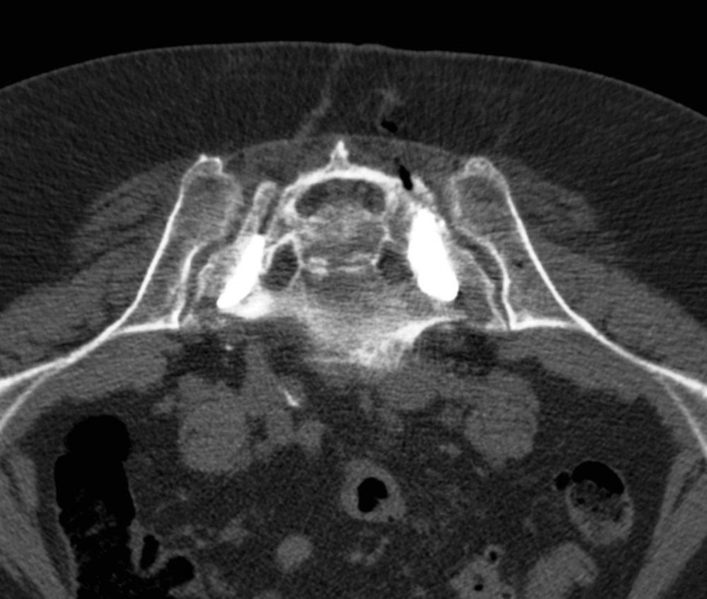
They were inflated and deflated stepwise from the central to peripheral under CT monitoring by means of single slice images (Figure 4). Cement (polymethyl methacrylate, PMMA) application was then performed in the previously prepared hollow space using the low-pressure method under CT monitoring (Figure 5). The final check by thin-slice spiral-CT of the pelvis showed sufficient distribution of the cement, without leakage into the neural foramina, the iliosacral joint or the visceral surface of the sacrum (Figure 6 and 7). In our institution, the cement distribution is usually observed by plain radiography (not shown) to have an initial image for potential follow-up examinations. Two days post-intervention, a pain reduction to VAS 3 was observed (Figure 8). The samples taken during the intervention showed no evidence of malignancy. The patient's previous pain-related restriction of mobility and her general condition improved markedly thereafter, so that the planned oncological therapy, including nephrectomy could be started.
3. Discussion
Percutaneous balloon sacroplasty already showed a marked reduction in pain and increased mobility in patients with insufficiency fractures in the first case reports published (6). By using balloon catheters and CT assistance, a controlled and reliable cement application can be achieved, so that cement leakages and neurological complications can be largely prevented. The compaction of the fractured and destroyed bone and the subsequent sealing with cement increases the stability of the posterior pelvis above all in the area of the neural foramina and appears to be the most important mechanism for prompt reduction of pain. In the meantime, the reliability and effectiveness of balloon sacroplasty in patients with insufficiency fractures has also been shown in a larger patient population (4). Apart from the general risk of anesthesia, and the potential risk of cement leakage into the neural foramina or the surrounding presacral soft-tissue structures, the case reports and studies published to date have not revealed any specific risks of this therapy. In a recent study, common sacroplasty in the vertebroplasty technique (sacrovertebroplasty) showed cement leakage rates of 27% into the fracture gap, which could potentially affect the lumbar nerve roots (7). Due to the use of balloons forming a presized hole and compacting the fracture lines, the rate of leakage into the fracture gap is probably lower in balloon sacroplasty (4). Nevertheless, the patient has to be informed about the risk of neurological impairments before intervention. To prevent any kind of cement leakage, it is important to choose the right strategy for the intervention regarding the access (long- or short-axis), the exact positioning of the balloon and the amount of cement. In the short-axis technique, it is sufficient to use single slice CT-images; whereas, in long-axis access an additional plain radiography using a c-arm turned out to be useful in our experience (4). As a result of the bilateral simultaneous and thus definitive treatment of the fractures, hospitalization of the patient can be kept to a minimum and any other therapies planned are not unnecessarily delayed. As interventional and minimally-invasive pain therapy, balloon sacroplasty also represents a promising treatment option for complex insufficiency fractures or malignant primary diseases within the context of multidisciplinary concepts. In addition, case reports have also shown the successful treatment of pathological fractures of the sacrum by means of CT-assisted balloon sacroplasty (8, 9). Thus, the indication for CT-assisted sacroplasty is not limited by metastatic infiltration that cannot be ultimately ruled out preinterventionally, as in our case. As a result of the greatest possible reduction in incapacitating pain, the planned nephrectomy could be performed promptly and without complications. At present, around 6 months after the intervention, the patient reports intermittent pain with a maximum severity of VAS 2 (Figure 8).
