Logo

A Primary Giant Hydatid Cyst of the Ovary

Author(s):
Abdurrahman KayaAbdurrahman Kaya1,*, Sibel YildizSibel Yildiz1, Reşat ÖzarasReşat Özaras1, Ali MertAli Mert1
1Department of Infectious Diseases, Medical School of Cerrahpasa, Istanbul University, Istanbul, Turkey


IJ Radiology:Vol. 9, issue 3; 165-166
Published online:Sep 17, 2012
Article type:Letter
Received:Jul 18, 2011
Accepted:Oct 16, 2011
How to Cite:Abdurrahman KayaSibel YildizReşat ÖzarasAli MertA Primary Giant Hydatid Cyst of the Ovary.I J Radiol.2012;9(3):165-166.https://doi.org/10.5812/iranjradiol.7955.
Keywords

Dear Editor,

A 38-year-old female patient was admitted with right lower abdominal pain of 4 months duration. On her clinical examination, right lower abdominal tenderness was noted. Complete blood count including the numbers of eosinophils (80/mm3) was normal. Biochemistry was normal. An abdominal MRI showed an 11 × 9 × 5.5 cm sized cystic lesion in the right ovary surrounded by loculated fluid collection with septations (Figure 1). Echinococcal serology was positive (hydatid cyst antibodies by ELISA and indirect hemagglutination test dilutions up to 1/1280 were positive). Pathological examination of the material had a cuticle and germinative membrane of the hydatid cyst and around it chronic nonspecific inflammation leading to the formation of lymphoid follicles was seen. No other organ involvement was detected. Albendazole (10mg/kg/day) was prescribed. The cyst was removed surgically on the 30th day of albendazole treatment. Pathological examination of the cyst was compatible with hydatid cyst. The patient had regular follow-ups, with no recurrence of the cyst. Echinococcal disease is endemic in many countries (1). It involves multiple organs particularly the liver (50-70%) and the lung (20-30%), while primary involvement of the ovary is very rare (2). The liver and spleen cysts can rupture into the peritoneal cavity which is the way of secondary involvement (3). In our case, there were no cysts in other organs. Although ovarian cysts are usually single, this case showed multilocular cysts the treatment is surgical removal and albendazole is recommended before surgery.
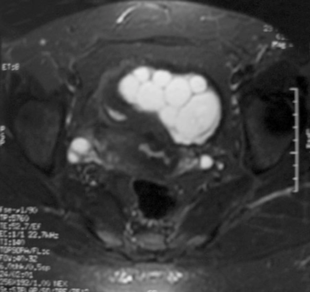
Pelvic MRI revealed a septated, lobulated and contrast enhancing cystic mass that is hyperintense on T2 and hypointense on T1 FLAIR sequences.
Figure 1

Pelvic MRI revealed a septated, lobulated and contrast enhancing cystic mass that is hyperintense on T2 and hypointense on T1 FLAIR sequences.

Footnotes

References

  • 1.
    Nakayama DK. Survival in a fetus with sacrococcygeal teratoma and hydrops. Am J Obstet Gynecol. 1990;163(2):682.
  • 2.
    Fahy AM, Peters EE. Eccrine tumours of the hand: two cases. J Hand Surg Br. 1987;12(1):120-2. https://doi.org/10.1016/0266-7681(87)90075-1.
  • 3.
    Sing P, Mushtaq D, Verma N, Mahajan NC. Pelvic hydatidosis mimicking a malignant multicystic ovarian tumor. Korean J Parasitol. 2010;48(3):263-5. [PubMed ID: 20877508]. https://doi.org/10.3347/kjp.2010.48.3.263.
comments

Leave a comment here


Crossmark
Crossmark
Checking
Share on
Metrics

Purchasing Reprints

  • Copyright Clearance Center (CCC) handles bulk orders for article reprints for Brieflands. To place an order for reprints, please click here (   https://www.copyright.com/landing/reprintsinquiryform/ ). Clicking this link will bring you to a CCC request form where you can provide the details of your order. Once complete, please click the ‘Submit Request’ button and CCC’s Reprints Services team will generate a quote for your review.
Search Relations

Author(s):

Related Articles