1. Introduction
Lipofibromatous hamartoma (LFH) is an extremely rare and slow-growing benign tumor, which is characterized by an excessive infiltration of the epineurium and perineurium by fibroadipose tissues (1-12). The entity was first described by Mason in 1952 (11) and a recent review of the literature revealed that 88 cases have been reported (11). This tumor had been known with many terms such as fatty infiltration of nerve, fatty or fibrous neoplasm of nerve, fibrofatty proliferation of nerve, fibrolipoma, perineurolipoma, intraneural lipoma, lipofibroma, and lipomatosis of nerve, until the consensus agreed upon the term of LFH (1-12). LFH has a congenital or developmental origin. Nonetheless, its symptoms typically present in the third or fourth decade of life. Because of abnormal enlargement of the involved nerve, compression neuropathy is a frequent finding (11). The median nerve is most commonly involved peripheral nerve, often with a predilection for the carpal tunnel (2). Separation of the individual nerve fascicles by fibro fatty infiltration of perineurium and epineurium produce remarkable changes in diagnostic imaging. This report presents a case of carpal tunnel syndrome (CTS) due to LFH of median nerve. By reporting this case of secondary CTS due to LFH, we want to emphasize on the role of ultrasonography, as an accurate and simple method in evaluation of patients with CTS, in preoperative diagnosis of secondary CTS.
2. Case Presentation
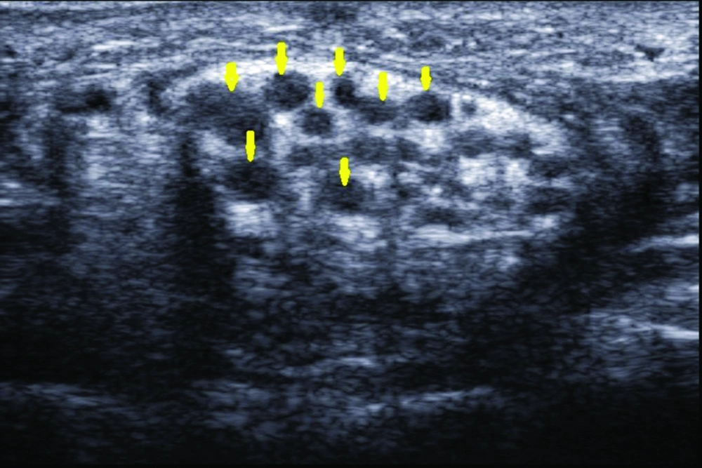
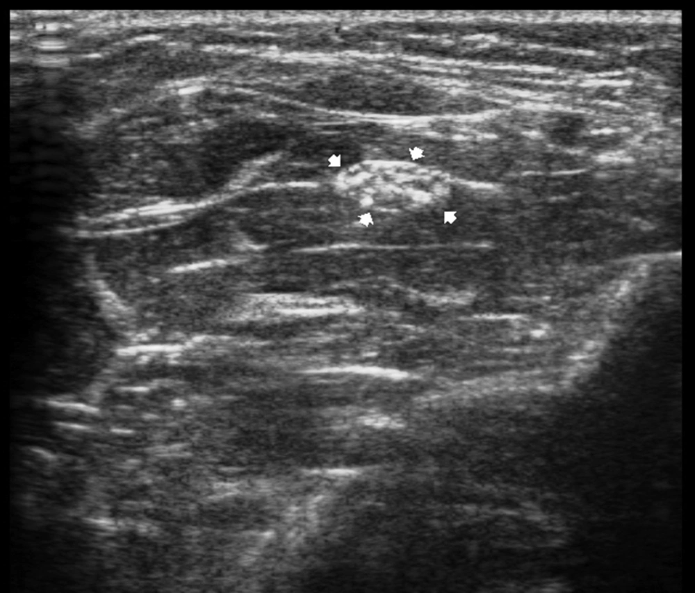
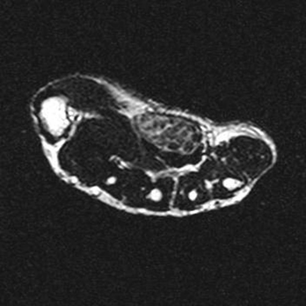
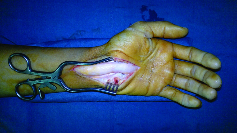
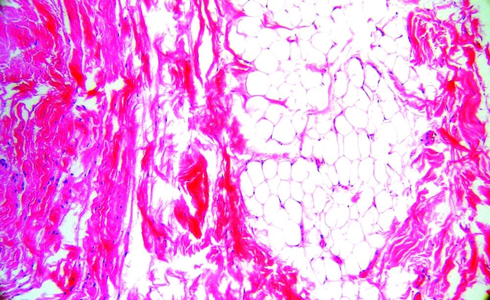
A 27-year-old woman presented with sensory disturbance along the radial three and half digits of her left hand. She had noticed a soft tissue swelling on volar surface of her wrist for six years and sometimes had experienced paresthesia and numbness in her radial three and half digits. Her symptoms worsened during the preceding year. On examination, results of Phalen’s test and Tinel’s sign were positive. Nerve conduction study revealed severe latency of the median nerve conduction velocity at carpal tunnel, consistent with severe CTS. Sonography of the swelling demonstrated enlarged cross-section of the median nerve (4.3 cm2; normal cross-section, 5.78 ± 0.9 mm2) under the flexor retinaculum with hypoechoic cable-like neural fascicle separated by hyperechoic fat (Figure 1). On ultrasonographic evaluation, the median nerve involvement was extended to the proximal forearm at the level of pronator teres muscle (Figure 2). Magnetic resonance imaging (MRI) of distal forearm demonstrated fusiform enlargement of the median nerve with low signal intensity of neural bundles in T1 and T2 sequences, surrounded by high signal intensity substratum representing fat. On axial image, the lesion demonstrated a characteristic “coaxial cable-like” appearance (Figure 3). She underwent carpal tunnel release because of severe secondary CTS. At surgery, enlargement of the median nerve under the flexor retinaculum in the distal forearm and carpal tunnel was observed (Figure 4). A small biopsy sample was obtained from the perineurium. No further treatment was performed. Histopathologic study demonstrated infiltration of perineurium with mature fibrous and adipose tissue (Figure 5). The patient had unremarkable postoperative course with complete resolution of her symptoms and remained asymptomatic at the four-month follow-up course.
3. Discussion
The vast majority of LFH cases occurs in infants and less commonly, in children and young adults and are commonly associated with macrodactyly (1, 4). Median nerve is the most commonly affected nerve (80% of cases). Ulnar and radial nerves, dorsum of the foot, and brachial plexus are the other less common sites of involvement (5). Involvement of the median nerve with LFH usually presents pain, paresthesia, and CTS (3). LFH is frequently associated with macrodystrophia lipomatosa (4). LFH can be diagnosed by ultrasonography, computed tomography, and MRI. MRI manifestation of LFH is pathognomonic (1-5). The radiologic manifestation of our case was consistent with previously reported descriptions. Toms et al. (3) in a series of 15 patients showed that LFH had characteristic MRI findings: the nerve looks enlarged with high signal intensity on T1-weighted and T2-weighted sequences due to fatty infiltration of median nerve, which consist of cable-like low signal intensity on T1 and T2 sequences due to enlarged axonal bundles. Al Jabri et al. (1) showed that typical "cable-like" appearance might dismiss the need for biopsy (1). Ultrasonographic feature of LFH is described only in few reports (3, 6). Toms et al described the ultrasonographic feature of LFH as “characteristic hypoechoic coaxial cabling encased by an echogenic substratum” (3). Agarwal et al. (11) also showed that hypoechoic bundle and echogenic substratum were the typical ultrasonographic appearance of LFH.
In the current case, the ultrasonographic appearance was similar to the few previous descriptions. The enlarged hyperechoic median nerve containing discrete hypoechoic cable-like bundles was well compatible with MRI signal patterns. In our case, ultrasonographic diagnosis of the extent of nerve involvement was accurate and compatible with surgical finding, which was very important for surgical planning. Our experience might suggests sonography as a more convenient technique in diagnosing and quantifying the extent of LFH. The surgical management of LFH remains highly controversial. Internal neurolysis is not feasible because fibrofatty tissues are infiltrated between the nerve fascicles and internal neurolysis would cause damage to the nerve function (7, 8). Resection of the abnormal segment of the median nerve and reconstruction by interposition nerve graft is not recommended because it would cause nerve deficit (7, 8). Therefore, some authors recommends observing the clinical course of LFH and when needed, treating the symptoms (9-11). LFH is a very rare nerve tumor and only 88 cases have been reported in the literature (11). The current case was the second case of LFH of the median nerve seen in our orthopedic department (12). We diagnosed the LFH of the median nerve by the characteristic MRI and sonography findings. In the current case, the median nerve was involved along a long course in the forearm, however; the patient needed carpal tunnel release because of severe compression of the median nerve under the flexor retinaculum.
