Primary melanocytic neoplasms are rare lesions which originate from normally existing leptomeningeal melanocytes (
3). Melanocytes arise from neural crest elements and are found within the basal layer of the epidermis and the leptomeninges that cover the base of brain and the brain stem (
4). Moreover, the highest concentration of melanocytes is seen ventrolateral to the medulla oblongata and upper cervical levels of spinal leptomeniges (
5,
6). In general, three main types are considered for these neoplasms, including diffuse melanosis, meningeal melanocytoma and primary malignant melanoma (
7). Nowadays, with improvements in neuroimaging and clarification of histological features, meningeal melanocytomas are being diagnosed with increased frequency. Clinical presentation of patients with these tumors typically occurs in their fifth decade. It is seen in women twice as often as men (
5). Unlike diffuse melanosis, it is not associated with skin pigmentation. Neurological and clinical features in meningeal melanocytoma is so varied, including the frequent occurrence of hydrocephalus, seizures, chronic basal meningitis, multiple cranial nerve palsies, psychiatric disturbances, intracranial hemorrhage of the meninges or subdural space and myeloradiculopathy. An important consideration is that intracranial and spinal melanocytomas have propensity to arise in proximity to cranial and spinal nerves as they exit the brainstem and spinal cord (
7-
9).
Biological behavior is variable; incomplete resection may yield in recurrence, an event that may occur because of extensive leptomeningeal involvement or persistence of neglected small foci of tumor. Some believe transition into malignant melanoma does not occur, however, we encountered to four cases of malignant transformation of meningeal melanocytoma, in the literature (
10-
13). We did not find any description for a case of melanocytoma with tonsillar hernaition; however, we presume that in a case with extensive meningeal involvement, an obstacle for complete debulking may increase the mass effect of the growing lesions consequently leading to this inevitable outcome. Generally, a good postoperative survival rate is expected for patients with meningeal melanocytoma. Hence, the surgeon should be advised of this possible diagnosis in suspected cases of meningioma, especially those involving the posterior fossa or Meckel’s cave, information that makes the surgeon accomplish maximal effort at tumor resection. However, preoperative diagnosis of meningeal melanocytoma is not invariably easy (
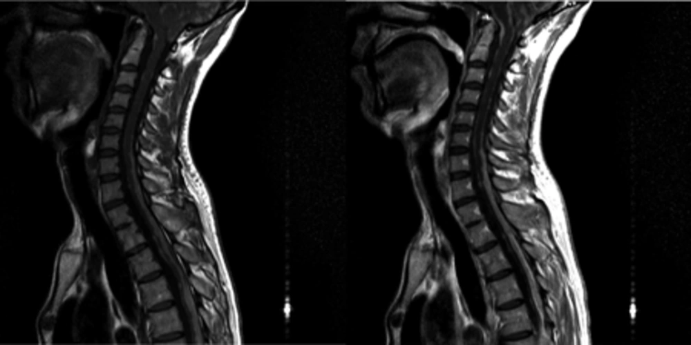
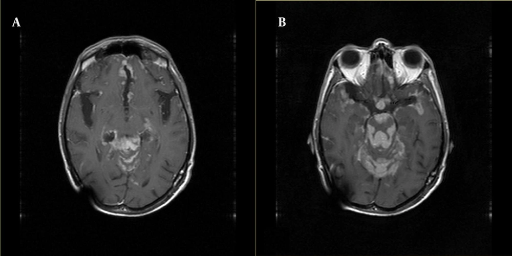
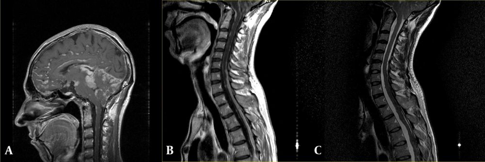
7). Fortunately, the characteristic shortening of T1 and T2 relaxation times in MR imaging , as a result of the paramagnetic stable free radicals that exist within melanin, may often suggest a diagnosis of a melanocytic leptomeningeal process (
7,
11). In addition, there is unique ultra-structural and immunohistochemical characteristics for these varied lesions (
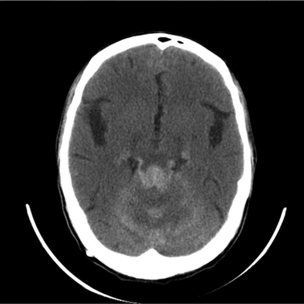
7). Another important hint is that the degree of melanization makes the imaging appearance of meningeal melanocytoma variable. Iso- to hyperattenuating lesions with variable contrast enhancement is detected in CT images. While, in MR images, these lesions generally show high signal intensity on T1-weighted images, diminished signal on T2-weighted images and diffuse enhancement after contrast administration (
7,
12). Here, an indispensible role is considered for immunohistochemical analysis, a study that can eventually make differentiation of meningeal melanocytoma from other similar pigmented lesions possible. Characteristic immunohistochemical reaction of meningeal melanocytoma is a positive response to antimelanoma (HMB-45), S-100 protein and vimentin (an indicator of cells with mesenchymal origin, which rarely appears in malignant melanoma) antibodies and a negative reaction to epithelial membrane antigen (EMA) (indicator of meningioma) and Leu7 (indicator of schwannoma) (
7,
13).