1. Background
Granulomatous mastitis (GM) is an uncommon benign inflammatory condition of the breast. Idiopathic GM, originally described by Kessler and Wolloch in 1972 (1), represents a sub-group of GM with unknown etiology. The remaining cases of GM are associated with infectious conditions, such as fungal infections, actinomycosis, histoplasmosis, brucellosis, and tuberculosis in particular, as well as with other conditions, such as Wegener’s granulomatosis and sarcoidosis (2). The real incidence of GM is unknown, with only a few hundred cases reported in the literature (3). Clinically, radiologically, and even cytologically, it can be confused with malignancy, requiring histopathological examination for a definitive diagnosis (4, 5). Conventional radiological findings are non-specific and exhibit wide variation. Magnetic resonance imaging (MRI) has emerged as an important diagnostic tool, providing certain advantages over other imaging modalities in the differential diagnosis of breast conditions.
2. Objectives
In this study, conventional radiological and MRI findings were evaluated in patients with GM, with an emphasis on the value of the imaging modality and the diagnostic role of radiology in GM.
3. Patients and Methods
This retrospective study involved a total of 29 GM patients between 20 and 69 years of age (mean ± SD: 35.14 ± 9.9) who were diagnosed with GM between 2009 and 2013 in our clinic. The study protocol was approved by the Institutional Ethics Committee (project number: 0545).
Mammography (MG) examinations were performed in all patients over the age of 35 years (14/29) in standard craniocaudal and mediolateral-oblique projections (Lorad M-IV, Hologic). In the remaining 15 patients (under age 35), MG was not performed.
All patients underwent ultrasound (US) and MRI examinations, and the results were documented. High-resolution US images (Xario SSA-660A, Toshiba) were obtained by a linear-array transducer with a center frequency of 7.5 MHz. MRI indications included exclusion of inflammatory cancer in treatment-resistant cases, further assessment in patients with inconclusive MG and/or sonography results, and determination of the extent of disease. MRI was performed after conventional examinations in all patients in a manner that would not result in any treatment delay. MRI examinations were performed with a 1.5-T whole-body imaging system (Signa Excite, GE Healthcare, Milwaukee, WI, USA). The patients were scanned in the prone position with the breast suspended in a four-channel breast coil. MR images were obtained in the transverse and sagittal planes with fat suppression. Pre-contrast transverse acquisitions were performed using a T1-weighted fast spin-echo sequence and transverse T2-weighted fast spin echo short-tau inversion recovery (STIR) imaging, and pre-contrast sagittal acquisitions were performed using a T2-weighted fast spin echo sequence with fat suppression. Sagittal pre and post-contrast dynamic imaging was performed using a 3D multi-phase fast gradient echo pulse sequence called VIBRANT (flip angle 10°; minimum echo time 2.4 msec; maximum echo time 14.0 msec; section thickness 3 mm with no intersection gap; field of view 20 cm; matrix size 256 × 256; NEX 1; one signal acquired; imaging time, 1 minute for each phase). Additionally, transverse post-contrast T1-weighted images were acquired using the fast spoiled gradient-recalled-echo sequence in the same manner used to acquire the pre-contrast images, without a change in the patient’s position. Subtraction images were done. The patients were given a bolus intravenous injection of gadolinium contrast (0.2 mmol/kg body weight) with a power injector. Both the morphological features and the kinetic characteristics of the lesions were examined. All MR images were reviewed on high-resolution PACS monitors (General Electric Medical Systems).
For a final differential diagnosis, biopsy was recommended in all cases, especially for treatment planning. Twenty-six patients underwent core biopsy, while three had excisional biopsy according to the surgeon’s preference. Core biopsy was performed under US guidance using a 14-G needle. Due to inconsistent radiological and core biopsy results in four patients, additional excisional biopsies were required. Among all 29 patients, seven required surgical excision of lesions.
A diagnosis of tuberculosis-associated GM was made in 10 patients, and cat-scratch disease-associated GM was diagnosed in one. No causative factors could be determined for the remaining 18 patients.
3.1. Statistical Methods
SPSS ver. 15 (SPSS Inc., Chicago, Illinois, USA) and Medcalc statistical software (Belgium) packages were used for statistical analysis. This study consisted of only GM patients and descriptions of their imaging features. Therefore, a percent calculation was performed. As the only statistical analysis, the chi-square (χ2) test was used to compare the ratios of the BI-RADS categories between the conventional methods and MRI. A value of P < 0.05 was accepted as statistically significant.
4. Results
4.1. Clinical Findings
The majority (26/29, 90%) of the patients were of reproductive age and none had a history of autoimmune disease, sarcoidosis, or systemic tuberculosis. Two patients had a family history of breast cancer. The symptoms had been present for 15 days to 4 months before a diagnosis was made, and included breast pain (26/29, 90%), palpable breast mass (23/29, 79%), and erythema and inflammation (9/29, 31%). Three (10.3%) patients had sinus tracts and one (3.4%) had nipple retraction. The condition was unilateral in all cases, with 20 on the right side (Table 1).
| Symptom | No. (%) |
|---|---|
| Breast pain | 26 (90) |
| Erythema | 9 (31) |
| Palpable lump | 23 (79) |
| Sinus tract | 3 (10.3) |
| Nipple retraction | 1 (3.4) |
| Axillary lymphadenopathy | 13 (45) |
4.2. Mammography Findings
Nine of the 14 (64.3%) patients who underwent MG had dense and heterogeneous dense parenchymal breast patterns. Nine (64.3%) patients had focal asymmetric densities, two (14.3%) had ill-defined nodular densities, one (7.1%) had diffuse increased density, and two (14.3%) had no pathological findings with dense patterns. No pathological calcifications were observed.
4.3. Ultrasonography Findings
Sixteen (55.2%) patients had ill-defined lesions with tubular extensions, eight (27.6%) had well-demarcated lesions with posterior acoustic enhancement, three (10.3%) had parenchymal edema-heterogeneity, one (3.4%) had a mass lesion with irregular borders, and one (3.4%) had normal results. All lesions exhibited heterogeneous hypoechogenicity. Three (10.3%) had fistula tracts. Twelve (41.4%) patients had axillary lymph nodes with mild or moderate enlargement, echogenic hila, and a symmetric or asymmetric thick cortex. One patient (3.4%) had markedly enlarged lymph nodes with fatty hila, which could not be clearly visualized. Color Doppler US findings were available for 16 patients, and increased arterial and venous vascularization was evident at the lesion level in all cases. Table 2 summarizes the mammography, US, and color Doppler US findings of the 29 GM patients.
| Findings | No. (%) |
|---|---|
| Mammography (n = 14) | |
| Normal | 2 (14.3) |
| Ill-defined nodular density | 2 (14.3) |
| Focal asymmetrically increased density | 9 (64.3) |
| Diffuse increased density | 1 (7.1) |
| Skin thickening | 3 (21.4) |
| Ultrasonography (n = 29) | |
| Normal | 1 (3.4) |
| Parenchymal heterogeneity | 3 (10.3) |
| Het. hypoechoic ill-defined lesion with tubular extensions | 16 (55.2) |
| Het. hypoechoic lesion with well-defined border | 8 (27.6) |
| Het. hypoechoic lesion with irregular border | 1 (3.4) |
| Fistulae | 3 (10.3) |
| Skin thickening | 5 (17.2) |
| Unilaterally enlarged axillary lymph nodes | 13 (44.8) |
| Color Doppler sonography (n = 16) | |
| Increased arterial and venous vascularization | 16 (100) |
Abbreviation: Het, Heterogeneous.
Conventional methods (US/US + MG) could identify 16 out of 29 (55.2%) cases with lesions of probably benign (BI-RADS 3) and inflammatory origin. However, these methods could not distinguish the remaining 13 (44.8%) cases from malignancy; 12 (41.4%) patients were categorized as having a suspicious abnormality (BI-RADS 4) and one (3.4%) patient’s lesion was highly suggestive of malignancy (BI-RADS 5).
4.4. MRI Findings
Twenty-five of the 29 (86.2%) participants had one or more separate or confluent lesions consistent with abscess formation, with regular or irregular borders and marked circumferential contrast enhancement following IV gadolinium injection. These lesions were hyperintense on T2-weighted images (T2WI) and hypointense on T1WI, with variable signal intensities depending on the protein content of the fluid. At the same level of these lesions or adjacent to them were areas with heterogeneous signal changes, without well-defined borders or mass effect, as well as segmental/regional heterogeneous non-mass-like contrast enhancement. The size of the MRI lesions ranged from 5 - 6 mm to 5 cm. In four patients (13.8%), no lesions were observed other than parenchymal heterogeneity and non-mass-like segmental contrast enhancement. Kinetic analyses of non-mass-like contrast-enhanced areas, lesions, and lesion walls showed type 1 kinetic curves in 24 (82.7%) patients at all levels, type 2 kinetic curves in the lesion walls in four (13.8%) patients, and type 3 kinetic curves in the lesion walls and non-mass-like contrast-enhanced areas in one (3.4%) patient. The patient with type 3 kinetic curves had a positive family history. MRI showed the presence of lesions within only one quadrant in 13 (44.8%) patients, while multiple quadrants were involved in 16 (55.2%). Twelve patients had peripheral involvement, two had retroareolar involvement, and seven had both peripheral and retroareolar involvement. Fistula tracts were present in three patients, and 16 (55.2%) patients had mildly or moderately enlarged axillary lymph nodes with symmetric or asymmetric thick cortex structures on MRI. A single (3.4%) patient had enlarged lymph nodes without fatty hila. Table 3 summarizes the MRI findings.
| MRI Findings | No. (%) |
|---|---|
| Focal lesion | 25 (86.2) |
| Solitary lesion with well-defined borders | 11 (44) |
| Multiple small lesions ( < 1 cm) with well-defined borders | 5 (20) |
| Confluent lesions with irregular margins | 8 (32) |
| Lesion signal intensity | |
| Hypointense on T1WI, hyperintense on T2WI | 16 (64) |
| Intermediate on T1WI, het. hyperintense on T2WI | 4 (16) |
| Hypointense on T1WI, het. hyperintense on T2WI | 4 (16) |
| Het. hypointense on T1WI, and het. hyperintense on T2WI | 1 (4) |
| Lesion enhancement | |
| Peripheral ring enhancement | 25 (100) |
| Parenchymal het. intensity changes | 29 (100) |
| Non-mass-like enhancement | 4 (13.8) |
| Type of enhancement curve | |
| Type 1 | 24 (82.7) |
| Type 2 | 4 (13.8) |
| Type 3 | 1 (3.4) |
| Other findings | |
| Fistulae | 3 (10.3) |
| Skin thickening | 12 (41.8) |
| Unilaterally enlarged axillary lymph nodes | 17 (58.6) |
Abbreviation: Het, Heterogeneous; WI, weighted imaging.
On the MRI examinations of the 29 cases, 24 (82.7%) were deemed probably benign (BI-RADS 3) and inflammatory in nature, four (13.8%) were considered suspicious for malignancy (BI-RADS 4), and one (3.4%) was considered highly suggestive of malignancy (BI-RADS 5).
Eleven cases interpreted as “suspicious” with conventional methods were considered probably benign based on MRI findings, while two cases assessed as “suspicious” on MRI were considered probably benign based on conventional methods. These two patients had clinical-radiological mismatches and the only MRI finding was non-mass-like segmental contrast enhancement. One patient with markedly enlarged lymph nodes without fatty hila was categorized based on conventional imaging and MRI as BI-RADS 5 and BI-RADS 4, respectively, and the reverse was true in another patient with a family history. The other cases of conventional and MRI findings were assessed as the same BI-RADS category (Table 4). Compared with conventional methods (55.17%), the rate of the BI-RADS 3 classification was significantly higher based on MRI (82.75%; P = 0.006).
| Patient | Age | US/US+MG | MRI |
|---|---|---|---|
| 1 | 29 | BI-RADS 4 | BI-RADS 3 |
| 2 | 35 | BI-RADS 4 | BI-RADS 3 |
| 3 | 39 | BI-RADS 3 | BI-RADS 4 |
| 4 | 41 | BI-RADS 3 | BI-RADS 3 |
| 5 | 42 | BI-RADS 3 | BI-RADS 4 |
| 6 | 34 | BI-RADS 3 | BI-RADS 3 |
| 7 | 34 | BI-RADS 4 | BI-RADS 3 |
| 8 | 30 | BI-RADS 4 | BI-RADS 3 |
| 9 | 25 | BI-RADS 3 | BI-RADS 3 |
| 10 | 35 | BI-RADS 3 | BI-RADS 3 |
| 11 | 41 | BI-RADS 3 | BI-RADS 3 |
| 12 | 34 | BI-RADS 3 | BI-RADS 3 |
| 13 | 45 | BI-RADS 4 | BI-RADS 3 |
| 14 | 41 | BI-RADS 5 | BI-RADS 3 |
| 15 | 69 | BI-RADS 5 | BI-RADS 4 |
| 16 | 35 | BI-RADS 4 | BI-RADS 3 |
| 17 | 24 | BI-RADS 3 | BI-RADS 3 |
| 18 | 45 | BI-RADS 3 | BI-RADS 3 |
| 19 | 40 | BI-RADS 4 | BI-RADS 5 |
| 20 | 34 | BI-RADS 3 | BI-RADS 3 |
| 21 | 39 | BI-RADS 3 | BI-RADS 3 |
| 22 | 29 | BI-RADS 4 | BI-RADS 3 |
| 23 | 34 | BI-RADS 3 | BI-RADS 3 |
| 24 | 23 | BI-RADS 3 | BI-RADS 3 |
| 25 | 24 | BI-RADS 3 | BI-RADS 3 |
| 26 | 48 | BI-RADS 4 | BI-RADS 4 |
| 27 | 24 | BI-RADS 3 | BI-RADS 3 |
| 28 | 20 | BI-RADS 4 | BI-RADS 3 |
| 29 | 26 | BI-RADS 4 | BI-RADS 3 |
Abbreviations: BI-RADS, Breast Imaging-Reporting and Data System; MG, mammography; MRI, magnetic resonance imaging; US, ultrasound.
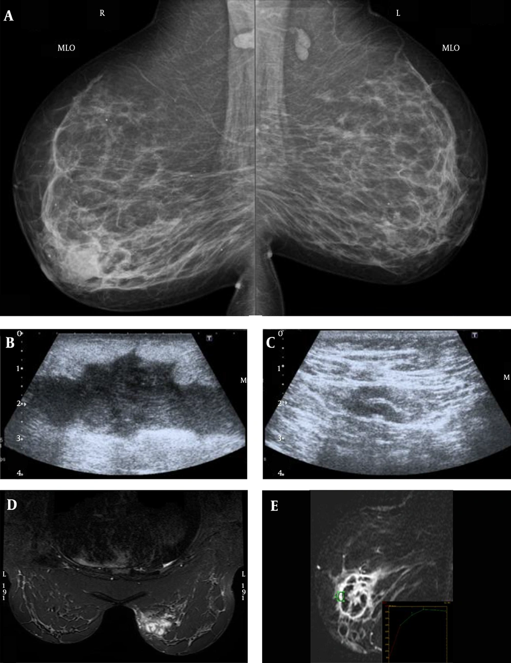
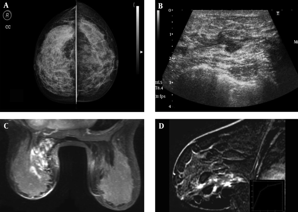
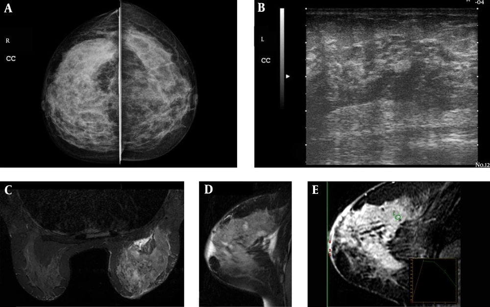
Figures 1 - 3 show examples of imaging findings from the study participants.
A 35-year old patient (patient no. 2), who presented with a palpable right breast mass. A, Bilateral mediolateral-oblique mammography showed a nodular density surrounded by peripheral fibroglandular tissue. B, Ultrasound showed a heterogeneous hypoechoic lesion with tubular extensions. C, Axillary examination showed moderately enlarged lymph node with thickened cortex. D, STIR axial and E, T1-weighted fat-suppressed post-contrast subtraction sagittal MR images showed a mass lesion with peripheral ring enhancement and irregular borders consistent with abscess formation in the same patient. Type 1 kinetic curve of the lesion wall is seen (E).
A 42-year-old patient who presented with breast pain (patient no. 5). A, Bilateral craniocaudal mammography showed asymmetric increased density, more prominent in the inner quadrant of the left breast. B, Ultrasound showed a hypoechoic heterogeneous ill-defined lesion, with tubular extensions. C, Post-contrast fat-suppressed maximum intensity projection reformatted axial and D, T1-weighted fat-suppressed post-contrast subtraction sagittal MR images showed non-mass-like segmental contrast enhancement. Type 1 kinetic curves at the level of contrast enhancement is seen (D).
A 40-year-old patient who presented with a palpable breast lesion (patient no. 19). A, Bilateral craniocaudal mammography showed asymmetric increased density, more prominent in the right lateral quadrant of the right breast. B, Ultrasound showed heterogeneous hypoechoic ill-defined lesions with tubular extensions. C, STIR axial , D, T2-weighted fat-suppressed sagittal and E, T1-weighted fat-suppressed post-contrast subtraction sagittal MR images showed parenchymal heterogeneous intensity changes, non-mass-like regional contrast enhancement, and lesions less than 1 cm in diameter with peripheral contrast enhancement, consistent with micro-abscess formation. Type 3 kinetic curves adjacent to the lesions are seen.
4.5. Treatment and Follow-Up
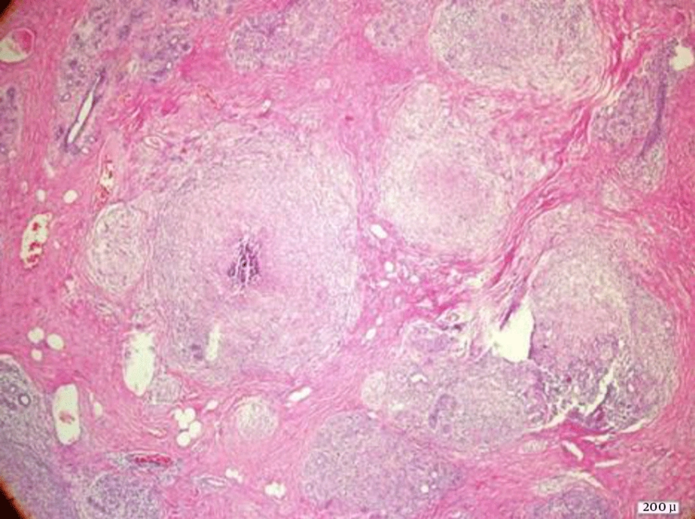
Most of the patients were on antibiotic therapy for findings of breast abscesses at the time of their referral to the radiology clinic. All of the patients consulted a general surgeon for a treatment plan. Before the treatment plan, all of the patients were evaluated for possible respiratory system illnesses (tuberculosis, sarcoidosis, etc.) with chest radiography and/or thoracic computerized tomography (CT). Tuberculin sensitivity tests were also performed on all patients. Biopsy and histopathological assessments were performed in all cases, especially for treatment planning (Figure 4). Ten patients were diagnosed with tuberculosis and received antituberculous treatment according to advice of the chest disease specialist. Three of the remaining 19 patients underwent complete excision, and the remainder (16/29) received low-dose steroid therapy (0.5 mg/kg/day) for 4 - 8 weeks. After the medical treatment, all of the patients were followed every three months for two years, then every six months for two years, then yearly. There were no recurrences requiring either medical or surgical therapy in our study group after 39 (15 - 73) months of follow-up. A complete response was observed after therapy; however, this does not ensure that no lesions are present histopathologically.
5. Discussion
GM is an uncommon inflammatory condition of the breast characterized by granuloma and abscess formation (2), generally affecting young women of reproductive age, mostly during the five years following childbirth (2-8). Until now, the largest patient series reported in the literature was that of Yildiz et al. (8), which involved 30 patients with a diagnosis of idiopathic GM. Thus, to our knowledge, our study represents the second-largest patient series in the literature with an evaluation of radiologic findings of GM.
The absence of complaints or clinical signs suggestive of inflammation despite the presence of a palpable mass in the majority of GM patients, and enlarged axillary lymph nodes in a certain proportion of these subjects, may lead to a suspicion of breast cancer. In addition, GM may not be readily differentiated from breast cancer radiologically (1-18). It generally involves one breast, although bilateral involvement has also been reported (2, 5, 7, 10). In our study, all of our cases had unilateral GM with a predominance of peripheral involvement, and the proportion of patients with retroareolar involvement was higher than that reported previously (6, 11, 12).
Several studies have reported on MG and US findings in GM patients, and a general consensus exists as to the conventional radiological signs of GM. MG generally shows non-specific signs, the most common of which is focal asymmetric increased density, ill-defined borders, and mass effect, or irregular masses with ill-demarcated borders (3, 8, 9, 11-15). MG has low sensitivity for this condition, particularly when considering the younger population that is affected, as the abovementioned findings may not be detected in women with dense breast tissue (3, 6, 13, 16). The MG findings of the current study are in line with previous reports, and the lesions could not be detected in two of our patients with dense breast tissue.
Since US is commonly used for the initial assessment of young females with palpable breast lesions, most GM patients undergo US examinations prior to diagnosis. The most common US findings in these patients are heterogeneous hypoechoic ill-defined lesions with tubular extensions, as in our study (3, 6, 11, 13, 15). However, other findings, such as hypoechoic lesions with regular or irregular contours, posterior acoustic enhancement or shadowing, parenchymal heterogeneity, and distortions may also be seen, in addition to skin thickening and diffuse parenchymal edema (13). One specific advantage of US is its ability to facilitate aspiration in cases of suspected fluid collection. However, it should be borne in mind that cytology alone may not be adequate to exclude underlying malignancy or to diagnose GM, necessitating a confirmatory histopathological diagnosis (3). In addition, a normal US examination cannot exclude a diagnosis of GM, as demonstrated by one of our cases with asymmetric densities on MG that were not detected by US.
MRI is an imaging modality with high sensitivity and specificity for breast lesions, allowing the detection and assessment of the extent of inflammatory breast conditions. The MRI characteristics of GM have been reported in only a few cases in the literature, and data on the use of MRI in GM are scarce, with a variety of definitions used for the description of MRI findings (2, 3, 5, 7, 8, 14, 16). In suspected cases of GM, MRI should not lead to delayed biopsy and may be used as a complementary diagnostic method in addition to US and MG in order to exclude a diagnosis of inflammatory breast cancer in cases with symptoms of treatment-resistant mastitis (17). However, MRI can help in detecting lesions that cannot be visualized by US and MG due to parenchymal edema. In addition, since GM generally affects younger females, MRI may also be useful in cases where MG or US is inconclusive due to the character of the breast parenchyma. The most common MRI findings in our study were solitary or multiple separate or confluent abscesses of different sizes, with marked peripheral ring enhancement, accompanied by intensity changes suggesting edematous inflammation in the peripheral parenchyma, as well as non-mass-like heterogeneous segmental and regional contrast enhancement. In some cases, annular enhancing nodules, probably reflecting micro-abscesses, were only a few millimeters in size. These observations are similar to those reported by Kocaoglu et al. and Gautier et al. (3, 16). Previous studies have reported variable kinetic curve analysis results between different patients and different lesions (2, 5, 16). In the study by Gautier et al., non-mass-like contrast-enhanced areas were generally associated with type 1 kinetic curves, while type 3 was the dominant kinetic curve in areas with annular contrast enhancement (3). In the present study, circumferential contrast enhancement was mostly associated with type 1 kinetic curves, while our findings were consistent with previous reports of non-mass-like contrast enhancement. In this study, MRI could more accurately and clearly identify the extent of disease and the typical findings of the inflammatory process, so it could more confidently classify lesions as BI-RADS 3, compared to conventional methods.
Several limitations of our study should be mentioned, including its retrospective and descriptive character. Second, the study group consisted only of patients with GM. Third, there were a limited number of patients who underwent MG and color Doppler US.
In conclusion, GM has no specific features on MG and US. MRI is a complementary diagnostic tool used with clinical and conventional radiological findings to assist in the diagnosis and differentiation of GM from malignant processes. A focal asymmetric density on MG associated with US findings of heterogeneous hypoechoic ill-defined lesions and tubular extensions, along with MRI findings of ring-enhancing lesions associated with type 1 kinetic curves, can suggest the diagnosis of GM. However, histopathology remains essential for a definitive diagnosis and appropriate management.


