Logo

Intraosseous Pleomorphic Adenoma of the Mandible

Author(s):
Leila AghaghazviniLeila Aghaghazvini1, 2,*, Shirin AghaghazviniShirin Aghaghazvini2
1Department of Radiology, Shariati Hospital, Tehran University of Medical Sciences, Tehran, Iran
2Advanced Diagnostic and Interventional Radiology Research Center (ADIR), Imam Khomeini Hospital, Tehran University of Medical Sciences, Tehran, Iran


IJ Radiology:Vol. 12, issue 1; e6921
Published online:Sep 30, 2014
Article type:Letter
Received:Jul 05, 2012
Accepted:Aug 07, 2013
How to Cite:Leila AghaghazviniShirin AghaghazviniIntraosseous Pleomorphic Adenoma of the Mandible.I J Radiol.12(1):e6921.https://doi.org/10.5812/iranjradiol.6921.

Dear Editor,

Salivary gland tumors consist of about 3% of all head and neck tumors. Benign tumors (mainly pleomorphic adenoma) are the most common type. About 80% of them occur in the parotid glands. Carcinomas are rare and more common in the smaller salivary glands (1, 2). Benign salivary gland tumors rarely develop in the jaw bones. They arise from either the ectopic salivary tissue or minor salivary glands that are implanted in the bones. Ex pleomorphic adenomas within the mandible are extremely rare and only a few cases have been reported to date (3, 4).

We present a rare case of intraosseous pleomorphic adenoma of the mandible mimicking a radicular cyst or a brown tumor with unusual presentation in a 38-year-old man with low socio-economic status and poor hygiene. He presented with a rapid enlargement of the left mandible bone during two months. He also had mild pain and paresthesis of the mental region. On physical examination, a bony non-tender mass lesion about 20 × 15 mm in size was detected in the left para-symphysial portion of the mandible. Multiple teeth caries and destruction were detected. On the orthopenthogram, there was a lobulated irregular border lytic lesion in the left parasymphyseal portion of the mandibular body with erosion of the overlying teeth roots. In addition, there were many other destructed and caried teeth together with destruction of a few lamina duras. Regarding imaging findings, large radicular cyst with periapical abscess was the first diagnosis. Brown tumor related to hyperparathyroidism and also other odontogenic cysts were our differential diagnoses.

The patient underwent surgery. A rubbery white-gray mass, 15 × 10 × 10 mm in size, was resected and sent for pathology. Pathology reported an infiltrative neoplasm exposed of epithelium with bland looking nuclei and acidophilic cytoplasm in a fibromyxoid stroma without necrosis. Pleomorphic adenoma was the diagnosis. There was no tumor recurrence during the one-year postoperative follow-up. This is an interesting case not only for rarity but also for the sudden onset presentation of the lesion and unusual diagnosis in underlying jaw and dental disease (Figure 1).

A 38-year-old man with rapid enlargement of the left mandible. A lobulated lytic bony lesion with a partially sclerotic border in the left parasymphyseal portion of the mandiblular alveolar ridge accompanied with destruction of the overlying teeth roots and lamina duras. There is a notable lack of a few other teeth with caries of the remaining in the maxilla and mandible.
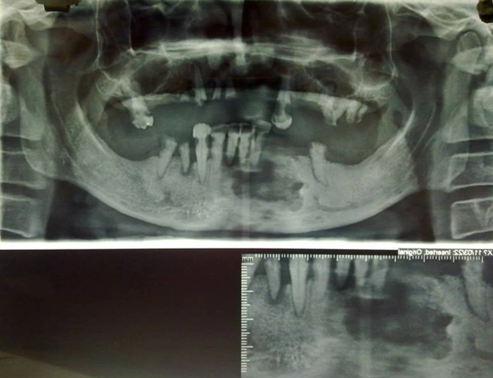
Figure 1.

A 38-year-old man with rapid enlargement of the left mandible. A lobulated lytic bony lesion with a partially sclerotic border in the left parasymphyseal portion of the mandiblular alveolar ridge accompanied with destruction of the overlying teeth roots and lamina duras. There is a notable lack of a few other teeth with caries of the remaining in the maxilla and mandible.

References

  • 1.
    Stodulski D, Rzepko R, Kowalska B, Stankiewicz C. [Carcinoma ex pleomorphic adenoma of major salivary glands--a clinicopathologic review]. Otolaryngol Pol. 2007;61(5):687-93. [PubMed ID: 18552001]. https://doi.org/10.1016/S0030-6657(07)70507-3.
  • 2.
    Mafee M. Imaging of the Head and Neck. 2 ed. Thieme; 2005.
  • 3.
    Ojha J, Bhattacharyya I, Islam MN, Manhart S, Cohen DM. Intraosseous pleomorphic adenoma of the mandible: report of a case and review of the literature. Oral Surg Oral Med Oral Pathol Oral Radiol Endod. 2007;104(2):e21-6. [PubMed ID: 17142071]. https://doi.org/10.1016/j.tripleo.2006.08.013.
  • 4.
    Zhao D, He H, Fang M, Liu M, Ji B, He Z. Primary central carcinoma ex pleomorphic adenoma of the mandible: report of a rare case and literature review. Oral Surg Oral Med Oral Pathol Oral Radiol Endod. 2011;112(2):e24-8. [PubMed ID: 21664840]. https://doi.org/10.1016/j.tripleo.2011.02.046.
comments

Leave a comment here


Crossmark
Crossmark
Checking
Share on
Metrics

Purchasing Reprints

  • Copyright Clearance Center (CCC) handles bulk orders for article reprints for Brieflands. To place an order for reprints, please click here (   https://www.copyright.com/landing/reprintsinquiryform/ ). Clicking this link will bring you to a CCC request form where you can provide the details of your order. Once complete, please click the ‘Submit Request’ button and CCC’s Reprints Services team will generate a quote for your review.
Search Relations

Author(s):

Related Articles