1. Background
Over the past decade, some non-contrast enhanced MR angiography (NCE-MRA) sequences such as spatial labeling with multiple inversion pulses (SLEEK) (1, 2), inflow inversion recovery (IFIR) (3, 4), time-spatial labeling inversion pulse (Time-SLIP) (5, 6), and steady-state free precession (SSFP) (7, 8) have been developed as an alternative to contrast-enhanced MRA (CE-MRA) and computer tomography angiography (CTA) for avoiding radically nephrogenic systemic fibrosis (NSF) or contrast-induced nephropathy (CIN) in renal function impairment patients. They can provide excellent results in the assessment of problematic vessels. Tang et al. (1) found that SLEEK NCE-MRA was better than ultrasound in displaying accessory renal arteries and there was an excellent relation of capacity in depicting transplant renal artery stenosis (TRAS) between SLEEK NCE-MRA and digital subtraction angiography (DSA). Lanzman et al. (9) stated that SSFP NCE-MRA was a reliable technique for evaluating TRAS in comparison with DSA. Mohrs et al. (10) found that the specificity and sensitivity of SSFP NCE-MRA were 99% and 75% in showing renal artery stenoses greater than 50%. However, the image quality of NCE-MRA still needs to improve in the visibility of renal artery, especially for displaying renal segmental branches in the renal parenchyma.
How to improve the NCE-MRA imaging quality for displaying the renal artery has become a hot research topic at present. For this purpose, we have performed SLEEK sequence for displaying the renal artery. In SLEEK, blood suppression inversion time (BSP TI) is defined as the duration from the start time point of the initial inversion pulse to the time point that longitudinal magnetization of blood reaches zero (null) point, which was also named as inversion time after the spatial-selective inversion-recovery pulse (ssTI) in Time-SLIP NCE-MRA (11), and TI (Time delay) in SSFP NCE-MRA (10). For distinctly showing the renal artery, Shonai et al. (11) believed that ssTI should be performed with 1200 - 1800 ms in Time-SLIP NCE-MRA, and Parienty et al. (8) applied 1100 - 1500 ms in SSFPNCE-MRA. Kurata et al. (12) held a TI with 1800 ms to visualize the renal artery using time-SLIP. In our previous study, we found that BSP TI which depended on the breathing rate and heart rate should be adopted with 800 - 1400 ms to clearly display the renal arteries (13). All the above-mentioned work indirectly suggested that BSP TI may be relative with the blood flow velocity (BFV). However, to the best of our knowledge, it is still unclear whether the applied BSP TI has a correlation with BFV for distinctly delineating renal artery, and even renal segmental branches in the renal parenchyma.
2. Objectives
The aim of our study was to disclose the relationship between BSP TI and BFV in SLEEK sequence in vitro, which will be confirmed in nine hypertensive subjects.
3. Patients and Methods
3.1. Simulated Vessel Device
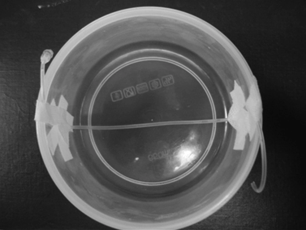
A round plastic container with a diameter of approximately 15 cm was taken as human tissue at the kidney level, in which a catheter with an interior diameter of 0.2 cm was installed as a simulated renal artery (Figure 1) and was imbedded with about 500 mL pork fat. A terminal of the catheter was connected to a high-pressure syringe (Stellant D, Medrad, Warrendale, PA, USA) for injecting saline with different flow velocity, and the other end was fixed in another 500 mL plastic container to gather the outflow saline. The catheter was placed along the longitudinal direction paralleled to the scanning bed in our research.
3.2. Hypertension Patients
This study was approved by the institutional review board and written informed consent was obtained from all participants prior to examination. Nine hypertensive male patients with similar height (165 - 170 cm), weight (60 - 65 Kg), and age (40 - 50 years) were selected from 93 consecutive hypertensive subjects diagnosed by a physician (systolic pressure ≥ 140 mmHg and diastolic pressure ≥ 90 mmHg), who were suspected of renal artery stenosis and SLEEK was carried out on them in our prior study (13).
3.3. MR Angiography
The vitro studies were performed with a superconductive 1.5T MRI scanner (EXCITE HD, GE Healthcare, Waukesha, WI, USA) using a dual channel phased-array knee coil. In SLEEK NCE-MRA, two orthogonal inversion bands were set to image the simulated vessel based during scanning on the in-flow effect. One 20 cm width vertical broad inversion band covered the whole container to invert all signals within the coil region to -Mz longitudinal magnetization. The other 10 cm width transversal inversion band was located on the end of the catheter connected with high-pressure syringe to bring the in-flow artery blood back to +Mz direction (2, 13). The diagrams of the SLEEK sequence and the signal acquisition are described in Figure 2.
Saline was injected into the catheter (simulated renal artery) by a high-pressure syringe with flow rates = 0.1, 0.2, 0.5, 1.0, 2.0, 3.0, and 4.0 mL/s. The corresponding simulated BFV was 3.2, 6.4, 16, 32, 64, 96, and 128 cm/s, respectively according to the formula: Simulated BFV (cm/s) = flow rate (mL/s)/πr2. Here πr2 was the cross-sectional area of the catheter, and r was the catheter radius. 1 mL = 1 cm3, π = 3.14 and r = 0.1 cm in our study. Four BSP TIs (200 ms, 500 ms, 800 ms, 1100 ms) were taken in our study. For each BSP TI, the SLEEK NCE-MRA was performed for the simulated renal arteries with different BFV. The specific image parameters included: Repetition time (TR) = 2.0 ms, echo time (TE) = 1.1 ms, Flip angle = 75, slice thickness = 0.8 mm, number of slices = 20, matrix = 128 × 128, field of view (FOV) = 20 cm × 16 cm, number of excitations (NEX) = 1, receiver bandwidth = ± 31.25 kHz. In addition, a false respiratory gating was triggered in whole SLEEK scanning. A SLEEK NCE-MRA with one BSP TI took approximately 40 - 50 seconds.
For hypertensive patients, SLEEK NCE-MRA with three various BSPTIs (800 ms, 1100 ms and 1400 ms) were carried out to display the renal artery. The scan parameters were identically as follows: TR = 3.9 ms, TE = 2.0 ms, slice thickness = 2 mm, matrix = 224 × 256, FOV = 38 cm × 30 cm, Flip angle = 75, NEX = 0.80, sense factor = 2, receiver bandwidth = ±125 kHz, respiratory interval = 1. A SLEEK NCE-MRA with one BSP TI was took about 3 - 4 minutes, the total SLEEK MR data acquisition time was approximately 10 - 12 minutes for each subject.
3.4. Image Analysis
The post-processing of all data of SLEEK including simulated vessel and renal artery in hypertensive patients were performed in an imaging workstation (Advantage Workstation 4.4, GE Healthcare, Buc, France). The main post-processing methods were multiple plane reconstruction and maximum intensity projection with the same thickness. The SLEEK images were assessed blindly and randomly.
For each simulated vessel, a region-of-interest (ROI: 8 - 12 mm2) was placed manually on the most hyper-intense point of the simulated renal artery to obtain signal intensity (SI), and the same ROI size was placed on the nearby background to gain standard deviation (SD) by two experienced radiologists in consensus. The ROIs were drawn three times in different days in a 1-week period and the mean SI and the mean SD were calculated. The signal-to-noise ratio (SNR) was calculated with the formula: SNR = SI/SD. For each BSP TI, the greatest SNR of catheter was decided in various BFVs with SLEEK scan.
For each hypertensive patient, the best image quality was determined by two experienced radiologists in consensus according to the following factors, including best renal artery’s SNR, homogeneous vessel signal intensity, sharp and complete delineation of vessel borders, and showing clearly segmental branches in the renal parenchyma. The optimal BSP TI was defined as BSP TI corresponding to the best image quality of the renal artery.
3.5. Statistical Analysis
The relationship between BSP TI and BFV was analyzed based on the best SNR by using Pearson’s correlation analysis. Statistical analysis was performed using commercially available software (SPSS for Windows, version 13.0; SPSS, Chicago, IL). P < 0.05 was considered to indicate a significant difference.
4. Results
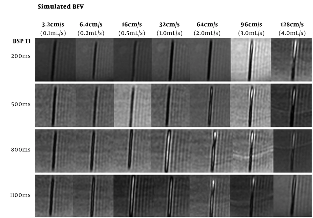
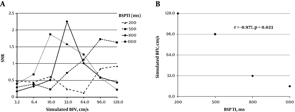
SLEEK NCE-MRA with various BSP TI was successfully performed and gained different SNR in the simulated renal artery with various BFVs. For SLEEK images with BSP TI = 200 ms, the simulated vessels appeared hyper-intense when BFVs were 96 cm/s and 128 cm/s, and the corresponding SNRs were 0.852 and 0.927, respectively. However, they presented hypo-intensity and SNRs were 0.476, 0.447, 0.616, 0.238, and 0.126, respectively when BFVs were 3.2 cm/s, 6.4 cm/s, 16 cm/s, 32 cm/s, and 64 cm/s. For BSP TI = 500 ms, the simulated vessels displayed high signal when BFVs were 64 cm/s, 96 cm/s, and 128 cm/s, the corresponding SNRs were 1.119, 1.732, and 1.633, respectively. In addition, the catheters presented low signal and SNRs were 0.296, 0.388, 0.232, and 0.725, respectively when BFVs were from 3.2 cm/s to 32 cm/s. For BSP TI = 800 ms, catheters showed hyper-intensity if BFVs were from 16 cm/s to 128 cm/s and the corresponding SNRs were 0.515, 2.256, 1.013, 0.592, and 0.442. On the other hand, catheters displayed hypo-intensity and SNRs were 0.178 and 0.327, respectively when BFVs were 3.2 cm/s and 6.4 cm/s. For SLEEK with BSP TI = 1100 ms, the simulated vessels presented high signal when BFVs were between 6.4 cm/s and 128 cm/s, the corresponding SNRs were 0.680, 1.875, 1.577, 1.274, 0.579, and 0.225, respectively. However, the only hypo-intensity presented and SNR was 0.401 when BFV was 3.2 cm/s (Figures 3 and 4A) (Table 1). A negative correlation between BFV and BSP TI was found in the in vitro study with SLEEK sequence (r = -0.977, P = 0.023) (Figure 4B).
The appearance of the imitated renal artery with different blood flow velocity (BFV) by spatial labeling with multiple inversion pulses (SLEEK) with various blood suppression inversion time (BSP TI). The maximum signal to noise ratio (SNR) was found when BFV = 128 cm/s for BSP TI = 200 ms, BFV = 96 cm/s for BSP TI = 500 ms, BFV = 32 cm/s for BSP TI = 800 ms, and BFV = 16 cm/s for BSP TI = 1100 ms. The BFV had a negative relation with BSP TI was implied.
A, The signal to noise ratio (SNR) of the simulated renal artery with different blood flow velocities (BFV) by spatial labeling with multiple inversion pulses (SLEEK) with various blood suppression inversion times (BSP TI); B, Finding of the relationship between BSP TI and BFV. In A, the greatest SNR was 0.927 when BFV = 128 cm/s for BSP TI = 200 ms, 1.732 when BFV = 96 cm/s for BSP TI = 500 ms, 2.256 when BFV = 32 cm/s for BSP TI = 800 ms, and 1.875 when BFV = 16 cm/s for BSP TI = 1100 ms. In B, based on the highest SNR for each BSPTI, a negative correction between BFV and BSP TI was found in SLEEK sequence (r = -0.977, P = 0.023).
| BSP TI | Simulated BFV | ||||||
|---|---|---|---|---|---|---|---|
| 3.2 cm/s | 6.4 cm/s | 16 cm/s | 32 cm/s | 64 cm/s | 96 cm/s | 128 cm/s | |
| 200 ms | 0.476 | 0.447 | 0.616 | 0.238 | 0.126 | 0.852 | 0.927 |
| 500 ms | 0.296 | 0.388 | 0.232 | 0.725 | 1.119 | 1.732 | 1.633 |
| 800 ms | 0.178 | 0.327 | 0.515 | 2.256 | 1.013 | 0.592 | 0.442 |
| 1100 ms | 0.401 | 0.680 | 1. 875 | 1. 577 | 1.274 | 0.579 | 0.225 |
Abbreviations: BFV, blood flow velocity; BSP TI, blood suppression inversion time; SLEEK, spatial labeling with multiple inversion pulses; SNR, signal-to-noise ratio.
a SNR was the mean value of the greatest SNR for three measurements in different time points. Simulated BFV was calculated with the formula: The simulated BFV (cm/s) = flow rate (mL/s)/πr2, 3.2 cm/s = 0.1 mL/s; 6.4 cm/s = 0.2 mL /s; 16 cm/s = 0.5 mL/s; 32 cm/s = 1.0 mL/s; 64 cm/s = 2.0 mL/s; 96 cm/s = 3.0 mL/s; 128 cm/s = 4.0 mL/s.
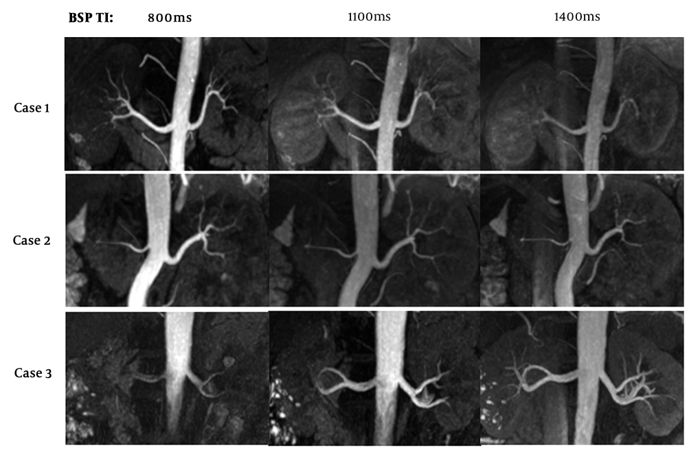
In nine hypertensive patients, all renal arteries could be displayed when BSP TI was 800 ms, 1100 ms, and 1400 ms with different SNRs, homogeneity, sharpness of vessel borders, and ability of delineating segmental branches in the renal parenchyma. An optimal BSP TI was found for each patient (optimal BSP TI was 800ms in two, 1100 ms in four, and 1400 ms in three patients) (Figure 5) (Table 2).
The optimal blood suppression inversion time (BSP TI) for best image quality of renal artery in spatial labeling with multiple inversion pulses (SLEEK) sequence in hypertension patients. In three hypertension male patients with similar height, weight and age, the best image quality was found when BSP TI was 800 ms in the first, 1100 ms in the second, and 1400 ms for the third patient, which suggested the optimal BSP TI was vital to improve the ability of presenting renal artery in SLEEK sequence.
| No. | Age (y) | Height (cm) | Weight (Kg) | The optimal BSP TI (ms) |
|---|---|---|---|---|
| 1 | 42 | 167 | 63 | 1400 |
| 2 | 49 | 166 | 61 | 1100 |
| 3 | 46 | 168 | 60 | 1100 |
| 4 | 40 | 166 | 65 | 800 |
| 5 | 44 | 168 | 63 | 1400 |
| 6 | 47 | 170 | 62 | 1400 |
| 7 | 41 | 170 | 60 | 800 |
| 8 | 47 | 167 | 63 | 1100 |
| 9 | 42 | 165 | 62 | 1100 |
Abbrevations: BSP TI, blood suppression inversion time; SLEEK, spatial labeling with multiple inversion pulses; SNR, signal-to-noise ratio.
a The optimal BSP TI (ms) was defined as the BSP TI with best renal artery image quality, which included best renal artery SNR, homogeneous vessel signal intensity, sharp and complete delineation of vessel borders, and showing clearly segmental branches in renal parenchyma.
5. Discussion
With development of MR technology and software, NCE-MRA has gradually obtained an excellent appearance for delineating large blood vessels, including intracranial arteries (14), thoracic aorta (15), renal artery (1, 2), and hepatic vessels (3, 16). In our study, the renal arteries were presented in coronal plane with SLEEK NCE-MRA sequence, which was a respiratory-triggered 3D fat saturation-fast imaging employing steady-state acquisition (FS-FIESTA) prepared with multiple spatial selective inversion recovery pulses. It is essential to boost the NCE-MRA image quality for those renal artery disease patients. Pei et al. (13) found that the heart and breathing rate can affect the presenting ability of the renal artery. Kurata et al. (12) deemed that patient age impacted optimal TI for the visualization of renal arteries. All the above pointed out that BSP TI may be related with the BFV.
In our results, a catheter was used to simulate the renal artery and carry out SLEEK with different BSP TIs and BFVs. To our knowledge, this is the first study that has evaluated the relationship between BFV and BSP TI in SLEEK in vitro. Meaningful results were found. First, the catheters appeared low signal when simulated BFV was 3.2 cm/s. The results illustrated that in SLEEK scan with any BSP TI, the simulated vessel presented hypo-intensity when BFV was so low that the simulated inflow blood could not arrive at the target vessel level. Second, for a given BSP TI, the simulated vessel with various BFVs can show hyper-intensity with SLEEK NCE-MRA. For example, the catheters presented high signal from BFV = 6.4 cm/s to 128 cm/s for BSP TI = 1100 ms. It pointed out that the simulated renal arteries with different BFVs could be shown by SLEEK in various SNRs, which can affect the homogeneity of vessel signal intensity and sharpness of renal artery borders. Third, the highest SNR was found when BFV was 128 cm/s for BSP TI = 200 ms, BFV was 96 cm/s for BSP TI = 500 ms, BFV was 32 cm/s for BSP TI = 800 ms, and 16 cm/s for 1100 ms. It suggested that the BFV has a negative relationship with BSP TI. The reason was that a fast BFV means shortened time of inflow blood from the heart to the renal artery level, which needs a short echo time to match for gaining a high signal in the renal artery. That means the optimal BSP TI must increase for obtaining the best SNR when BFV decreases. So, adopting an optimal BSP TI can improve the ability of clearly displaying the renal artery.
In nine hypertensive patients, the distances between heart and renal artery level were almost identical due to their similar height, weight and ages. However, the optimal BSP TI was different for each subject, which hinted that the labeled blood’s arriving time was different from heart to renal artery level due to various BFVs. For those subjects with BSP TI = 1400 ms, their BVF should be the lowest, and vice versa. Therefore, the choice of optimal BSP TI could be determined by the patients’ BFV in SLEEK for distinctly displaying the renal artery, and even evaluating renal artery diseases especially renal artery branch disease in the renal parenchyma. We recommend choosing optimal BSP TI based on BFV to avoid the repeat SLEEK scan with different BSP TIs and to improve work efficiency.
There were several limitations in our study. First, a 0.2 cm diameter catheter was used to simulate the renal artery, which is smaller and less stretched than the real renal artery. Second, the simulated device was not complex enough to represent the vessel in the individual’s body. In conclusion, BFV has a negative relationship with BSP TI in SLEEK NCE-MRA. It is very vital to choose the optimal BSP TI based on BFV for improving the ability of delineating renal artery and to assess renal artery disease, which can enhance the diagnostic confidence and make a further treatment plan for patients.


