Full Text
Full text is available in PDF
An Innovative Journal in the Field of Radiology
Authors
Recently, association between the length of abdominal esophagus and increased risk for gastro-esophageal reflux disease has been hypothesized.
The aim of the present study was to provide sonographic measurements of the abdominal esophagus length in premature neonates with and without GERD and to investigate its diagnostic value to assess GERD in comparison with clinical diagnosis of disease.
In a cross-sectional study, 75 consecutive premature neonates aged less than 30 days with birth weights of less than 2000 grams hospitalized in NICU ward of the Mahdieh hospital in Tehran in 2013 were included in the study. The certain diagnosis of GERD was based on clinical manifestations. The presence of GERD and also measurement of the abdominal esophageal length was assessed by portable sonography using SIUI sonography device.
Clinically, reflux was diagnosed in 15 neonates (20.0%). It was also diagnosed in 20 cases (26.7%) by sonography assessment yielding a sensitivity of 86.7%, a specificity of 88.3%, a positive predictive value of 65.0%, a negative predictive value of 96.4%, and an accuracy of 88% for this diagnostic device. The mean of the length of abdominal esophagus was 15.19 + 4.10 mm (ranging from 8 to 27 mm). There was a strong positive association of the length of abdominal esophagus with neonatal birth weight (r = 0.553, P < 0.001), and also with gestational age (r = 0.491, P < 0.001). Furthermore, those neonates with underlying jaundice and those with sepsis had significantly shorter esophagus than those with RDS. Those with clinical diagnosis of reflux had significantly shorter abdominal esophagus than neonates without reflux (11.47 + 1.36 mm versus 16.12 + 4.03 mm, P < 0.001). In a multivariate linear regression model, shorter abdominal esophagus was shown to be related to the presence of reflux (beta = 2.764, SE = 1.068, P = 0.012).
Shorter abdominal esophagus in premature neonates is associated with increased risk for GERD that is more highlighted in those neonates with lower birth weight and lower gestational age. Sonography has a high value for assessment of both GERD and also anatomical state of abdominal esophagus in premature neonates.
© 2014, Author(s). This open-access article is available under the Creative Commons Attribution 4.0 (CC BY 4.0) International License (https://creativecommons.org/licenses/by/4.0/), which allows for unrestricted use, distribution, and reproduction in any medium, provided that the original work is properly cited.
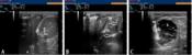
Chen J, Liu L, Huang C, Dai Y. An Investigation of Ultrasonography as a Method for Determining Orogastric Tube Placement in Newborns. Inn J Pediatr. 2024;34(2):e143559. doi: https://doi.org/10.5812/ijp-143559
Mehdizadeh M. Diagnosis of Peptic Esophagitis in Children; Ultrasound versus Endoscopy . I J Radiol. 2003;1(1&2):e79273. doi:
Fathi M, Bagheri B, Razavi S, Mahdavi A, Salarian S. Duration of Intubation After Gastric Pull up Operation in Young Children. Arch Pediatr Infect Dis. 2015;3(3):e24165. doi: https://doi.org/10.5812/pedinfect.24165v2
Karamimagham S, Poursadeghfard M, Hemmati F. Normal Reference Range of Lateral Ventricle Parameters in Preterm Neonates by Ultrasonography. Shiraz E-Med J. 2017;18(8):e57569. doi: https://doi.org/10.5812/semj.57569
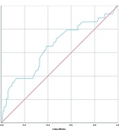
Fallahi M, Babaei G, Kazemian S, Tajalli S, Noripour S, et al. Predictors of Early Mortality in Esophageal Atresia: A 12-year Cohort Study in Tehran, Iran. Inn J Pediatr. 2022;32(6):e129266. doi: https://doi.org/10.5812/ijp-129266
Last Update: 3 weeks ago
Last Update: 3 weeks ago
Last Update: 3 weeks ago
Last Update: 6 days ago