

1. Background
Because of the rapid development of medical technology and computers, computed tomography (CT) tests play an important role in diagnostic imaging. In clinical practice, CT scanners are used for the rapid treatment of emergency patients and multiple-trauma patients. CT scans have been used to diagnose and treat unstable patients with multiple injuries (1).
Optimization efforts are needed for active radiological protection in abdominal CT examinations in which organs with a high-radiation tissue-weighting factor (WT) are gathered to reduce radiation exposure. Optimization techniques include tube-current modulation techniques using selected reference images, tube-current modulation along the z-axis of a patient, or tube-current modulation according to an angle due to elliptical cross-section of the human body. Abdominal CT has been applied in clinical practice through tube-current modulation techniques. However, abdominal CT using a tube-current modulation technique may result in artifacts due to changes in the position of the patient’s arms, resulting in poor image quality and excessive radiation exposure. To solve these problems, the patient’s arm is positioned mainly on the head or outside the examination area. However, when the position of the patient’s arm is not in the normal position, the problem of deterioration in image quality arises and it is difficult to diagnose the artifact due to the linear hardening phenomenon and photon shortage in the CT image.
Analysis of the existing quantitative images for CT image evaluation involves the evaluation of the image using noise, contrast noise ratio (CNR), signal-to-noise ratio (SNR ), and coefficient of variation (COV) of the image (2, 3). In the analysis of CT images according to the position of the arm, images were quantitatively analyzed by using noise, CNR, and SNR (4). In an emergency CT scan, trauma patients cannot lift their arms over their heads due to trauma injuries. The position under the arm has been reported to reduce the diagnostic value of artifacts due to hardening of the spine in the liver, spleen, and back of the kidney (4). In addition, the location of the upper arms with the chest has been reported to degrade image quality due to beam hardening artifacts in the liver, spleen, and kidney (5, 6).
2. Objectives
The purpose of this study is to evaluate the effect of arm position on the images in an anthropomorphic CT phantom. We propose an optimization method to acquire high-quality images in CT examination by comparing the radiation dose and image according to the change in arm position and the use of the tube-current modulation method in CT examination.
3. Materials and Methods
3.1. In vitro Study and RPL Measurement
We scanned an anthropomorphic phantom (PBU-60, Kyoto Kagaku Co., Ltd., Kyoto, Japan) with a body weight of 50 kg and a height of 165 cm by using a CT system (Brilliance CT 64- channel, Philips, The Netherlands) (7). The phantom was used to evaluate the radiation dose and image according to the position of the arm. The scan parameters applied to the CT scan are given in Table 1. The following scan parameters were kept identical for all anthropomorphic phantom data acquisitions in multidetector computed tomography (MDCT) examinations: tube voltage 120 kV; reference tube current-time product of 188 mAs, using attenuation-based tube-current modulation [z-axis thickness modulation (Z-DOM); Philips]; pitch 0.89; slice collimation 64 mm × 0.625 mm, gantry rotation time 0.75 seconds; slice thickness, 3 mm; reconstruction increment, 3 mm.
| Parameters | Description or value |
|---|---|
| Tube voltage, kV | 120 |
| Tube-current modulation | Z-DOM |
| Slice thickness, mm | 3 |
| Beam collimation, mm | 64 × 0.625 |
| Image matrix (rows and columns) | 512 × 512 |
| Pitch | 0.89 |
| Field of view, mm | 350.0 |
| Rotation time, s | 0.75 |
| Table feed, mm/s | 47.5 |
| Slice increment, mm | 3.00 |
| Filter type | Y-sharp (YA) |
| Convolution kernel | Y-sharp (YA) |
Abbreviation: Z-DOM, z-axis thickness modulation
The radiophotoluminescent (RPL) glass dosimetry system (ATGC, Asahi Techno Glass Corporation, Tokyo, Japan) is an activated phosphate glass plate. In this measurement study, the RPL dosimeter was investigated by using a 137Cs source, and the dose correction factor of the RPL was determined by comparing the RPL dose of the measured dose with the standard exposure of 60Co gamma radiation. The results for energy dependence have shown that only GD-352M (Element Holder: ATGC, Asahi Techno Glass Corporation, Tokyo, Japan) is suitable for use in medical exposures. The RPL dose measurement system is a Dose Ace FGD-1000 (ATGC, Asahi Techno Glass Corporation, Tokyo, Japan) used for measuring the radiation dose of DGD-1000 (ATGC, Asahi Techno Glass Corporation, Tokyo, Japan).
3.2. Image Quality Analysis
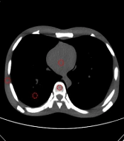
To analyze the CT images, digital imaging and communications in medicine (DICOM) image-processing software (ImageJ Version 1.43u; National Institutes of Health, Bethesda, MD, USA) was used to measure the various pixel values of the images. The mean Hounsfield unit (HU), standard deviation (SD) values and COV were analyzed by measuring the pixel values of anthropomorphic phantom heart, chest, lung, and bone by setting the same size region of interest (ROI) of 1 cm2 using ImageJ’s ROI manage (Figure 1). The radiological technologist who carried out the research drew the ROI directly from the anthropomorphic phantom image. The examiner measured the 4 portions (heart, chest, lung, and bone) of each 3 times on each of the anthropomorphic phantom.
 position in the heart, chest, axilla, lung, and bone region. Both arms raised position (A) and z-axis thickness modulation (Z-DOM) dose modulation (B) and arms down of CT image (C).")
Examples of region of interest (ROI) position in the heart, chest, axilla, lung, and bone region. Both arms raised position (A) and z-axis thickness modulation (Z-DOM) dose modulation (B) and arms down of CT image (C).
We calculated the SD of the HU of the CT representing the noise in the image of the anthropomorphic phantom. The COV was then estimated as the ratio of SD (σ) to the mean value (µ). The mean of the CT HUs of all anthropomorphic phantoms is the SD of the measured SD (σ) values. The COV was calculated by using Equation 1 to evaluate whether the CT image noise was more uniform when the automatic exposure control (AEC) (Z-DOM) system was deactivated by comparison.
3.3. CTDIvol (Computed Tomography Dose Index) and Radiation Dose Measurement
Volume CT dose index (CTDIvol) is an index that can measure the irradiation dose per tissue section without depending on the scan length. The dose length product (DLP), which is the total amount of irradiation dose, can be calculated through CTDIvol. DLP is useful in assessing the effective dose of the entire area, which the patient is receiving. The dose information of the DICOM file was analyzed to evaluate the CTDIvol in the scan image of the anthropomorphic phantom. The CT dose was calculated by multiplying the DLP provided by the scanner console with the conversion factor (k ≤ 0.017 mSv mGy-1 cm-1) (8, 9).
3.4. Statistical Analysis
Statistica analyses were performed by SPSS for windows ver. 20 (IBM Corp. Released 2011. IBM SPSS Statistics for Windows, version20.0. Armonk, NY). An unpaired Student’s t-test was used to compare the continuous values such as CTDIvol, DLP between both arms and raised in the anthropomorphic phantom. Comparisons between eye, thyroid, breast and humerus for four radiation dose datasets was performed using one-way analysis of variance (ANOVA) and compared to the 95% confidence interval for the HU of the standardized CT, the difference was analyzed. Post analysis was performed without assuming the same distribution using the Dunnett’s post-hoc test. Statistical significance was considered when the P value was less than 0.05.
4. Results
The noise in the anthropomorphic phantom CT image was analyzed according to the change in the position of upper arms and Z-DOM application. Standard CT scanning parameters for the abdomen were used to perform image quality measurements by evaluating the HU of the CT, SD of the noise, and COV of the available ROIs in the scanner. The dose of CTDI and DLP according to arm position increased with a CTDIvol of 7 mGy when the arm was raised and 116 mGy when it was lowered. The DLP was 246.61 mGy·cm and the effective dose was 4.4 mSv, when the arm was raised, and when the hand was down the corresponding values were 436.39 mGy·cm and 7.4 mSv, respectively, higher than when the hand was raised (Table 2). We found a significant radiation dose between both arms raised and down (P < 0.05).
| Both arms raised | Both arms down | P value | |
|---|---|---|---|
| CTDIvol, mGy | 7 | 11.6 | 0.000 |
| DLP, mGy·cm | 264.61 | 436.39 | 0.000 |
Abbreviations: CTDIvol, volume CT dose index; DLP, dose length product.
The radiation dose in the CT scans using an anthropomorphic phantom with the arm raised measured by using a glass dosimeter is shown in Table 3. Radiation dose in different anatomical locations based on the arm position with thyroid gland were 16.185 - 22.775 µGy, breast were 15.545 - 18.005 µGy and humerus position calculated were 862 - 1.428 µGy.
| No. | Region | Position | Both arms down | Both arms raised |
|---|---|---|---|---|
| 1 | Eye | Left | 856 | 421 |
| 2 | 1.022 | 538 | ||
| 3 | Right | 791 | 445 | |
| 4 | 768 | 643 | ||
| 5 | Thyroid | Left | 21.595 | 20.895 |
| 6 | 24.455 | 16.185 | ||
| 7 | Right | 20.855 | 18.455 | |
| 8 | 24.685 | 22.775 | ||
| 9 | Breast | Left | 18.855 | 15.545 |
| 10 | 21.045 | 17.425 | ||
| 11 | Right | 19.365 | 17.555 | |
| 12 | 20.765 | 18.005 | ||
| 13 | Humerus | Left | 12.405 | 862 |
| 14 | 12.455 | 1.060 | ||
| 15 | Right | 9.197 | 1.235 | |
| 16 | 10.535 | 1.428 |
The dose comparison when the arm is raised and lowered is shown in Figure 2. The changes in tube currents in the Z-DOM are shown in Figure 3; the tube current in the shoulder part increased, that in the lung decreased, and that in the abdomen increased.

Radiation dose according to scan length for anthropomorphic phantom position with both arms in down and raised position

Radiation dose of scan length for anthropomorphic phantom position with both arms raised according to X-ray tube current
When the anthropomorphic phantom was raised, the artifact was not present (Figure 4A), but when the upper arms were lowered, the artifact was present (Figure 4B). The peak signal-to-noise-ratio (PSNR) was measured as 13.24 dB in the ImageJ program with the fixed tube current technique (Figure 5A) and Z-DOM (Figure 5B) for AEC application. The PSNR in the CT scan of the anthropomorphic phantom calculated by using AEC was 38.21 dB (Table 4).
| SNR | PSNR, dB | RMSE | MAE | |
|---|---|---|---|---|
| Both arms raised vs. both arms down | 12.59 | 13.24 | 202.85 | 71.10 |
| Z-DOM vs. fixed tube current | 37.56 | 38.21 | 11.44 | 7.04 |
Abbreviations: db, decibel; MAE: mean absolute error; PSNR, peak signal-to-noise ratio; RMSE, root mean square error; SNR, signal-to-noise ratio; Z-DOM, z-axis thickness modulation
 and down (B) manipulated multidetector CT scans obtained in anthropomorphic phantom imaged with both arms alongside the torso")
Both arms raised position (A) and down (B) manipulated multidetector CT scans obtained in anthropomorphic phantom imaged with both arms alongside the torso
 and z-axis thickness modulation (Z-DOM) dose modulation of CT image (B)")
Original of both arms raised position (A) and z-axis thickness modulation (Z-DOM) dose modulation of CT image (B)
There was no significant difference in the mean, SD, and COV of the heart, chest, lung, and bone using the Z-DOM and fixed tube current method in the anthropomorphic phantom images (P > 0.05). However, there was a significant difference (P < 0.05) when the arm was lowered and CT scan was performed (Table 5). Noise in the image increased when the arm was raised to a height and the dose increased in the thyroid and upper chest.
| Parameter/position | Mean ± SD | Minimum | Maximum | COV, % |
|---|---|---|---|---|
| Both arms raised (Z-DOM) | ||||
| Heart | -6.14 ± 8.39 | -25 | 16 | -136.64 |
| Chest | -20 ± 9.3 | -45 | 3 | -46.5 |
| Lung | -993.68 ± 5.48 | -1008 | -980 | -0.55 |
| Bone | 341.92 ± 148.91 | 215 | 8610 | 43.55 |
| Both arms raised (fixed tube current) | ||||
| Heart | -2.58 ± 4.81 | -19 | 9 | -186.43 |
| Chest | -21 ± 9.4 | -44 | -4 | -44.76 |
| Lung | -994.24 ± 4.14 | -1005 | -983 | -0.41 |
| Bone | 393.98 ± 151.17 | 18 | 810 | 38.36 |
| Both arms down | ||||
| Heart | -13.73 ± 51.82 | -132 | 55 | -377.42 |
| Chest | -13.9 ± 22.96 | -62 | 27 | -165.17 |
| Lung | -995.66 ± 10.52 | -1019 | -967 | -1.05 |
| Bone | 342.5 ± 163.87 | -5 | 885 | 47.84 |
Abbreviations: AEC, Automatic exposure control; COV, Coefficient of variation; SD, Standard deviation; Z-DOM, z-axis thickness modulation
5. Discussion
The purpose of this study was to compare the radiological dose and image quality of the arms with the anthropomorphic phantom and glass dosimeter. Whole-body MDCT testing can provide accurate and quick results for the diagnosis and treatment of traumatic injuries. In general, additional abdominal injury can be prevented by bringing the two arms to the side of the body rather than lifting them above the head (10) before performing abdominal CT. The disadvantage of positioning the arm with the patient’s torso is that the image quality of the posterior part of the liver and spleen of the abdominal organs is reduced (6, 11, 12).
According to the results of this study, when the arm was not lifted above the head, the image quality with of the liver and spleen was significantly better than side-by-side with the body when the arm was placed on the chest because there was no beam hardening artifact.
The number of patients suffering from multiple injuries is increasing, as is the importance of CT examinations (13, 14). A major disadvantage of CT scan is that the patient’s radiation exposure is three to five times higher than that in conventional radiology. On the other hand, systemic CT has been reported to make clinically relevant diagnosis much more frequently than general CT, and the use of whole-body CT has been shown to increase survival probability after multiple injuries (15). For these reasons, there is a continuing effort to perform CT screening faster and at a lower dose.
In our study, a CTDIvol was 7 mGy when the arm was raised and 116 mGy when the hand was lowered. When the arm was raised DLP was 246.61 mGy·cm and the effective dose was 4.4 mSv. When the hand was lowered DLP was 436.39 mGy·cm and the effective dose was 7.4 mSv, which was higher than when the hands were raised.
In the study of existing effective doses, the effective dose in this study was included in 5 - 7 mSv in the chest CT, and 7.4 mSv in 8 mSv to 15 mSv in the abdomen. The amount of radiation was determined (16). Brink et al. (5) suggested using a tool to optimize the arm of the trauma patient in a CT scan, compared to increasing the arm above the 8 mSv head when the arm was lowered.
In the case of using the Z-DOM in the measurement of the radiation dose with the glass dosimeter, the tube current increased in the shoulder part when the hand was raised, decreased in the lung, and increased in the abdomen. When the upper body of the anthropomorphic phantom was raised, there was no artifact, but when the upper body was lowered, an artifact occurred. This is consistent with the results of a study in which patients were placed on the upper arm and the dose was decreased and the quality of the image was higher than that of the lower position (4, 10). PSNR was measured as 13.24 dB in the fixed-tube current technique and Z-DOM image comparison for AEC application. AEC was applied to the A PSNR of 38.21 dB was calculated for the Z-DOM application in the CT scan of the anthropomorphic phantom. This means that higher PSNR values provide better picture quality, better quality at 40 dB, slightly lower image quality at 30 dB, and increased noise at 20 dB. A noise increase of 10 dB continues, and 0 dB image for strong noise intensity (17).
The Philips CT AEC system applied to the study consists of three parts. Patient-based AEC, D-DOM according to angle provision, and longitudinal Z-DOM (18, 19). In this study, the dose was calculated by applying Z-DOM. There was no significant difference in the mean, SD, and COV of the heart, chest, lung, and bone using Z-DOM and fixed tube current technique (P > 0.05) (P < 0.05). Previous studies have shown no significant difference in imaging and dose reduction has been reported by the AEC technique (20). When the arm was raised, the noise of the anthropomorphic phantom image increased, and the dose increased at the thyroid and chest.
Our study did not extend to the neck and lower abdomen in the chest and abdominal examinations of anthropomorphic phantoms. However, there is a growing need for additional studies on the lower abdomen. In the future, there is an increasing need for studies involving clinical patients with scan range, various tube-current settings, AEC application, and trauma patients (1).
The limitations of the study are as follows: First, an anthropomorphic phantom was used and the study was not applied to real patient. Second, the AEC was applied to the anthropomorphic phantom and the arm was not scanned. Third, a variety of MDCT models and AEC applications are needed. Fourth, the quality and dose of images are measured in the lung and abdomen area. Finally, the dose was measured with a glass dosimeter. However, various measurements and further studies are needed to measure the dose of the surface without applying the internal dose.
In conclusion, the position of the upper arms directly affects the image in the MDCT imaging of the anthropomorphic phantom. Therefore, placing the position of the arm above the head can improve the image quality by reducing artifacts and radiation dose. The results of this study could be applied in clinic using this optimal method as a method to achieve high image quality and low noise in MDCT of trauma patients.
