Primary thymic carcinoma accounts for 14.1% of thymic epithelial tumors. It has more than 10 subtypes (
8). Although there are some differences according to race and geography, squamous cell carcinoma is the most common, accounting for about 61.8% - 84.07% and thymic adenocarcinoma is relatively rare, accounting for 1.6% - 3.98% of all cases of primary thymic carcinomas (
1,
2,
8). According to the World Health Organization (WHO) classification, thymic adenocarcinoma can be divided into four subtypes: papillary adenocarcinoma, thymic carcinoma with adenoid cystic carcinoma-like features, mucinous adenocarcinoma, and not otherwise specified (NOS) (
9). Mucinous adenocarcinoma is a rare subtype. It was first reported by Choi et al. (
10) in 2003. To date, 27 cases of mucinous adenocarcinoma have been reported in the English literature.
Patients with primary thymic mucinous adenocarcinoma most commonly complain of respiratory symptoms such as coughing and dyspnea. Sometimes, they have chest pain or dysphagia (
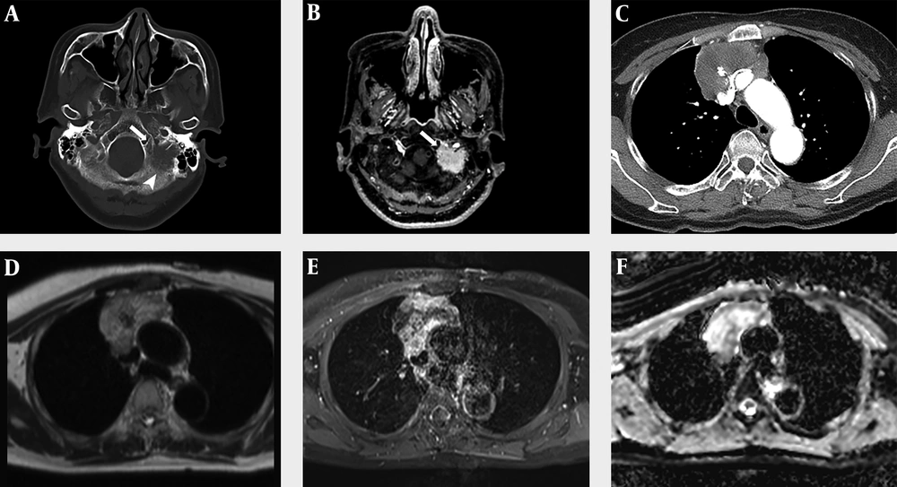
7). However, symptoms of patients with primary thymic mucinous adenocarcinoma are often nonspecific. For this reason, it is difficult to diagnose primary thymic mucinous adenocarcinoma based on clinical findings. Radiologic imaging findings are helpful for its diagnosis. Our patient had no respiratory symptoms but showed dysarthria, and a physical examination revealed the left-sided deviation of the tongue. This was thought to be the result of the metastatic mass in the occipital bone and petrous apex invading the left hypoglossal canal.
Up to date, there were very few radiologic reviews of primary thymic mucinous adenocarcinoma. Most cases of primary thymic mucinous adenocarcinoma are reported in pathology journals. CT figures and descriptions were found in 11 of 27 cases reported in the English literature (
3-
7,
11-
16). We analyzed a total of 12 cases of CT images, including the previous 11 cases and our case. Information that could not be identified with a given CT image such as tumor size or component was analyzed based on the description shown in the literature. We classified CT images by tumor composition (solid predominancy: > 50% of solid portion; cystic predominancy: > 50% of cystic portion), enhancement pattern (homogeneous or heterogeneous), presence of calcification, and adjacent invasion (present or not).
For a total of 12 cases, their mean age was 59 years (range, 28 - 79 years). The male-to-female ratio was 1: 2 (4 men and 8 women). The average maximal diameter of tumor was 9.2 cm (range, 5.6 - 14.5 cm). Eight (67%) of these 12 cases showed solid predominancy while four (33%) cases showed cystic predominancy. All (100%) tumors had a heterogeneous enhancement pattern. No calcification was observed in seven (58%) cases. Calcification was observed in five (42%) cases. Adjacent tissue invasion was observed in seven (58%) cases at the time of diagnosis. It was not observed in five (42%) cases. The association between thymic mucinous adenocarcinoma and thymic cyst has been observed in several reports (
4-
6,
9,
10,
17-
19). Thymic cyst was identified in six (50%) of 12 cases. Clinical and radiologic findings of these 12 cases of primary thymic mucinous adenocarcinoma are shown in
Table 1.
| Case | Age/sex | Size, cm | CT findings | Prognosis |
|---|
| Composition | Enhancement | Calcification | Thymic cyst | Adjacent invasion |
|---|
| 1) Takahashi et al. (11) | 59/M | > 10 | Solid | Heterogeneous | - | - | - | DOD after 11 mon |
| 2) Ra et al. (3) | 82/F | 14.5 | Cystic | Heterogeneous | - | + | + (aortic arch) | Passed away from complications of surgery |
| 3) Seki et al. (12) | 49/M | 8.5 | Solid | Heterogeneous | + | - | + (pericardium, left innominate vein) | Recurrence (pleural dissemination) AWD for 11 mon |
| 4) Maeda et al. (4) | 52/F | 7.5 | Solid | Heterogeneous | - | + | + (sternum) | Recurrence (lung, cervical LN) AWD for 11 mon |
| 5) Seon et al. (13) | 66/F | 12.6 | Cystic | Heterogeneous | + | + | - | AWD for 60 mon |
| 6) Abdul-Ghafar et al. (5) | 36/F | 5.6 | Cystic | Heterogeneous | - | + | - | Recurrence (lung, pleural dissemination) DOD after 15 mon |
| 7) Maghbool et al. (6) | 28/F | - | Solid | Heterogeneous | - | + | + (pericardium) | AWD for 30 mon |
| 8) Lee et al. (14) | 31/M | 11.9 | Solid | Heterogeneous | + | - | - | |
| 9) Sakanoue et al. (15) | 39/F | 6.5 | Solid | Heterogeneous | - | + | + (pericardium, left innominate vein) | |
| 10) Kinoshita et al. (7) | 79/F | 7 | Cystic | Heterogeneous | + | - | - | AWD for 14 mon |
| 11) Liu et al. (16) | 50/M | 10 | Solid | Heterogeneous | - | - | + (superior vena cava) | DOD after 3.5 mon |
| 12) Current case | 60/F | 6.9 | Solid | Heterogeneous | + | - | + (superior vena cava, left innominate vein) | AWD for 15 mon |
Abbreviations: AWD, alive with the disease; DOD, dead because of the disease; F, female; M, male; mon, months.
MRI findings were rarely described in previous literature. In our patients, there was an intermediate SI in the T1-weighted image and a heterogeneous high SI in the T2-weighted image, which were similar to SI of the mucinous component of mucinous adenocarcinoma occurring in the abdomen or pelvic cavity. This correlated with the low attenuation area observed in CT. However, this finding is not specific for mucin. So more research is needed on the findings of MRI images, including image-pathology correlation.
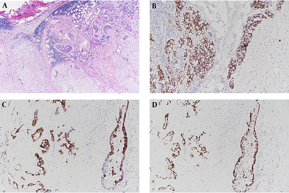
Histologically, primary thymic mucinous adenocarcinoma has malignant tumor cells in nests, tubules, and cribriform structures floated in pools of extracellular mucin (
4). Immunohistochemical staining is important for determining the origin of the tumor and for distinguishing it from thymic mucinous adenocarcinoma and other thymic carcinomas. CD5 is a widely known thymic origin marker. About 70% of thymic carcinomas are positive for CD5 (
11). CK7, CK20, and CDX-2 are useful markers for distinguishing mucinous and other subtypes of thymic carcinoma (
5). However, they cannot distinguish between primary and metastatic lesions. They are also positive in tumors of pulmonary, gastrointestinal tract, and ovary origins. MUC (Mucin) family proteins are evaluated as markers for thymic mucinous adenocarcinoma in limited studies. In addition, markers such as TTF-1 can be used. TTF-1 is a primary pulmonary adenocarcinoma-specific marker. It is negative in thymic tumors. Our case showed positive staining for CDX2, CK7, CK20, and MUC2, indicating mucinous type of tumor. Our case showed negative staining for TTF-1, meaning that the tumor was not of pulmonary origin.
Primary thymic mucinous adenocarcinoma has a poor prognosis, similar to thymic carcinoma. Although there is no absolute guideline for treating primary thymic mucinous adenocarcinoma, surgical resection is usually performed in the absence of adjacent tissue infiltration or insignificant adjacent tissue infiltration without distant metastasis. Although it has been reported that patients without adjacent tissue invasion can survive more than 5 years after surgical resection, many patients require additional chemotherapy and radiotherapy after surgery due to infiltration into adjacent tissues at the time of diagnosis (
7). In case of severe infiltration into adjacent tissues and metastatic lesions, palliative chemotherapy is used. With incomplete resection or palliative treatment, the prognosis is known to be very poor (
7).
In conclusion, although primary thymic mucinous adenocarcinoma is a rare disease, it should be considered if a large mass with a heterogeneous enhancement pattern is accompanied by adjacent tissue involvement, associated thymic cyst, or calcification. Histologically abundant extracellular mucins and immunohistochemical stains are important for its diagnosis. The prognosis differs depending on whether complete resection is possible. In general, the prognosis is poor. Therefore, early diagnosis is important. Further studies on radiology findings including MRI findings are needed.