1. Background
Among various cancers, lung cancer (early malignant pulmonary nodules) has the highest morbidity and mortality rates in Iran and other countries (1), with nearly 600,000 new cases around the world annually (2, 3). Chest computed tomography (CT) scan, as a non-invasive tool which yields high-resolution cross-sectional images without interference from the adjacent structures and detects foci hidden on X-ray films, is used as a routine screening approach to detect early lung cancer. Due to the use of this modality, lung cancer mortality has reduced by 20% (3, 4) Overall, contrast-enhanced chest CT scan can clearly represent the blood supply and small lesions. The iodine concentration in pulmonary nodules directly reflects the blood supply to nodules (5), which can be helpful for the clinical diagnosis and timely treatment of lung cancer (6).
Although great scientific advances have been made in human healthcare, contrast-enhanced chest CT scan faces challenges in radiation dose and iodine contrast safety. According to previous studies, due to the increased number of CT scans, the risk of ionizing radiation-related cancer has increased from 0.4% to 1.5 - 2.0% in the United States (7). Besides, application of a high concentration of contrast medium causes various problems, such as contrast agent nephropathy, which is the third major cause of hospital-acquired renal failure with an incidence of about 11% (8). Therefore, research has focused on reducing X-ray radiation dose and contrast agent concentration while maintaining the diagnostic image quality.
The radiation dose is proportional to the square of tube voltage. A reduction in the tube voltage can effectively decrease the radiation dose received by the patient. In this regard, Gang et al. (9) used a tube voltage of 100 kVp in coronary CT angiography and reported a 50% decline in the radiation dose compared to the 120-kVp group. It is known that the filtering back projection (FBP) reconstruction algorithm provides high-quality, normal-dose CT images; however, its ability to reduce the radiation dose is limited by the loss of image quality when the tube voltage decreases. With technological advances, iterative reconstruction (IR) has gradually assumed essential roles in CT imaging. As a new-generation IR algorithm, the multi-model adaptive statistical iterative reconstruction (ASIR, GE Healthcare, USA) yields high-quality images via iterative filtering in the original data space and suppressing noise following backward projection. Consequently, ASIR can be a suitable option for low-tube-voltage CT scanning by reducing the radiation dose and maintaining the image quality.
Some studies have focused on double-low (i.e., a low tube voltage and a low iodine dose) CT scan protocols. In a previous study (10), ASIR was employed in double low-dose pulmonary artery CT imaging to diagnose pulmonary embolism. Chen et al. also applied the IR algorithm from another vendor for contrast-enhanced chest CT imaging and acquired acceptable CT images using the double-low technique (11). However, further research is needed to explore the potential of ASIR in low-dose contrast-enhanced chest CT imaging for the detection of early malignant pulmonary nodules at a low tube voltage and a low concentration of contrast agent.
2. Objectives
This study aimed to explore the value of ASIR at a low tube voltage (100 kVp) and a low concentration of isotonic contrast agent (270 mgI/L of iodixanol) in contrast-enhanced chest CT imaging by assessing the image quality, radiation dose, and iodixanol content.
3. Patients and Methods
3.1. Patients
This prospective study was carried out according to the Declaration of Helsinki (revised in 2013) and approved by Wuhan Third Hospital Ethics Committee (Wuhan Third Hospital Ethics Approval No.: 2015-008). All patients signed an informed consent form before the examinations. This study was performed on 80 patients diagnosed with nodules or masses, who required contrast-enhanced chest CT imaging. Forty patients were allocated to the experimental group (using a conventional-dose hypertonic contrast medium) and 40 patients to the comparison group (using a low-dose isotonic contrast medium).
Similar to previous research (12, 13), the imaging data comprised of retrospective and prospective information. The imaging data of the experimental group were retrospectively selected from the hospital database before June 1, 2015, and the imaging data of patients in the comparison group were collected between February 2018 and October 2020. The inclusion criteria were as follows: (1) 18 kg/m2 < body mass index (BMI) < 26 kg/m2; (2) stable breathing; and (3) ability to hold breathing. On the other hand, the exclusion criteria were as follows: (1) iodine allergy; (2) metformin use; (3) history of thyroid disease; (4) a poor general condition and inability to coordinate breathing; (5) renal dysfunction; and (6) pregnancy.
3.2. Equipment and Methods
A 64-row CT scanner (Discovery CT 750 HD, GE Healthcare, Waukesha, WI, USA) was used in this study. All 80 patients were placed in the supine position with hands up and were trained to breathe in and hold their breath before scanning. The scanning field extended from the tip of the lung to below the diaphragm. The scanning and reconstruction parameters were set as follows: Layer thickness, 5 mm; interlayer spacing, 5 mm; pitch, 1.375; tube rotation speed, 0.7 s/r; auto mA with a tube current of 80 - 450 mA; matrix, 512 × 512; and reconstructed layer thickness, 1.25 mm.
For the experimental group, a tube voltage of 120 kVp and FBP were applied, while a tube voltage of 100 kVp and 60% FBP + 40% ASIR 40% (14, 15) were utilized for the comparison group. The images were reconstructed on the control computer. A non-ionic contrast agent, that is, iodixanol (270 mgI/mL), was used for the comparison group, while iohexol (350 mgI/mL) was used for the experimental group. The contrast agent (80 mL) and normal saline (30 mL) were subsequently injected through the median cubital vein at a flow rate of 3 mL/s. The contrast-enhanced chest CT mode was adopted, and scanning was initiated 30 seconds after drug injection.
3.3. Image Quality Analysis
The reconstructed images were transmitted to a GE Advanced Workstation 4.6, and chest CT scans were evaluated at a fixed window width and window level: Lung window image (window width, 1200 Hounsfield unite (HU); window position, -700 HU) and mediastinal window image (window width, 350 HU; window position, 40 HU).
The subjective evaluation of images was based on the CT image quality-related scoring criteria, recommended by the European Radiology guidelines (16). Two radiologists with more than five years of experience, who were blinded to the groups, read the images using the same window width and window level in the same range. In the lung window images, the following structures were evaluated: (1) lung texture; (2) bronchus; (3) proximal bronchus and the adjacent vessels; (4) peripheral bronchus and the adjacent vessels; and (5) clarity of the margins of lung lesions. In the mediastinal window images, the following structures were evaluated: (1) trachea and the surrounding soft tissue at the aortic arch level; (2) hilar protuberances and the surrounding lymph nodes; (3) three levels of the thoracic segment of the esophagus; (4) pericardium at the level of the right ventricle; and (5) chest wall and muscle tissue.
Each of the abovementioned structures was scored according to the following five-point visual scoring method (14): 5 (excellent): Clear lesions, a complete and clear tissue structure, good contrast, and no artifacts; 4 (good): Clear lesions, a complete tissue structure, good contrast, and slight artifacts; 3 (moderate): Clear lesions, partially good tissue structures, moderate contrast, and slight artifacts; 2 (insufficient): Fuzzy lesions and tissue structure, insufficient contrast, and moderate artifacts; 1 (poor): Unclear lesions and tissue structure and heavy artifacts. In case of disagreement, consensus was reached through consultation and discussion. The average score of the abovementioned 10 structures was calculated, and the integer was rounded as the subjective score of overall image quality. Therefore, a score of 3 - 5 meant acceptable quality for diagnosis, while a score of 1 - 2 indicated the quality did not meet the requirement for diagnosis.
The objective evaluation of images was carried out according to the CT image quality criteria, recommended by the European Radiology guidelines (16). The CT values and standard deviations (SD) of the pulmonary trunk, left pulmonary artery, right pulmonary artery, and thoracic paravertebral erector spinae after enhancement were measured. The contrast-to-noise ratio (CNR) of vessels was also calculated as follows:
The CTvessel indicated mean CT value on vessels and was calculated based on the CT values of the arcusaortae, right pulmonary artery, and left pulmonary artery. The CTmuscle was the mean CT value after enhancement of the erector spinae muscle on both sides of the thoracic spine. SD pointed to image noise which was defined as the mean SD of ROI measured at the abovementioned five locations; the ROI needed to be as close as possible to the vessel size (17).
3.4. Assessment of Radiation Dose and Total Iodine Content
After scanning, the volume CT dose index (CTDIvol, mGy) and CT dose-length product (DLP, mGy.cm) were recorded for each patient. The CT effective radiation dose (ED, mSV) was also calculated, using the chest ED calculation formula (18):
Where K represents the conversion factor, and the mean chest value was 0.014 according to the standard European CT quality guidelines. The total iodine content for each patient was calculated as follows: Total iodine (g) = [Contrast agent concentration (mgI/L) × Contrast agent volume (mL)]/1000
3.5. Statistical Analysis
SPSS version 22.0 (IBM SPSS Statistics for Windows, released in 2013, IBM Corp., Armonk, NY, USA) was used for statistical analysis. The normal distribution of all continuous data was evaluated using Kolmogorov-Smirnov test. Continuous data with a normal distribution are expressed as mean ± SD, and categorical data are shown as number and percentage. The sex ratio was compared using chi-square test. For the objective parameters (CT values, SD, CNR) and radiation dose (CTDIvol, DLP and ED), two-sample independent t-test was used to compare the groups. The total iodine dose and subjective scores were compared using non-parametric Mann-Whitney U test. The level of statistical significance was set at P < 0.05.
4. Results
4.1. Population
The characteristics of the participants were analyzed in this study. In the comparison group, there were 21 males (52.5%) and 19 females (47.5%), with a mean age of 62.88 ± 12.10 years (age range, 34 - 86 years) and a mean BMI of 22.66 ± 1.25 kg/m2. In the experimental group, there were 22 males (55%) and 18 females (45%), with a mean age of 63.25 ± 11.04 years (age range, 36 - 85 years) and a mean BMI of 23.02 ± 1.01 kg/m2. The two groups were not significantly different in terms of sex, age, and BMI (P > 0.05 for all).
4.2. Subjective Evaluation of Images
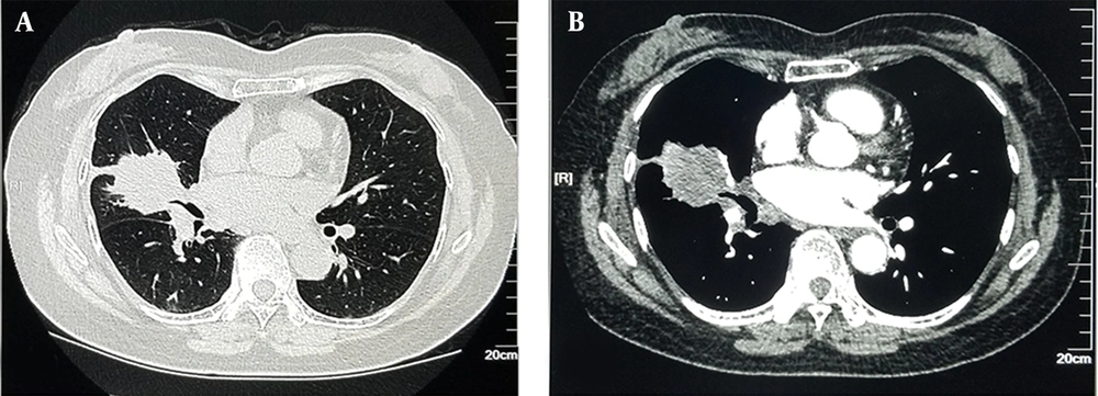
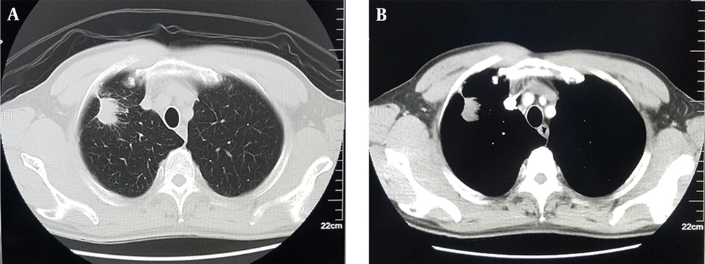
The subjective findings are summarized in Table 1. For all 10 structures and the overall image, the scores of the two groups were diagnostic (≥ 3 points). For the structures observed in the lung window (i.e., lung texture, bronchus, proximal bronchus and adjacent vessels, peripheral bronchus and adjacent vessels, and clarity of the margins of lung lesions) and anatomies detected in the mediastinal window (trachea and surrounding soft tissue at the aortic arch level, hilar protuberances and surrounding lymph nodes, three levels of the thoracic segment of the esophagus, pericardium at the level of the right ventricle, chest wall, and muscle tissue), the subjective scores were not significantly different between the experimental and comparison groups (P > 0.05 for all). Also, the two groups were not significantly different regarding the overall subjective image quality (P > 0.05). Figures 1 and 2 represent the contrast-enhanced chest CT images acquired based on different scan and reconstruction protocols. The comparison group exhibited excellent image quality similar to the experimental group with clear lesions and no artifacts.
| Variables | Experimental group (120 kVp) | Comparison group (100 kVp) | Z | P | ||||||||
|---|---|---|---|---|---|---|---|---|---|---|---|---|
| 5 | 4 | 3 | 2 | 1 | 5 | 4 | 3 | 2 | 1 | |||
| Lung window | ||||||||||||
| Lung texture | 32 | 6 | 2 | 0 | 0 | 35 | 3 | 2 | 0 | 0 | -0.855 | 0.508 |
| Bronchus | 31 | 7 | 2 | 0 | 0 | 35 | 3 | 2 | 0 | 0 | -1.107 | 0.355 |
| Proximal bronchus and adjacent vessels | 34 | 6 | 0 | 0 | 0 | 35 | 2 | 3 | 0 | 0 | -0.177 | 1.000 |
| Peripheral bronchus and adjacent vessels | 32 | 7 | 1 | 0 | 0 | 34 | 3 | 3 | 0 | 0 | -0.452 | 0.764 |
| Clarity of the margins of lung lesions | 37 | 7 | 1 | 0 | 0 | 34 | 4 | 2 | 0 | 0 | -0.51 | 0.748 |
| Mediastinal window | ||||||||||||
| Trachea and surrounding soft tissue at the aortic arch level | 32 | 7 | 1 | 0 | 0 | 34 | 5 | 1 | 0 | 0 | -0.569 | 0.705 |
| Hilar protuberances and surrounding lymph nodes | 32 | 6 | 2 | 0 | 0 | 34 | 3 | 3 | 0 | 0 | -0.495 | 0.745 |
| Three levels of the thoracic segment of the esophagus | 31 | 5 | 4 | 0 | 0 | 34 | 4 | 2 | 0 | 0 | -0.892 | 0.424 |
| Pericardium at the level of the right ventricle | 33 | 6 | 1 | 0 | 0 | 34 | 4 | 2 | 0 | 0 | -0.24 | 0.968 |
| Chest wall and muscle tissue | 32 | 6 | 2 | 0 | 0 | 34 | 4 | 2 | 0 | 0 | -0.553 | 0.701 |
| Overall | 32 | 7 | 1 | 0 | 0 | 34 | 4 | 2 | 0 | 0 | -0.51 | 0.748 |
A 61-year-old female patient with a body mass index (BMI) of 24.3 kg/m2. Enhanced chest CT scan (100 kVp, 270 mgI/mL of contrast agent, 40% ASIR) scored five points in the subjective evaluation of image quality. A, Lung window: The lesions showed lumpiness and high density with rough edges and multiple short burrs; B, Mediastinal window: Heterogeneous enhancement (ASIR, adaptive statistical iterative reconstruction).
A 66-year-old male patient with a body mass index (BMI) of 23.7 kg/m2. The enhanced chest CT scan (120 kVp, 350 mgI/mL of contrast agent, FBP) scored five points in the subjective evaluation of image quality. A, Lung window: The lung window clearly shows the edge of the nodule lesion which is not smooth, along with the burr sign and the pleural depression sign; B, Mediastinal window: The lesion is significantly enhanced with a pleural depression sign (FBP, filter back projection).
4.3. Objective Evaluation of Images
There were no significant differences in the mean CT values of enhanced tissues (muscles and vessels) between the two groups (P > 0.05 for all). There were also no significant differences in terms of noise and CNR of the images between the groups (P > 0.05 for all) (Table 2).
| Parameters | Comparison group (100 kVp) | Experimental group (120 kVp) | t | P |
|---|---|---|---|---|
| CTmuscle (HU) | 45.72 ± 2.38 | 45.55 ± 2.36 | -0.318 | 0.751 |
| CTvessel (HU) | 314.90 ± 23.42 | 308.93 ± 21.40 | -1.191 | 0.237 |
| SD (HU) | 13.03 ± 0.88 | 12.83 ± 0.90 | -1.039 | 0.302 |
| CNR | 20.74 ± 2.22 | 20.60 ± 1.94 | -0.306 | 0.760 |
Abbreviations: CNR, contrast-to-noise ratio; CTmuscle, mean CT value of the muscle; CTvessel, mean CT value for vessels; SD, mean standard deviation of muscles and vessels, image noise; HU, Hounsfield unite.
4.4. Radiation Dose and Total Iodine Content
As shown in Table 3, the mean CTDIvoI (5.84 ± 1.76 vs. 8.06 ± 2.49 mGy; P < 0.05), the mean DLP (199.08 ± 57.84 vs. 314.24 ± 69.99 mGy/cm; P < 0.05), and the average ED (2.79 ± 0.81 vs. 4.40 ± 0.98 mSV; P < 0.05) in the comparison group were lower by 27.58%, 36.65%, and 36.59%, respectively compared to the experimental group, with significant differences between the groups (P < 0.05). The total iodine content was 21.6 ± 0.0 g in the comparison group and 28 ± 0.0 g in the experimental group; the iodine content in the comparison group was lower than that of the experimental group (Z = -0.8888, P < 0.001), and the total iodine content reduced by 22.86% in comparison group.
| Parameters | Comparison group (100 kVp) | Experimental group (120 kVp) | t | P |
|---|---|---|---|---|
| CTDIvol (mGy) | 5.84 ± 1.76 | 8.06 ± 2.49 | 4.609 | < 0.001 |
| DLP (mGy.cm) | 199.08 ± 57.84 | 314.24 ± 69.99 | 8.022 | < 0.001 |
| ED (mSV) | 2.79 ± 0.81 | 4.40 ± 0.98 | 8.028 | < 0.001 |
Abbreviations: CTDIvoI (mGy), volumetric CT dose index; DLP (mGy.cm), CT dose-length product; ED (mSV), effective dose.
5. Discussion
In the present study conducted on 80 patients receiving contrast-enhanced chest CT scan with different radiation and contrast doses, the application of ASIR technology could significantly reduce the effective radiation dose and total iodine content, while maintaining the diagnostic image quality. As lung cancer accounts for the highest morbidity and mortality rates among different cancers (4), contrast-enhanced chest CT scan has been widely applied in follow-up visits as an effective diagnostic tool, as patients often undergo multiple CT scans. For this population, the risk of ionizing radiation-related cancer and contrast-induced nephropathy increases (19-21). In this study, the effective radiation dose reduced by 36.59%, and the total iodine content decreased by 22.86%, which could be beneficial.
Overall, reduction of tube voltage can significantly decrease the radiation dose of patients. However, the main consequence of reducing the tube voltage in FBP-constructed images is an increase in image noise and a decrease in density and spatial resolution, affecting the imaging of low-contrast tissue lesions. In a previous study, the FBP reconstructed image quality of the brain and liver was significantly decreased when decreasing tube voltage (22). Except for the change of tube voltage, efforts have been made to modulate the tube current and pitch to reduce the radiation dose. Considering the tube current, Naidich et al. (23) first proposed the concept of low-dose CT scan in 1990. In this technique, when other scanning parameters remain unchanged, a lower tube current can meet the diagnostic requirements, because the radiation dose is linear with the tube current, and the radiation dose decreases accordingly (24).
However, reduction of tube current has limited effects on the radiation dose, and the influence of tube current reduction on the signal-to-noise ratio is significant. Based on the findings, an increase in pitch can effectively shorten the scanning time and reduce the radiation dose. However, an increase in pitch increases the effective layer thickness of the reconstructed image and diminishes the spatial resolution in the z-axis direction, resulting in the generation of artifacts. The present study investigated the feasibility of using a low-concentration isotonic contrast agent, a low tube voltage (100 kVp), and the ASIR technique in contrast-enhanced chest CT scan (lung nodules/masses).
Besides the image quality, enhancement is an important index for diagnosis. Accordingly, contrast enhancement was evaluated by measuring the CT value of pulmonary arteries. The results indicated that CT attenuation of pulmonary arteries was not significantly different between the images of the two groups (314.90 ± 23.42 HU in the comparison group vs. 308.93 ± 21.40 HU in the experimental group; P > 0.05) (Table 2); therefore, the scanning and injection protocols did not influence contrast enhancement. In this study, the radiation dose and total iodine content reduced, while the high quality of images with low noise was maintained, leading to a balance between strong diagnostic confidence and proper patient care.
In this study, the benefits of reducing the radiation dose and iodine content while maintaining the image quality of double-low scans were derived from the ASIR algorithm. Generally, the FBP reconstruction algorithm provides normal-dose high-quality CT images, whereas its capability to reduce dose radiation is limited by the loss of image quality when the tube voltage decreases. Compared to the traditional FBP reconstruction technology, ASIR (25, 26) is used in the original data space to carry out operations using a precise noise model and introduce statistical iterative information to carry out repeated corrections and obtain high-quality images.
In a study by Manousaki et al. (27), it was feasible to reduce the radiation dose and ensure the image quality by lowering the tube voltage, whereas Hara et al. (28) found that ASIR effectively reduced the radiation dose compared to the conventional FBP reconstruction. When the radiation dose is reduced, the images contain higher noise levels; ASIR can maintain excellent image quality, even under a low tube voltage (29). The dose advantage is achieved by reducing the image noise; therefore, the scanning dose can be significantly reduced while maintaining the same noise level and image quality. Overall, ASIR provides a good solution for low-tube-voltage CT scanning by reducing the radiation dose and maintaining the image quality (30).
Additionally, Xuemei et al. (10) applied the ASIR iterative algorithm and explored the feasibility of double-low dose pulmonary artery imaging. Chen et al. (11) also applied an IR algorithm from another vendor for enhanced chest CT imaging and obtained acceptable CT images using the double-low technique; their results are consistent with the present findings. There are several studies comparing the image quality and radiation dose between low-kVp CTA examinations and FBP and iterative algorithms (31). They found that a lower kVp led to a lower radiation dose; however, when using FBP, low-kVp images had high noise levels and lower subjective scores, while the application of iterative algorithms led to improved diagnostic image quality. Besides, in double-low scanning (9), images with different reconstruction algorithms had different qualities, and iterative reconstructed images were superior to FBP images. Therefore, the iterative algorithm can greatly reduce the image noise and compensate for the reduction of image quality caused by reduced X-ray dose and contrast concentration, which is not satisfactory in FBP.
This study had some limitations. First, differences in the BMI distribution were not controlled, and the effects of BMI on the results need to be further explored. Second, a fixed, but not customized contrast injection protocol was applied in this study, without considerations for BMI. Third, regarding uncontrolled lesion types, patients should be carefully selected, and contrast enhancement on lesions should be further evaluated. Fourth, the impact of ASIR on the differentiation of benign and malignant thoracic mass lesions should be further explored. Fifth, in this study, a single blending factor of ASIR was used, while different blending factors need to be further studied. Sixth, an advanced generation of adaptive statistical iterative reconstruction (ASIR-V) was not applied in this study, and further exploration should be conducted with ASIR-V, where lower radiation and iodine doses might be achieved compared to ASIR. Finally, this study was not a randomized trial.
In conclusion, based on the present findings, the use of ASIR maintained the diagnostic CT image quality, while it significantly reduced the effective radiation dose by 36.59% and the total iodine content by 22.86% compared to FBP in contrast-enhanced chest CT scans for lung cancer detection.


